

Karyomegalic interstitial nephritis as a rare cause of kidney graft dysfunction: case report and review of literature
- PMID: 37208636
- PMCID: PMC10199555
- DOI: 10.1186/s12882-023-03185-3
Karyomegalic interstitial nephritis as a rare cause of kidney graft dysfunction: case report and review of literature
Abstract
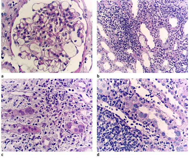
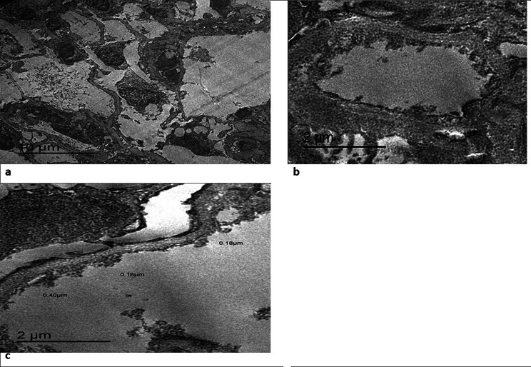
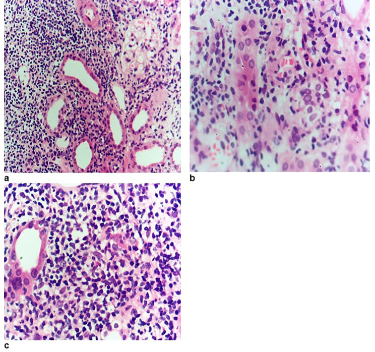
Karyomegalic interstitial nephritis (KIN) is a rare cause of chronic interstitial nephritis characterized by enlarged renal tubular epithelial nuclei. The first case of KIN reported in a kidney graft was in 2019. Here, we report the first case of KIN in 2 brothers receiving kidneys from 2 different unrelated living donors. A male kidney transplant recipient with focal segmental glomerulosclerosis as the original kidney disease presented with graft impairment and proteinuria, and graft biopsy revealed KIN. This patient had a brother who was also a kidney transplant recipient and had one episode of graft impairment and was diagnosed with KIN as well.
Keywords: Interstitial nephritis; Karyomegalic interstitial nephritis; Kidney transplantation.
© 2023. The Author(s).
Conflict of interest statement
No competing interests
Figures

References
-
- Monga G, Banfi G, Salvadore M, Amatruda O, Bozzola C, Mazzucco G. Karyomegalic interstitial nephritis: report of 3 new cases and review of the literature. Clin Nephrol. 2006;65(5). - PubMed
-
- Mihatsch M, Gudat F, Zollinger H, Heierli C, Thölen H, Reutter F. Systemic karyomegaly associated with chronic interstitial nephritis. A new disease entity? Clin Nephrol. 1979;12(2):54–62. - PubMed
Publication types
MeSH terms
LinkOut - more resources
Full Text Sources
Medical
