. 2023 May 19;20(1):117.
doi: 10.1186/s12974-023-02782-1.

Dysbiosis of gut microbiota inhibits NMNAT2 to promote neurobehavioral deficits and oxidative stress response in the 6-OHDA-lesioned rat model of Parkinson's disease

Affiliations

Dysbiosis of gut microbiota inhibits NMNAT2 to promote neurobehavioral deficits and oxidative stress response in the 6-OHDA-lesioned rat model of Parkinson's disease

Jianjun Yu et al. J Neuroinflammation. .

Abstract

Background: New data are accumulating on gut microbial dysbiosis in Parkinson's disease (PD), while the specific mechanism remains uncharacterized. This study aims to investigate the potential role and pathophysiological mechanism of dysbiosis of gut microbiota in 6-hydroxydopamine (6-OHDA)-induced PD rat models.

Methods: The shotgun metagenome sequencing data of fecal samples from PD patients and healthy individuals were obtained from the Sequence Read Archive (SRA) database. The diversity, abundance, and functional composition of gut microbiota were further analyzed in these data. After the exploration of the functional pathway-related genes, KEGG and GEO databases were used to obtain PD-related microarray datasets for differential expression analysis. Finally, in vivo experiments were performed to confirm the roles of fecal microbiota transplantation (FMT) and upregulated NMNAT2 in neurobehavioral symptoms and oxidative stress response in 6-OHDA-lesioned rats.

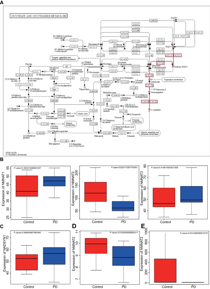
Results: Significant differences were found in the diversity, abundance, and functional composition of gut microbiota between PD patients and healthy individuals. Dysbiosis of gut microbiota could regulate NAD+ anabolic pathway to affect the occurrence and development of PD. As a NAD+ anabolic pathway-related gene, NMNAT2 was poorly expressed in the brain tissues of PD patients. More importantly, FMT or overexpression of NMNAT2 alleviated neurobehavioral deficits and reduced oxidative stress in 6-OHDA-lesioned rats.

Conclusions: Taken together, we demonstrated that dysbiosis of gut microbiota suppressed NMNAT2 expression, thus exacerbating neurobehavioral deficits and oxidative stress response in 6-OHDA-lesioned rats, which could be rescued by FMT or NMNAT2 restoration.

Keywords: Dysbiosis of gut microbiota; Fecal microbiota transplantation; NMNAT2; Neurobehavioral symptoms; Oxidative stress response; Parkinson’s disease.


Conflict of interest statement

The authors declare no competing interests.

Figures

Fig. 1
Fig. 1
Comparison of species diversity of gut microbiota between PD patients and healthy individuals. A Alpha diversity analysis of gut microbiota in samples from PD patients (n = 31) and healthy individuals (n = 28). B Beta diversity analysis of gut microbiota in samples from PD patients (n = 31) and healthy individuals (n = 28). C Quantitative analysis of relative abundance at the phylum level with the group as the horizontal axis. Different colors represent the gut microbiota of different phyla. D Quantitative analysis of the relative abundance of the main samples in PD patients (n = 31) and healthy individuals (n = 28). Different colors represent the gut microbiota of different phyla. *p < 0.05
Fig. 2
Fig. 2
Difference in gut microbiota composition between PD patients and healthy individuals. A Branch plot showing the classification of species abundance of gut microbiota in PD patients (n = 31) and healthy individuals (n = 28). The circle radiating from inside to outside represents the classification level from phyla to genus, and the diameter represents the size of relative abundance. The yellow nodes indicate species without significant differences, the red nodes indicate species with higher abundance in healthy individuals, and the blue nodes indicate species with higher abundance in PD patients. B Histogram showing LDA distribution of species abundance of gut microbiota in PD patients (n = 31) and healthy individuals (n = 28)
Fig. 3
Fig. 3
Functional analysis of the gut microbiota in PD patients and healthy individuals. A Functional analysis of gut microbiota in PD patients (n = 31) and healthy individuals (n = 28). B Difference in superpathway of arginine and polyamine biosynthesis in the samples from PD patients (n = 31) and healthy individuals (n = 28). C Difference in NAD biosynthesis I (from aspartate) in the samples from PD patients (n = 31) and healthy individuals (n = 28). D Difference in the abundance of Alistipes shahii and Coprococcus in the samples from PD patients (n = 31) and healthy individuals (n = 28). * p < 0.05
Fig. 4
Fig. 4
Expression of NADSYN1 and NMNAT in different regions in PD patients. A Nicotinic acid and nicotinamide metabolic pathways in KEGG database (map00760, NAD+ anabolic pathway in red, M00115). B Differential mRNA expression of NMNAT1, NMNAT2, and NMNAT3 in PD patients (n = 16) and healthy individuals (n = 9) in microarray dataset GSE7621. C Differential expression of NADSYN1 mRNA in PD patients (n = 16) and healthy individuals (n = 9) in microarray dataset GSE7621. D Differential expression of NMNAT2 mRNA in PD patients (n = 14) and healthy individuals (n = 15) in microarray dataset GSE20168. E Differential expression of NMNAT2 mRNA of in PD patients (n = 15) and healthy individuals (n = 20) in microarray dataset GSE20291
Fig. 5
Fig. 5
Effects of FMT on neurobehavioral symptoms and oxidative stress response in 6-OHDA-lesioned rats. PD rat models were induced by 6-OHDA, followed by treatment with FMT (n = 8). A Expression of NMNAT2 in brain tissues of 6-OHDA-lesioned rats detected by ELISA. B Number of rotations of 6-OHDA-lesioned rats measured by apomorphine-induced rotation test. C Total distance of movement of 6-OHDA-lesioned rats measured by open field test. D Average speed of movement of 6-OHDA-lesioned rats measured by open field test. E Latency period of 6-OHDA-lesioned rats falling from the rotating rod measured by rotation test. F Dopaminergic neurons in the SN of 6-OHDA-lesioned rats detected by IHC. G NMNAT2 protein expression in the SN of 6-OHDA-lesioned rats detected by IHC. H MDA content in the brain tissues of 6-OHDA-lesioned rats. I GSH content in the brain tissues of 6-OHDA-lesioned rats. J GSH-Px activity in the brain tissues of 6-OHDA-lesioned rats. K SOD activity in the brain tissues of 6-OHDA-lesioned rats. *p < 0.05; **p < 0.01; ***p < 0.001
Fig. 6
Fig. 6
Effects of upregulation of NMNAT2 on neurobehavioral symptoms and oxidative stress response in 6-OHDA-lesioned rats. PD rat models were induced by 6-OHDA, followed by treatment with oe-NMNAT2, FMT or sh-NMNAT2 + FMT (n = 8). A Expression of NMNAT2 in brain tissues of 6-OHDA-lesioned rats detected by ELISA. B Number of rotations of 6-OHDA-lesioned rats measured by apomorphine-induced rotation test. C Total distance of movement of 6-OHDA-lesioned rats measured by open field test. D Average speed of movement of 6-OHDA-lesioned rats measured by open field test. E Latency period of 6-OHDA-lesioned rats falling from the rotating rod measured by rotation test. F Dopaminergic neurons in the SN of 6-OHDA-lesioned rats detected by IHC. G NMNAT2 protein expression in the SN of 6-OHDA-lesioned rats detected by IHC. H MDA content in the brain tissues of 6-OHDA-lesioned rats. I GSH content in the brain tissues of 6-OHDA-lesioned rats. J GSH-Px activity in the brain tissues of 6-OHDA-lesioned rats. K SOD activity in the brain tissues of 6-OHDA-lesioned rats. *p < 0.05; **p < 0.01; ***p < 0.001
Fig. 7
Fig. 7
Schematic illustration of the mechanisms of dysbiosis of gut microbiota in PD. Dysbiosis of gut microbiota aggravates neurobehavioral symptoms and oxidative stress responses in 6-OHDA-lesioned PD rat models via regulation of NMNAT2, which is a key gene of the NAD+ anabolic pathway

References

    1. Lin CH, Chen CC, Chiang HL, Liou JM, Chang CM, Lu TP, et al. Altered gut microbiota and inflammatory cytokine responses in patients with Parkinson’s disease. J Neuroinflammation. 2019;16(1):129. doi: 10.1186/s12974-019-1528-y. - DOI - PMC - PubMed
    1. Blauwendraat C, Nalls MA, Singleton AB. The genetic architecture of Parkinson’s disease. Lancet Neurol. 2020;19(2):170–178. doi: 10.1016/S1474-4422(19)30287-X. - DOI - PMC - PubMed
    1. Tolosa E, Garrido A, Scholz SW, Poewe W. Challenges in the diagnosis of Parkinson’s disease. Lancet Neurol. 2021;20(5):385–397. doi: 10.1016/S1474-4422(21)00030-2. - DOI - PMC - PubMed
    1. Vijiaratnam N, Simuni T, Bandmann O, Morris HR, Foltynie T. Progress towards therapies for disease modification in Parkinson’s disease. Lancet Neurol. 2021;20(7):559–572. doi: 10.1016/S1474-4422(21)00061-2. - DOI - PubMed
    1. Pajares M, Rojo AI, Manda G, Bosca L, Cuadrado A. Inflammation in Parkinson’s disease: mechanisms and therapeutic implications. Cells. 2020;9(7):1687. doi: 10.3390/cells9071687. - DOI - PMC - PubMed