

Sex differences in the effects of high fat diet on underlying neuropathology in a mouse model of VCID
- PMID: 37208759
- PMCID: PMC10199629
- DOI: 10.1186/s13293-023-00513-y
Sex differences in the effects of high fat diet on underlying neuropathology in a mouse model of VCID
Abstract
Background: Damage to the cerebral vasculature can lead to vascular contributions to cognitive impairment and dementia (VCID). A reduction in blood flow to the brain leads to neuropathology, including neuroinflammation and white matter lesions that are a hallmark of VCID. Mid-life metabolic disease (obesity, prediabetes, or diabetes) is a risk factor for VCID which may be sex-dependent (female bias).
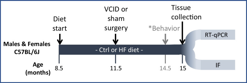
Methods: We compared the effects of mid-life metabolic disease between males and females in a chronic cerebral hypoperfusion mouse model of VCID. C57BL/6J mice were fed a control or high fat (HF) diet starting at ~ 8.5 months of age. Three months after diet initiation, sham or unilateral carotid artery occlusion surgery (VCID model) was performed. Three months later, mice underwent behavior testing and brains were collected to assess pathology.
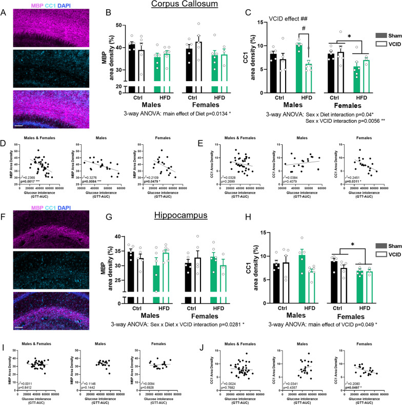
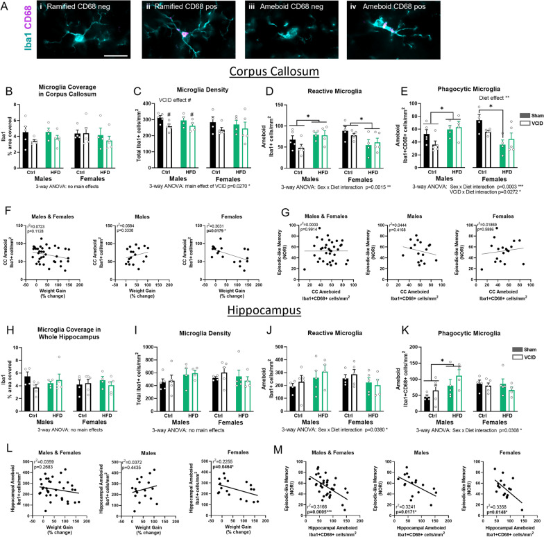
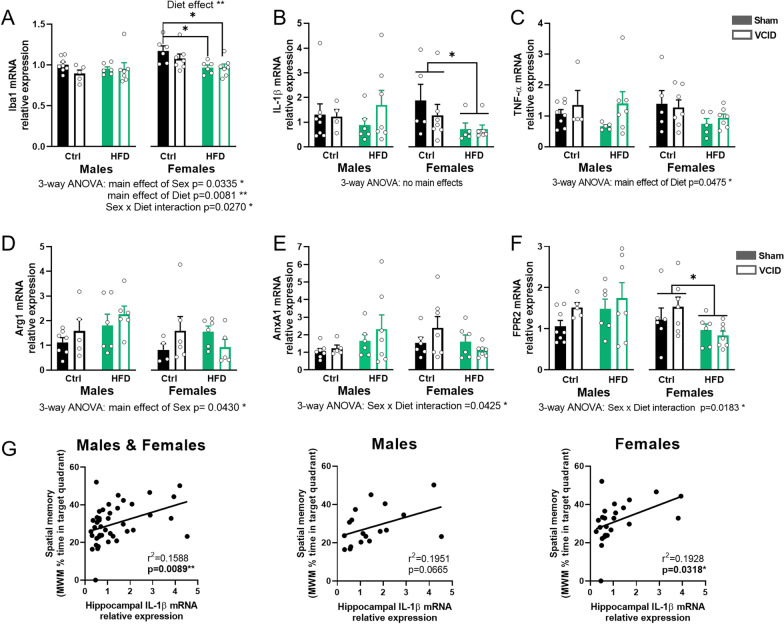
Results: We have previously shown that in this VCID model, HF diet causes greater metabolic impairment and a wider array of cognitive deficits in females compared to males. Here, we report on sex differences in the underlying neuropathology, specifically white matter changes and neuroinflammation in several areas of the brain. White matter was negatively impacted by VCID in males and HF diet in females, with greater metabolic impairment correlating with less myelin markers in females only. High fat diet led to an increase in microglia activation in males but not in females. Further, HF diet led to a decrease in proinflammatory cytokines and pro-resolving mediator mRNA expression in females but not males.
Conclusions: The current study adds to our understanding of sex differences in underlying neuropathology of VCID in the presence of a common risk factor (obesity/prediabetes). This information is crucial for the development of effective, sex-specific therapeutic interventions for VCID.
Keywords: Diet-induced obesity; High fat diet; Neuroinflammation; Prediabetes; Sex; Vascular contributions to cognitive impairment and dementia; White matter.
Plain language summary
Reduced blood flow to the brain resulting from damaged blood vessels can lead to vascular dementia. Neuroinflammation and white matter damage are characteristics of vascular dementia. Middle-age is a time when obesity and prediabetes can increase risk for vascular dementia. This increase in risk is greater for women. A high fat diet causes obesity and prediabetes in mice. We compared the effects of diet-induced obesity in middle-age between males and females in a mouse model of vascular dementia. We have previously shown that a high fat diet causes greater obesity and prediabetes and a wider array of learning and memory problems in females compared to males. Here, we report on sex differences in the damage to the brain. White matter was negatively impacted by vascular dementia in males and high fat diet in females, with more severe prediabetes correlating with less white matter markers in females only. High fat diet led to an increase in activation of microglia (immune cells in the brain) in males but not in females. High fat diet also led to a decrease in pro-inflammatory and pro-resolving mediators expression in females but not males. The current study adds to our understanding of sex differences in underlying damage to the brain caused by vascular dementia in the presence of common risk factors (obesity and prediabetes). This information is needed for the development of effective, sex-specific treatments for vascular dementia.
© 2023. The Author(s).
Conflict of interest statement
The authors have no conflicts to disclose.
Figures
References
-
- Gorelick PB, Scuteri A, Black SE, Decarli C, Greenberg SM, Iadecola C, et al. Vascular contributions to cognitive impairment and dementia: a statement for healthcare professionals from the American heart association/American stroke association. Stroke. 2011;42(9):2672–2713. doi: 10.1161/STR.0b013e3182299496. - DOI - PMC - PubMed
Publication types
MeSH terms
Grants and funding
LinkOut - more resources
Full Text Sources
Medical
Research Materials
Miscellaneous
