. 2023 May 30;42(5):112524.
doi: 10.1016/j.celrep.2023.112524. Epub 2023 May 18.

Structural conservation of Lassa virus glycoproteins and recognition by neutralizing antibodies

Affiliations

Structural conservation of Lassa virus glycoproteins and recognition by neutralizing antibodies

Hailee R Perrett et al. Cell Rep. .

Abstract

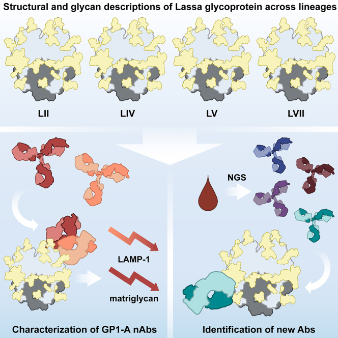
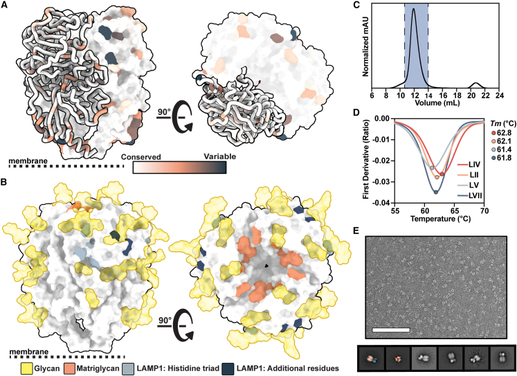
Lassa fever is an acute hemorrhagic fever caused by the zoonotic Lassa virus (LASV). The LASV glycoprotein complex (GPC) mediates viral entry and is the sole target for neutralizing antibodies. Immunogen design is complicated by the metastable nature of recombinant GPCs and the antigenic differences among phylogenetically distinct LASV lineages. Despite the sequence diversity of the GPC, structures of most lineages are lacking. We present the development and characterization of prefusion-stabilized, trimeric GPCs of LASV lineages II, V, and VII, revealing structural conservation despite sequence diversity. High-resolution structures and biophysical characterization of the GPC in complex with GP1-A-specific antibodies suggest their neutralization mechanisms. Finally, we present the isolation and characterization of a trimer-preferring neutralizing antibody belonging to the GPC-B competition group with an epitope that spans adjacent protomers and includes the fusion peptide. Our work provides molecular detail information on LASV antigenic diversity and will guide efforts to design pan-LASV vaccines.

Keywords: CP: Immunology; Lassa fever; Lassa mammarenavirus; arenavirus; cryo-EM; neutralizing antibody; prefusion glycoprotein; structure-based vaccine design.


Conflict of interest statement

Declaration of interests The authors declare no competing interests.

Figures

None
Graphical abstract
Figure 1
Figure 1
Biophysical characterization of LASV GPCs derived from diverse lineages and scaffolded on I53-50A (A) LASV GPC sequence conservation mapped on ribbon and surface representation of the LIV GPC (PDB: 8EJD). Residues with increasing sequence variability are depicted in orange and dark blue. (B) Glycans modeled from experimental density (gold; PDB: 8EJD), residues involved in matriglycan binding (orange), and residues suspected in LAMP-1 binding (histidine triad in gray, additional residues in navy blue40,41) mapped on the surface representation of the LIV GPC. (C) Representative size-exclusion chromatogram (SEC) of GPC-I53-50A. Fractions containing GPC-I53-50A trimer are shown in blue. (D) Thermostability of GPC-I53-50As assessed by the inflection point of the ratio of signal at 350 and 330 nm, as measured by nanoDSF. Circles mark the midpoint of thermal denaturation, or melting temperature (Tm), of each protein, with values listed on the right of the graph. Each melting curve is a representative of triplicate curves with Tm within ±0.1°C. (E) Raw negative stain EM image (top) of the SEC-purified LIV GPC-I53-50A. Scale bar represents 200 nm. 2D class averages (bottom) of the GPC-I53-50A are shown with the left two classes pseudocolored to represent the GPC (orange) and I53-50A scaffold (blue).
Figure 2
Figure 2
Site-specific glycosylation and structural analysis of LASV GPCs from different lineages (A) Relative quantification of distinct glycan types of GPC-I53-50As determined by LC-MS describe the relative glycan processing state at a particular PNGS. Oligomannose-type glycans are shown in green, hybrid in dashed pink, and complex glycans in pink. Unoccupied sites are shown in gray. (B) Representative micrograph of ligand-free GPC-I53-50A. Sample 2D classes are shown below, with the leftmost class pseudocolored to indicate the GPC (orange) and I53-50A trimerization scaffold (blue). Scale bar represents 100 nm. (C) Refined atomic models of ligand-free LASV GPC structures of LIV (strain Josiah), LII (strain NIG08-A41), LV (strain Soromba-R), and LVII (strain Togo/2016/7082). Glycans are shown as colored surfaces according to their oligomannose content. Though MS data show N394 on the LVII GPC as primarily unoccupied, it is colored according to its glycan identity when present since the PNGS site was observed in the EM data. Access codes are as follows: LIV, PDB: 8EJD, EMDB: EMD-28178; LII, PDB: 8EJE, EMDB: EMD-28179; LV, PDB: 8EJF, EMDB: EMD-28180; and LVII, PDB: 8EJG, EMDB: EMD-28181. (D) Comparison of models in (C). (E) Comparison of fusion peptides (LIV and LV residues 260–299; LII and LVII residues 259–298) of models in (C) with PDB: 6P91, which features the LIV GPC in complex with 18.5C Fab.
Figure 3
Figure 3
Characterization of the neutralizing GP1-A-specific mAbs 12.1F and 19.7E (A) Summary of mAb binding to GPCs by BLI (raw data in Figure S7). Binding efficiency is based on the relative on-rate of IgG to immobilized GPCs and is indicated as follows: +++, very strong binding; ++ strong binding; +, moderate binding; −, minimal binding. Proposed IgG stoichiometry per GPC is estimated based on relative Rmax values under the assumption that the highest Rmax indicates full occupancy and that 37.7H has a preferred occupancy of 3 Fabs per trimer, as in the crystal structure. (B) mAb neutralization of pseudoviruses derived from LASV LIV (strain Josiah), LII (strain NIG08-A41), and LIII (strain CSF). Dotted lines indicate 50% neutralization. Data points represent the mean with error bars indicating the SEM of three technical replicates. (C) Thermostability of LIV GPC-I53-50A in complex with indicated Fabs assessed by nanoDSF. Points represent the Tm of each complex. Each melting curve is a representative of triplicate curves with Tm within ±0.1°C. (D) Synthetic matriglycan competition microarray measuring StrepTagged GPC-I53-50A binding to matriglycan with and without pretreatment with 12.1F and 19.7E IgG. GPC-I53-50A bound to matriglycan was detected using StrepMAB Ab (Figure S9E). Column height reflects the mean RFU with error bars indicating standard deviation. Statistical differences between the groups (n = 4 technical replicates) were determined using two-tailed Mann-Whitney U tests (p < 0.05). (E) BLI competition analysis of immobilized GPC bound to indicated IgG and then exposed to recombinant LAMP-1 at a pH of 5 (Figure S9F). Presented data indicate representative curves from three technical replicates.
Figure 4
Figure 4
Structural description of the GP1-A epitope cluster (A) Atomic model of LIV GPC (gray) bound to 12.1F Fab (red) determined by cryo-EM. Inset depicts key interactions between GP1 and 12.1F Fab at the epitope-paratope interface. Glycans within close proximity (<4 Å) shown in gold. More details can be found in Table S2. (B) Atomic model of LIV GPC (gray) bound to 19.7E Fab (orange) determined by cryo-EM. Inset depicts key interactions between GP1 and 19.7E Fab at the epitope-paratope interface. Glycans within close proximity (<4 Å) shown in gold. More details can be found in Table S3. (C) The GP1-A antigenic landscape mapped on LIV GPC and colored according to the 12.1F (red), 19.7E (orange), or shared (yellow) Ab footprint. Glycan contacts are noted as transparent surfaces colored according to Fab interaction. (D) Overlaid, Gaussian-filtered maps showing the angle of approach taken by 12.1F (red) and 19.7E (orange) Fabs to engage LIV GPC (gray). (E) Analysis of the residues at the 19.7E binding site for LIV and LV GPCs. The gold star indicates the loop in the inset panels (right). The top panel shows the LIV GPC conformation when bound to 19.7E with the rotameric shift of LIV’s N114 shown. The bottom panels shows both LIV and LV GPCs in their unliganded conformation with 19.7E shown in translucent orange to indicate its positioning when bound to the LIV GPC. Marked residues indicate differences in the amino acid sequences of LIV and LV. (F) BLI binding analysis of immobilized 19.7E IgG binding to 140 nM of the following LV GPC-I53-50As: native strain Soromba-R (orange) or Soromba-R featuring a D114N mutation (teal; left). Presented data indicate representative curves from three technical replicates.
Figure 5
Figure 5
Isolation of a mAb using GPC-I53-50A (A) BLI sensorgrams depicting immobilized GPC-I53-50A binding to S370.7 IgG in a dose-dependent manner. IgG concentrations used were 50, 25, and 12.5 nM. KD value determined using a 1:1 binding profile and assuming partial dissociation. Further details in Figure S11A. (B) LIV LASV pseudovirus neutralization by S370.7. Dotted line indicates 50% neutralization. Data points represent the mean with error bars indicating the SEM of three technical replicates. (C) BLI sensorgram comparing immobilized GPC binding by S370.7 IgG and Fab. IgG and Fab were added at an equimolar concentration of 400 nM. Presented data indicate representative curves from three technical replicates. (D) Thermostability of LIV GPC-I53-50A in complex with S370.7 assessed by nanoDSF. Points represent the Tm. Each melting curve is a representative of triplicate curves with Tm within ±0.1°C. (E) Synthetic matriglycan binding microarray of StrepTagged GPC-I53-50A bound to S370.7 IgG and detected using StrepMAB Ab. Column height reflects the mean with error bars indicating standard deviation. Statistical differences between the groups (n = 4 technical replicates) were determined using two-tailed Mann-Whitney U tests (p < 0.05). (F) BLI analysis of immobilized GPC bound to S370.7 or 25.10C IgG and then exposed to recombinant LAMP-1 at a pH of 5. Presented data indicate representative curves from three technical replicates.
Figure 6
Figure 6
Structural characterization of the trimer-preferring mAb S370.7 (A) Atomic model of LIV GPC (gray) bound to S370.7 Fab (teal) determined by cryo-EM. (B) Key interactions between S370.7 LC (top) and HC (bottom) residues with GPC. More detailed information can be found in Table S4. (C) BLI sensorgram showing the binding profile of immobilized S370.7 IgG to GPC trimer or GPC monomer in equal protomer concentrations. Presented data indicate representative curves from three technical replicates. (D) S370.7 Ab footprint. HC interactions are shown in dark teal and LC interactions in light teal. Inset image shows the overlap and distinctions with known GPC-B-binding NAb 37.2D. Further comparisons can be drawn between Tables S4 and S5. (E) Comparison of the fusion peptides of S370.7-bound LIV GPC (teal) with unbound LIV GPC (PDB: 8EJD; yellow).

Similar articles

Cited by

References

    1. Carlson C.J., Albery G.F., Merow C., Trisos C.H., Zipfel C.M., Eskew E.A., Olival K.J., Ross N., Bansal S. Climate change increases cross-species viral transmission risk. Nature. 2022;607:555–562. doi: 10.1038/s41586-022-04788-w. - DOI - PubMed
    1. Gebreyes W.A., Dupouy-Camet J., Newport M.J., Oliveira C.J.B., Schlesinger L.S., Saif Y.M., Kariuki S., Saif L.J., Saville W., Wittum T., et al. The global one health paradigm: challenges and opportunities for tackling infectious diseases at the human, animal, and environment interface in low-resource settings. PLoS Negl. Trop. Dis. 2014;8:e3257. doi: 10.1371/journal.pntd.0003257. - DOI - PMC - PubMed
    1. McCormick J.B., King I.J., Webb P.A., Johnson K.M., O’Sullivan R., Smith E.S., Trippel S., Tong T.C. A case-control study of the clinical diagnosis and course of Lassa fever. J. Infect. Dis. 1987;155:445–455. doi: 10.1093/infdis/155.3.445. - DOI - PubMed
    1. Monath T.P. A short history of Lassa fever: the first 10–15 years after discovery. Curr. Opin. Virol. 2019;37:77–83. doi: 10.1016/j.coviro.2019.06.005. - DOI - PubMed
    1. Akpede G.O., Asogun D.A., Okogbenin S.A., Dawodu S.O., Momoh M.O., Dongo A.E., Ike C., Tobin E., Akpede N., Ogbaini-Emovon E., et al. Caseload and case fatality of Lassa fever in Nigeria, 2001–2018: a specialist center’s experience and its implications. Front. Public Heal. 2019;7:170. doi: 10.3389/fpubh.2019.00170. - DOI - PMC - PubMed

Publication types