Magnetic resonance bone imaging: applications to vertebral lesions
- PMID: 37209299
- PMCID: PMC10613598
- DOI: 10.1007/s11604-023-01449-4
Magnetic resonance bone imaging: applications to vertebral lesions
Abstract
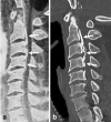
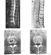
MR bone imaging is a recently introduced technique, that allows visualization of bony structures in good contrast against adjacent structures, like CT. Although CT has long been considered the modality of choice for bone imaging, MR bone imaging allows visualization of the bone without radiation exposure while simultaneously allowing conventional MR images to be obtained. Accordingly, MR bone imaging is expected as a new imaging technique for the diagnosis of miscellaneous spinal diseases. This review presents several sequences used in MR bone imaging including black bone imaging, ultrashort/zero echo time (UTE/ZTE) sequences, and T1-weighted 3D gradient-echo sequence. We also illustrate clinical cases in which spinal lesions could be effectively demonstrated on MR bone imaging, performed in most cases using a 3D gradient-echo sequence at our institution. The lesions presented herein include degenerative diseases, tumors and similar diseases, fractures, infectious diseases, and hemangioma. Finally, we discuss the differences between MR bone imaging and previously reported techniques, and the limitations and future perspectives of MR bone imaging.
Keywords: Bone imaging; CT-like MRI; MR imaging; Spine.
© 2023. The Author(s).
Conflict of interest statement
The authors declare that they have no conflict of interest with regard to this publication.
Figures


Similar articles
-
Diagnostic value of water-fat-separated images and CT-like susceptibility-weighted images extracted from a single ultrashort echo time sequence for the evaluation of vertebral fractures and degenerative changes of the spine.Eur Radiol. 2023 Feb;33(2):1445-1455. doi: 10.1007/s00330-022-09061-2. Epub 2022 Aug 18. Eur Radiol. 2023. PMID: 35980430 Free PMC article.
-
CT-like images based on T1 spoiled gradient-echo and ultra-short echo time MRI sequences for the assessment of vertebral fractures and degenerative bone changes of the spine.Eur Radiol. 2021 Jul;31(7):4680-4689. doi: 10.1007/s00330-020-07597-9. Epub 2021 Jan 14. Eur Radiol. 2021. PMID: 33443599 Free PMC article.
-
Clinical application of ultrashort echo time (UTE) and zero echo time (ZTE) magnetic resonance (MR) imaging in the evaluation of osteoarthritis.Skeletal Radiol. 2023 Nov;52(11):2149-2157. doi: 10.1007/s00256-022-04269-1. Epub 2023 Jan 6. Skeletal Radiol. 2023. PMID: 36607355 Free PMC article. Review.
-
Comparing CT-Like Images Based on Ultra-Short Echo Time and Gradient Echo T1-Weighted MRI Sequences for the Assessment of Vertebral Disorders Using Histology and True CT as the Reference Standard.J Magn Reson Imaging. 2024 May;59(5):1542-1552. doi: 10.1002/jmri.28927. Epub 2023 Jul 27. J Magn Reson Imaging. 2024. PMID: 37501387
-
3D MRI with CT-like bone contrast - An overview of current approaches and practical clinical implementation.Eur J Radiol. 2021 Oct;143:109915. doi: 10.1016/j.ejrad.2021.109915. Epub 2021 Aug 19. Eur J Radiol. 2021. PMID: 34461599 Review.
Cited by
-
Ultrashort Echo Time and Fast Field Echo Imaging for Spine Bone Imaging with Application in Spondylolysis Evaluation.Computation (Basel). 2024 Aug;12(8):152. doi: 10.3390/computation12080152. Epub 2024 Jul 24. Computation (Basel). 2024. PMID: 40735498 Free PMC article.
-
Assessing the Volume of the Head of the Mandibular Condyle Using 3T-MRI-A Preliminary Trial.Dent J (Basel). 2024 Jul 16;12(7):220. doi: 10.3390/dj12070220. Dent J (Basel). 2024. PMID: 39057007 Free PMC article.
-
Bone injury imaging in knee and ankle joints using fast-field-echo resembling a CT using restricted echo-spacing MRI: a feasibility study.Front Endocrinol (Lausanne). 2024 Jul 11;15:1421876. doi: 10.3389/fendo.2024.1421876. eCollection 2024. Front Endocrinol (Lausanne). 2024. PMID: 39072275 Free PMC article.
-
Diagnosing Ankylosing Spondylitis via Architecture-Modified ResNet and Combined Conventional Magnetic Resonance Imagery.J Imaging Inform Med. 2025 Mar 3. doi: 10.1007/s10278-025-01427-4. Online ahead of print. J Imaging Inform Med. 2025. PMID: 40032762
-
Cartilaginous endplate coverage of developmental Schmorl's node and the relevance of this in Schmorl's node etiology-based classification.Quant Imaging Med Surg. 2024 Jun 1;14(6):4288-4303. doi: 10.21037/qims-24-335. Epub 2024 May 9. Quant Imaging Med Surg. 2024. PMID: 38846309 Free PMC article. No abstract available.
References
Publication types
MeSH terms
LinkOut - more resources
Full Text Sources
Medical

