. 2023 May 20;23(1):165.
doi: 10.1186/s12906-023-03994-x.

Origanum majorana L. protects against neuroinflammation-mediated cognitive impairment: a phyto-pharmacological study

Affiliations

Origanum majorana L. protects against neuroinflammation-mediated cognitive impairment: a phyto-pharmacological study

Reham Wagdy et al. BMC Complement Med Ther. .

Abstract

Background: Neuroinflammation and oxidative stress are critical players in the pathogenesis of numerous neurodegenerative diseases, such as Alzheimer's disease (AD) which is responsible for most cases of dementia in the elderly. With the lack of curative treatments, natural phenolics are potential candidates to delay the onset and progression of such age-related disorders due to their potent antioxidant and anti-inflammatory effects. This study aims at assessing the phytochemical characteristics of Origanum majorana L. (OM) hydroalcohol extract and its neuroprotective activities in a murine neuroinflammatory model.

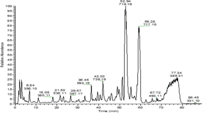
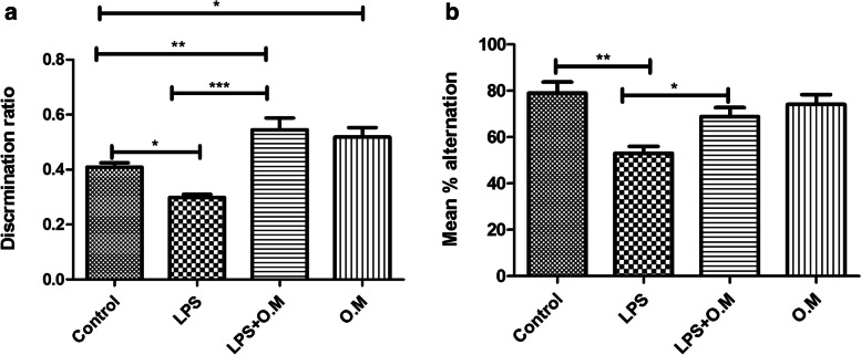
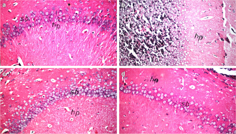
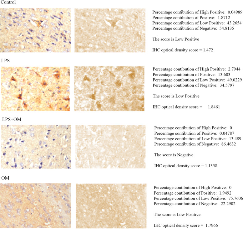
Methods: OM phytochemical analysis was done by HPLC/PDA/ESI-MSn. Oxidative stress was induced in vitro by hydrogen peroxide and cell viability was measured using WST-1 assay. Swiss albino mice were injected intraperitoneally with OM extract at a dose of 100 mg/kg for 12 days and with 250 μg/kg LPS daily starting from day 6 to induce neuroinflammation. Cognitive functions were assessed by novel object recognition and Y-maze behavioral tests. Hematoxylin and eosin staining was used to assess the degree of neurodegeneration in the brain. Reactive astrogliosis and inflammation were assessed by immunohistochemistry using GFAP and COX-2 antibodies, respectively.

Results: OM is rich in phenolics, with rosmarinic acid and its derivatives being major constituents. OM extract and rosmarinic acid significantly protected microglial cells against oxidative stress-induced cell death (p < 0.001). OM protected against the LPS-induced alteration of recognition and spatial memory in mice (p < 0.001) and (p < 0.05), respectively. Mice that received OM extract prior to the induction of neuroinflammation showed comparable histology to control brains, with no overt neurodegeneration. Furthermore, OM pre-treatment decreased the immunohistochemistry profiler score of GFAP from positive to low positive and COX-2 from low positive to negative in the brain tissue, compared to the LPS group.

Conclusion: These findings highlight the potential preventive effects of OM phenolics against neuroinflammation and pave the way toward drug discovery and development for neurodegenerative disorders.

Keywords: Cognition; LPS; Neurodegeneration; Neuroinflammation; Origanum majorana; Phenolics.


Conflict of interest statement

The authors declare no competing interests.

Figures

Fig. 1
Fig. 1
HPLC/PDA/ESI/MSn of metabolites detected in hydroalcoholic extract of leaves of Origanum majorana L. (negative mode)
Fig. 2
Fig. 2
Antioxidant effect of OM extract and RA. BV2 cells were pre-treated with OM extract (a) or RA (b) using several safe concentrations for one day prior to H2O2 (IC50) stimulation at 100 µM. OM extract and RA protected BV2 cells from H2O2-induced cell death. Data are represented as mean ± SEM (n = 3). Significance from control is represented as: *** at p < 0.001. Significance from 100 µM H2O2 is represented as: ### at p < 0.001
Fig. 3
Fig. 3
Effect of Origanum majorana on cognitive functions. LPS administration affected mean discrimination ratio (a) and alternation percentage (b). LPS + OM showed a significant increase in the mean discrimination ratio and alternation percentage compared to the LPS group. Mean discrimination ratio of LPS + OM and OM groups was higher than the control group. Data are expressed as mean ± SEM (n = 8), *P ≤ 0.05, **P ≤ 0.01, ***P ≤ 0.001
Fig. 4
Fig. 4
Photomicrograph of the hippocampus stained with H&E (× 40). LPS group (b) shows nuclear pyknosis and degeneration of the neurons compared to the control group (a). Pre-treatment with OM (c) shows comparable histology to the control group. OM (d) group presents normal histology (n = 3)
Fig. 5
Fig. 5
Immunohistochemical analysis of GFAP in the cerebral cortex. Deconvoluted images of DAB staining (second row) was produced by IHC profiler, and the percentage contributions was calculated relative to each image pixel intensity as high positive, positive, low positive, and negative pixels. Automated score of low positive is found for the control group and LPS + OM group, while LPS group presented as positive. The generated automated score for OM group is negative. For further quantitative analysis, calculation of IHC optical density score was done. LPS group scored 2.154 compared with 1.583 for control group, 1.5626 for LPS + OM group, and 1.4937 for OM group. Our results show that GFAP expression was enhanced in the LPS model and was attenuated under the effect of OM extract (COX-2 immunostaining, × 80) (n = 3)
Fig. 6
Fig. 6
Immunohistochemical analysis of COX-2 in the cerebral cortex. Deconvoluted images of DAB staining (second row) was produced by IHC profiler, and the percentage contributions was calculated relative to each image pixel intensity as high positive, positive, low positive, and negative pixels. An automated score of low positive is found for the control group, LPS group, and OM group. However, the generated automated score for LPS + OM group is negative. For further quantitative analysis, calculation of IHC optical density score was done. LPS group scored 1.8461 compared with 1.472 for control group, 1.1358 for LPS + OM group, and 1.7966 for OM group. Our results show that COX-2 expression was attenuated under the effect of OM extract (COX-2 immunostaining, × 80) (n = 3)

References

    1. Zhang B, Gaiteri C, Bodea L-G, et al. Integrated Systems Approach Identifies Genetic Nodes and Networks in Late-Onset Alzheimer’s Disease. Cell. 2013;153:707–720. doi: 10.1016/j.cell.2013.03.030. - DOI - PMC - PubMed
    1. Griffin WS, Stanley LC, Ling C, et al. Brain interleukin 1 and S-100 immunoreactivity are elevated in Down syndrome and Alzheimer disease. Proc Natl Acad Sci. 1989;86:7611–7615. doi: 10.1073/pnas.86.19.7611. - DOI - PMC - PubMed
    1. Sheng JG, Mrak RE, Griffin WS. Glial-neuronal interactions in Alzheimer disease: progressive association of IL-1alpha+ microglia and S100beta+ astrocytes with neurofibrillary tangle stages. J Neuropathol Exp Neurol. 1997;56:285–290. doi: 10.1097/00005072-199703000-00007. - DOI - PubMed
    1. Lyra e Silva NM, Gonçalves RA, Pascoal TA, et al. Pro-inflammatory interleukin-6 signaling links cognitive impairments and peripheral metabolic alterations in Alzheimer’s disease. Transl Psychiatry. 2021;11:251. 10.1038/s41398-021-01349-z. - PMC - PubMed
    1. Álvarez A, Cacabelos R, Sanpedro C, et al. Serum TNF-alpha levels are increased and correlate negatively with free IGF-I in Alzheimer disease. Neurobiol Aging. 2007;28:533–536. doi: 10.1016/j.neurobiolaging.2006.02.012. - DOI - PubMed