. 2023 Jul;22(7):100579.
doi: 10.1016/j.mcpro.2023.100579. Epub 2023 May 20.

A New Cellular Interactome of SARS-CoV-2 Nucleocapsid Protein and Its Biological Implications

Affiliations

A New Cellular Interactome of SARS-CoV-2 Nucleocapsid Protein and Its Biological Implications

Yuan-Qin Min et al. Mol Cell Proteomics. 2023 Jul.

Abstract

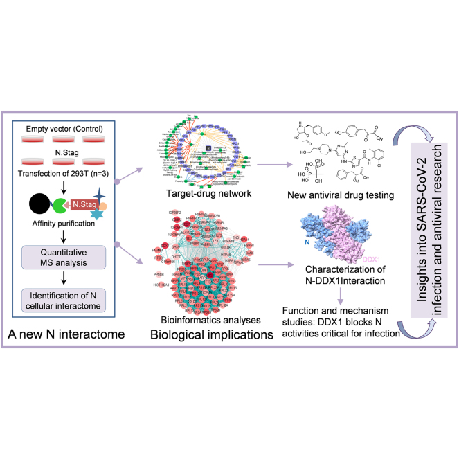
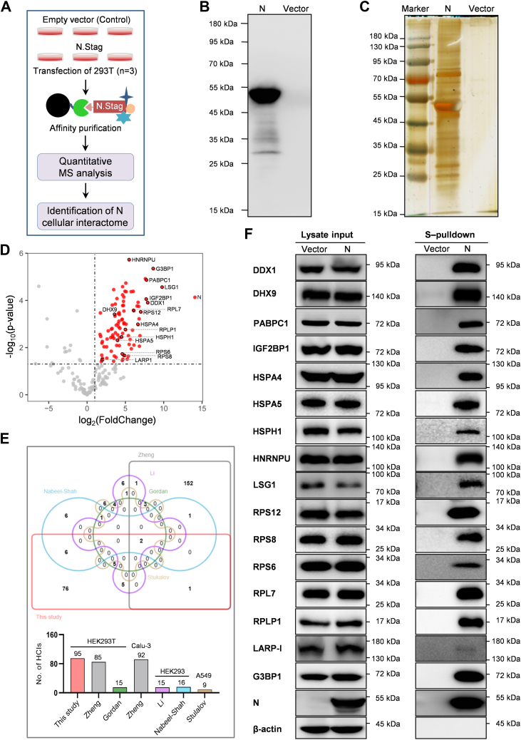
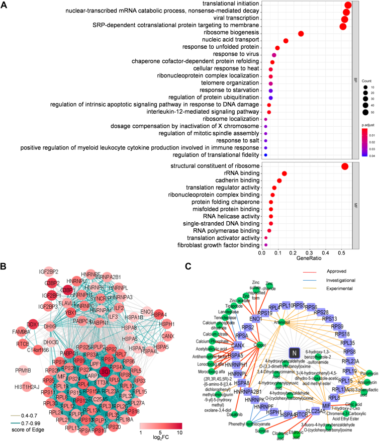
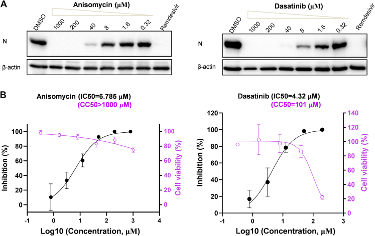
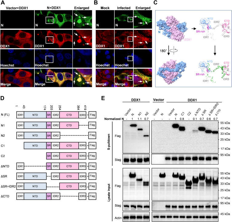
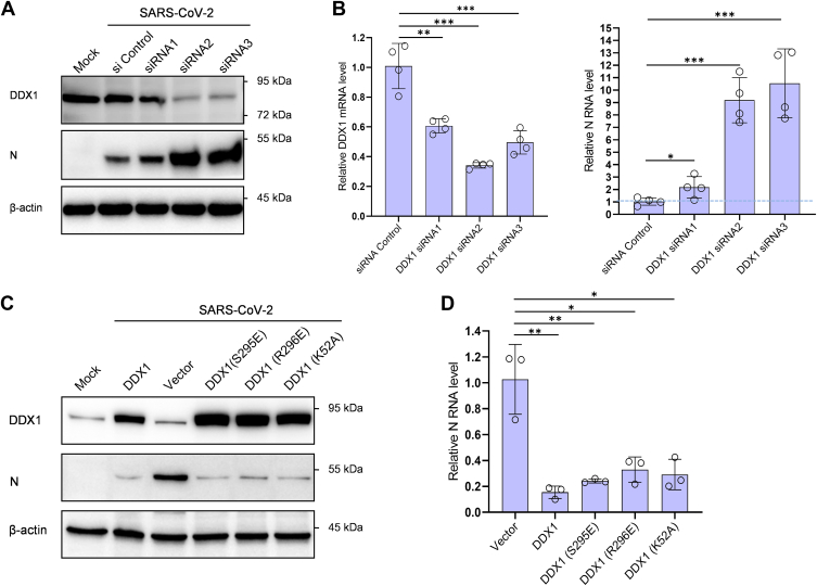
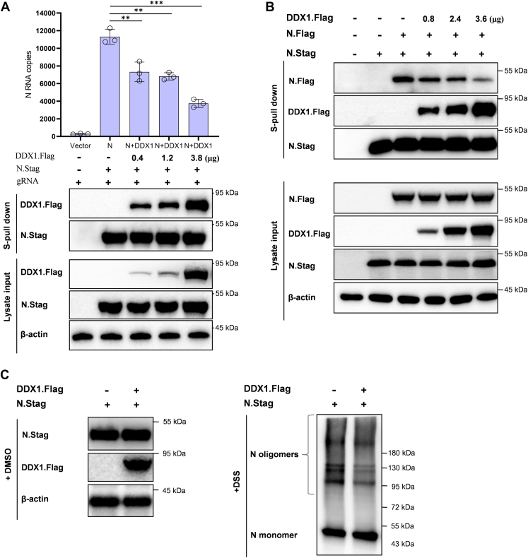
There is still much to uncover regarding the molecular details of severe acute respiratory syndrome coronavirus 2 (SARS-CoV-2) infection. As the most abundant protein, coronavirus nucleocapsid (N) protein encapsidates viral RNAs, serving as the structural component of ribonucleoprotein and virion, and participates in transcription, replication, and host regulations. Virus-host interaction might give clues to better understand how the virus affects or is affected by its host during infection and identify promising therapeutic candidates. Considering the critical roles of N, we here established a new cellular interactome of SARS-CoV-2 N by using a high-specific affinity purification (S-pulldown) assay coupled with quantitative mass spectrometry and immunoblotting validations, uncovering many N-interacting host proteins unreported previously. Bioinformatics analysis revealed that these host factors are mainly involved in translation regulations, viral transcription, RNA processes, stress responses, protein folding and modification, and inflammatory/immune signaling pathways, in line with the supposed actions of N in viral infection. Existing pharmacological cellular targets and the directing drugs were then mined, generating a drug-host protein network. Accordingly, we experimentally identified several small-molecule compounds as novel inhibitors against SARS-CoV-2 replication. Furthermore, a newly identified host factor, DDX1, was verified to interact and colocalize with N mainly by binding to the N-terminal domain of the viral protein. Importantly, loss/gain/reconstitution-of-function experiments showed that DDX1 acts as a potent anti-SARS-CoV-2 host factor, inhibiting the viral replication and protein expression. The N-targeting and anti-SARS-CoV-2 abilities of DDX1 are consistently independent of its ATPase/helicase activity. Further mechanism studies revealed that DDX1 impedes multiple activities of N, including the N-N interaction, N oligomerization, and N-viral RNA binding, thus likely inhibiting viral propagation. These data provide new clues to better depiction of the N-cell interactions and SARS-CoV-2 infection and may help inform the development of new therapeutic candidates.

Keywords: DDX1; SARS-COV-2; antiviral drug; host restriction factor; interactome; mass spectrometry; nucleocapsid (N) protein; virus–host interaction.


Conflict of interest statement

Conflict of interest The authors declare no competing interests.

Figures

None
Graphical abstract
Fig. 1
Fig. 1
Establishment of a new cellular interactome of SARS-CoV-2 N.A, schematic representation of the strategy for analyzing cellular interacting proteins of SARS-COV-2 N via S-pulldown. HEK293T cells were transfected with plasmid DNA encoding N.STAG or the empty vector (control). Thirty-six hours post-transfection, supernatants of the cell lysates were harvested and subjected to S-pulldown affinity purification assays, followed by quantitative mass spectrometry (MS) analysis and protein identification. B and C, immunoblot and silver staining analyses of N and the coprecipitated host proteins. S-pulldown products from (A) were also delivered to Western blotting with anti-N monoclonal antibody (mAb) (B) and silver staining of SDS-polyacrylamide gel (C). D, Volcano diagram of the identified protein targets. Points with fold change (FC) >2 and p value <0.05 are highlighted in red as N-interacting targets of high confidence. Targets marked with a black circle represent those validated by immunoblotting in the following. E, Venn diagram analysis of potential N interactors identified from different studies. Size of each list together with the cell types used in the studies was indicated in the histogram below. See also supplemental Fig. S1. F, validation of the SARS-CoV-2 N interactome data obtained in this study with immunoblotting. The interactions of N with representative host proteins identified here were confirmed by independent pulldown assays and Western blotting analyses with specific antibodies against the indicated proteins. HEK293T, human embryonic kidney 293 cell line; N protein, nucleocapsid protein; SARS-CoV-2, severe acute respiratory syndrome coronavirus 2.
Fig. 2
Fig. 2
Gene Ontology (GO), interaction network, and druggable target analyses of the N interactome.A, GO analysis was used to determine the biological process (BP) and molecular function (MF) attributes of the identified N-interacting cellular proteins by clusterProfiler and significant GO terms (adjusted p < 0.05). B, interaction network analysis of the identified cellular proteins interacting with N. Interaction networks of the cellular proteins (fold change [FC] >2, p < 0.05) were analyzed by STRING and visualized with Cytoscape. Proteins were clustered into five units through Markov clustering based on their molecular action. Disconnected nodes (HST1H2AJ and PPM1B) are also displayed in the network. The edge color is based on confidence (score) cutoff values (light gray, 0.4–0.7; blue, >0.7). The node color proportional to the rank of log2(FC) values. C, target–drug network. Pharmacological targets of the N-interacting host factors were identified by data mining. The druggable proteins along with the targeting drugs were then visualized in the network by Cytoscape. See also Figure 3 for the data-guided drug testing. N protein, nucleocapsid protein.
Fig. 3
Fig. 3
Anisomycin and dasatinib, identified with the N interactome, exhibit notable anti-SARS-CoV-2 activities.A and B, Huh7-ACE2 cells were pretreated with different doses of the indicated drugs for 1 h, followed by SARS-CoV-2 infection (0.05 moi) in the presence of the drugs. At 24 h postinfection, cells were collected for Western blot (WB) analyses of the expression levels of SARS-CoV-2 N proteins by using antibodies against the indicated proteins (A) or for the following quantitative PCR (qPCR) analyses of viral RNA (B, black curves), respectively. Remdesivir (10 μM) was used as a control. The left Y-axes in (B) represent mean percent inhibition of virus yield determined by qPCR (black curves). The right Y-axes represent mean percent cell viability of Huh7-ACE2 cells treated with different doses of the indicated drugs for 24 h, which was determined by CCK8 kit (purple curves). Graphs show mean ± SD, n ≥ 3 biological replicates. The IC50 and half-cytotoxic concentration (CC50) were calculated by GraphPad Prism 8.3. ACE2, angiotensin-converting enzyme 2; CCK8, Cell Counting Kit-8; SARS-CoV-2, severe acute respiratory syndrome coronavirus 2.
Fig. 4
Fig. 4
The colocalization and interaction of DDX1 with SARS-CoV-2 N.A, colocalization of ectopically expressed DDX1 with N. Huh7 cells were transfected with the DDX1 and N expression plasmids for 24 h, followed by fixation and immunofluorescence assay (IFA) analysis. B, colocalization of endogenous DDX1 with N upon SARS-CoV-2 infection. CaCo-2 cells were infected with SARS-COV-2 for 24 h, followed by fixation and IFA analysis. Nuclei were stained with Hoechst. Arrows indicate some typical N-condensates. Consistent with the previous studies (6), N-condensates could be more notable in some cells. Colocalization of DDX1 with N could be also observed in these condensate puncta. See also supplemental Fig. S3. C, predication of the DDX1–N interaction by AlphaFold2. AlphaFold2 predication reveals that DDX1 (purple) and N (blue) may form an intertwined dimer (left panels). The potential interaction interfaces on N were marked in green (right panels). D, linear representation of the domain organization for FLAG-tagged full-length, N-terminal- or C-terminal-truncated, or specific domain-deleted N, named as indicated. E, mapping of N domains targeted by DDX1. Huh7-ACE2 were cotransfected with the DDX1.STAG expression plasmid and the plasmids encoding full-length or truncated N proteins fused with FLAG or the corresponding control vectors as indicated. At 36 h post-transfection, protein interactions were analyzed by S-pulldown assays. Cell lysates (input) and pulldown products (S-pulldown) were subjected to Western blot (WB) analyses with specific antibodies against the indicated proteins. Relative coprecipitation ratios of N were calculated by normalization of the band intensities to those of the inputs and precipitated DDX1. The coprecipitation ratio of the full-length N in the same membrane was set as 1. Band intensities were measured using ImageJ. CTD, C-terminal oligomerization domain; IDR, putatively intrinsically disordered region; N, nucleocapsid protein; NTD, N-terminal domain; SARS-CoV-2, severe acute respiratory syndrome coronavirus 2; SR, serine/arginine-rich region.
Fig. 5
Fig. 5
DDX1 acts as a cellular restriction factor against SARS-CoV-2 infection, independently of its ATPase/helicase activities.A and B, DDX1 knockdown promotes SARS-CoV-2 infection. Huh7-ACE2 cells were transfected with specific DDX1 siRNAs or siRNA control. At 48 h post-transfection, cells were infected with SARS-CoV-2 (0.05 moi) for 24 h, followed by Western blot (WB) or quantitative PCR (qPCR) analyses of the expression levels of DDX1 or SARS-CoV-2 N proteins (A) or RNAs (B), respectively. Cells that were not transfected or infected (mock) were also analyzed by WB as additional controls. See also supplemental Fig. S4 for the similar results obtained in HEK293 cells with DDX1 knockdown or KO using CRISPR–Cas9 editing. C and D, DDX1 or mutant overexpression inhibits SARS-CoV-2 infection. Huh7-ACE2 cells were transfected with DDX1 or DDX1 mutants or the control vector. At 24 h post-transfection, cells were infected with SARS-CoV-2 (0.05 moi) for 24 h, followed by WB or qPCR analyses of the expression levels of the cellular or viral proteins or RNAs, respectively. WB analyses were conducted by using antibodies against the indicated proteins. Graphs show mean ± SD. ∗p < 0.05; ∗∗p < 0.01; ∗∗∗p < 0.001. See also supplemental Fig. S4 for the similar results obtained with reconstitution of DDX1 and its mutants in DDX1-KO HEK293 cells and supplemental Fig. S4E showing that the targeting of N by DDX1 is consistently independent of the ATPase/helicase activities of DDX1. HEK293, human embryonic kidney 293 cell line; SARS-CoV-2, severe acute respiratory syndrome coronavirus 2.
Fig. 6
Fig. 6
DDX1 can inhibit the N–genome RNA (gRNA) binding, N–N interaction, and N oligomerization, the critical activities of N in SARS-CoV-2 propagation.A, the effect of DDX1 on the N–gRNA interaction. Huh7-ACE2 cells were transfected with the N.Stag-expressing plasmid, along with different amounts of the DDX1.Flag expression plasmid or control vector. At 48 h post-transfection, the supernatants of the cell lysates were incubated with RNase inhibitor (1 U/μl) and viral gRNA (∼2 × 105 copies) extracted from virus stock, followed by S-pulldown assays. The N coprecipitates of RNAs and proteins were respectively analyzed by quantitative PCR (qPCR) or Western blot (WB) using antibodies against the indicated tags (or β-actin protein). Graphs show mean ± SD. ∗∗p < 0.01; ∗∗∗p < 0.001. B, DDX1 impedes the N–N interaction. Huh7-ACE2 cells were cotransfected with the plasmids expressing N proteins fused with Stag or Flag (N.Stag or N.Flag) together with different amounts of the plasmid encoding Flag-tagged DDX1 (DDX1.Flag) or the corresponding control vectors. At 48 h post-transfection, cells were lysed for S-pulldown assays. The cell lysates and N.Stag coprecipitates were respectively analyzed by Western blot (WB) using antibodies against the indicated tags (or β-actin protein). C, DDX1 weakens the N oligomerization. Huh7-ACE2 cells were transfected with the DDX.Flag-expressing plasmid or control vector, together with the plasmid encoding N. At 48 h post-transfection, cells were harvested and washed by PBS, followed by crosslinking with disuccinimidyl suberate (DSS, 0.5 mM) that enables the fixation of oligomerized N in situ. After crosslinking at 37 °C for 30 min, the reaction was quenched by 1 M Tris buffer (pH 7.5), and samples were delivered to SDS-PAGE and WB analyses of the N oligomerization and protein expression. ACE2, angiotensin-converting enzyme 2; N, nucleocapsid protein; SARS-CoV-2, severe acute respiratory syndrome coronavirus 2.

Similar articles

Cited by

References

    1. Wu F., Zhao S., Yu B., Chen Y.M., Wang W., Song Z.G., et al. A new coronavirus associated with human respiratory disease in China. Nature. 2020;579:265. - PMC - PubMed
    1. Klein S., Cortese M., Winter S.L., Wachsmuth-Melm M., Neufeldt C.J., Cerikan B., et al. SARS-CoV-2 structure and replication characterized by in situ cryo-electron tomography. Nat. Commun. 2020;11:5885. - PMC - PubMed
    1. Yao H., Song Y., Chen Y., Wu N., Xu J., Sun C., et al. Molecular architecture of the SARS-CoV-2 virus. Cell. 2020;183:730–738.e13. - PMC - PubMed
    1. Cao C., Cai Z., Xiao X., Rao J., Chen J., Hu N., et al. The architecture of the SARS-CoV-2 RNA genome inside virion. Nat. Commun. 2021;12:3917. - PMC - PubMed
    1. Cubuk J., Alston J.J., Incicco J.J., Singh S., Stuchell-Brereton M.D., Ward M.D., et al. The SARS-CoV-2 nucleocapsid protein is dynamic, disordered, and phase separates with RNA. Nat. Commun. 2021;12:1936. - PMC - PubMed

Publication types