

Control of atypical PKCι membrane dissociation by tyrosine phosphorylation within a PB1-C1 interdomain interface
- PMID: 37211093
- PMCID: PMC10333572
- DOI: 10.1016/j.jbc.2023.104847
Control of atypical PKCι membrane dissociation by tyrosine phosphorylation within a PB1-C1 interdomain interface
Abstract
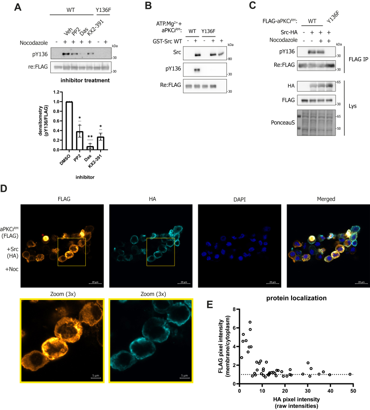
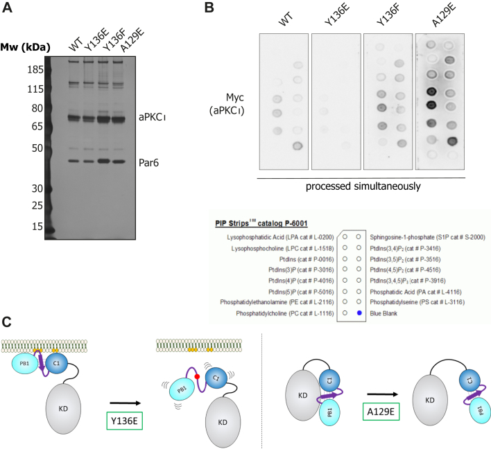
Atypical PKCs are cell polarity kinases that operate at the plasma membrane where they function within multiple molecular complexes to contribute to the establishment and maintenance of polarity. In contrast to the classical and novel PKCs, atypical PKCs do not respond to diacylglycerol cues to bind the membrane compartment. Until recently, it was not clear how aPKCs are recruited; whether aPKCs can directly interact with membranes or whether they are dependent on other protein interactors to do so. Two recent studies identified the pseudosubstrate region and the C1 domain as direct membrane interaction modules; however, their relative importance and coupling are unknown. We combined molecular modeling and functional assays to show that the regulatory module of aPKCι, comprising the PB1 pseudosubstrate and C1 domains, forms a cooperative and spatially continuous invariant membrane interaction platform. Furthermore, we show the coordinated orientation of membrane-binding elements within the regulatory module requires a key PB1-C1 interfacial β-strand (beta-strand linker). We show this element contains a highly conserved Tyr residue that can be phosphorylated and that negatively regulates the integrity of the regulatory module, leading to membrane release. We thus expose a hitherto unknown regulatory mechanism of aPKCι membrane binding and release during cell polarization.
Keywords: atypical protein kinase C; cell polarity; cell signaling; membrane recruitment; tyrosine phosphorylation.
Copyright © 2023. Published by Elsevier Inc.
Conflict of interest statement
Conflict of interest The authors declare that they have no conflicts of interest with the contents of this article.
Figures


Similar articles
-
Src-dependent aprotein kinase C iota/lambda (aPKCiota/lambda) tyrosine phosphorylation is required for aPKCiota/lambda association with Rab2 and glyceraldehyde-3-phosphate dehydrogenase on pre-golgi intermediates.J Biol Chem. 2006 Mar 31;281(13):8436-42. doi: 10.1074/jbc.M513031200. Epub 2006 Feb 1. J Biol Chem. 2006. PMID: 16452474 Free PMC article.
-
Pleckstrin Homology (PH) Domain Leucine-rich Repeat Protein Phosphatase Controls Cell Polarity by Negatively Regulating the Activity of Atypical Protein Kinase C.J Biol Chem. 2016 Nov 25;291(48):25167-25178. doi: 10.1074/jbc.M116.740639. Epub 2016 Oct 19. J Biol Chem. 2016. PMID: 27760826 Free PMC article.
-
PDK1 in apical signaling endosomes participates in the rescue of the polarity complex atypical PKC by intermediate filaments in intestinal epithelia.Mol Biol Cell. 2012 May;23(9):1664-74. doi: 10.1091/mbc.E11-12-0988. Epub 2012 Mar 7. Mol Biol Cell. 2012. PMID: 22398726 Free PMC article.
-
Structure and function of the PB1 domain, a protein interaction module conserved in animals, fungi, amoebas, and plants.Sci STKE. 2007 Aug 28;2007(401):re6. doi: 10.1126/stke.4012007re6. Sci STKE. 2007. PMID: 17726178 Review.
-
The C2 domains of classical and novel PKCs as versatile decoders of membrane signals.Biofactors. 2010 Jan-Feb;36(1):1-7. doi: 10.1002/biof.68. Biofactors. 2010. PMID: 20049899 Review.
Cited by
-
Cooperative regulation of C1-domain membrane recruitment polarizes atypical protein kinase C.J Cell Biol. 2023 Oct 2;222(10):e202112143. doi: 10.1083/jcb.202112143. Epub 2023 Aug 17. J Cell Biol. 2023. PMID: 37589718 Free PMC article.
-
The Drosophila neuroblast polarity cycle at a glance.J Cell Sci. 2024 Mar 1;137(5):jcs261789. doi: 10.1242/jcs.261789. Epub 2024 Mar 11. J Cell Sci. 2024. PMID: 38465513 Free PMC article.
-
Into the fold: advances in understanding aPKC membrane dynamics.Biochem J. 2023 Dec 20;480(24):2037-2044. doi: 10.1042/BCJ20230390. Biochem J. 2023. PMID: 38100320 Free PMC article.
-
Capture, mutual inhibition and release mechanism for aPKC-Par6 and its multisite polarity substrate Lgl.Nat Struct Mol Biol. 2025 Apr;32(4):729-739. doi: 10.1038/s41594-024-01425-0. Epub 2025 Jan 6. Nat Struct Mol Biol. 2025. PMID: 39762628 Free PMC article.
-
Rare variants in PRKCI cause Van der Woude syndrome and other features of peridermopathy.medRxiv [Preprint]. 2025 Jan 17:2025.01.17.25320742. doi: 10.1101/2025.01.17.25320742. medRxiv. 2025. PMID: 39867391 Free PMC article. Preprint.
References
-
- Parker P.J., Brown S.J., Calleja V., Chakravarty P., Cobbaut M., Linch M., et al. Equivocal, explicit and emergent actions of PKC isoforms in cancer. Nat. Rev. Cancer. 2021;21:51–63. - PubMed
-
- Leroux A.E., Schulze J.O., Biondi R.M. AGC kinases, mechanisms of regulation and innovative drug development. Semin. Cancer Biol. 2018;48:1–17. - PubMed
-
- Hirano Y., Yoshinaga S., Takeya R., Suzuki N.N., Horiuchi M., Kohjima M., et al. Structure of a cell polarity regulator, a complex between atypical PKC and Par6 PB1 domains∗. J. Biol. Chem. 2005;280:9653–9661. - PubMed
Publication types
MeSH terms
Substances
Grants and funding
LinkOut - more resources
Full Text Sources
Miscellaneous
