. 2023 May 21;21(1):335.
doi: 10.1186/s12967-023-04187-3.

IL-17A-mediated mitochondrial dysfunction induces pyroptosis in colorectal cancer cells and promotes CD8 + T-cell tumour infiltration

Affiliations

IL-17A-mediated mitochondrial dysfunction induces pyroptosis in colorectal cancer cells and promotes CD8 + T-cell tumour infiltration

Wen-Qing Feng et al. J Transl Med. .

Abstract

Background: Interleukin-17A (IL-17A), a proinflammatory cytokine primarily secreted by Th17 cells, γδT cells and natural killer T (NKT) cells, performs essential roles in the microenvironment of certain inflammation-related tumours by regulating cancer growth and tumour elimination proved in previous literature. In this study, the mechanism of IL-17A that induces mitochondrial dysfunction promoted pyroptosis has been explored in colorectal cancer cells.

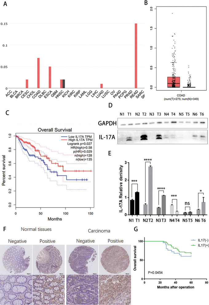
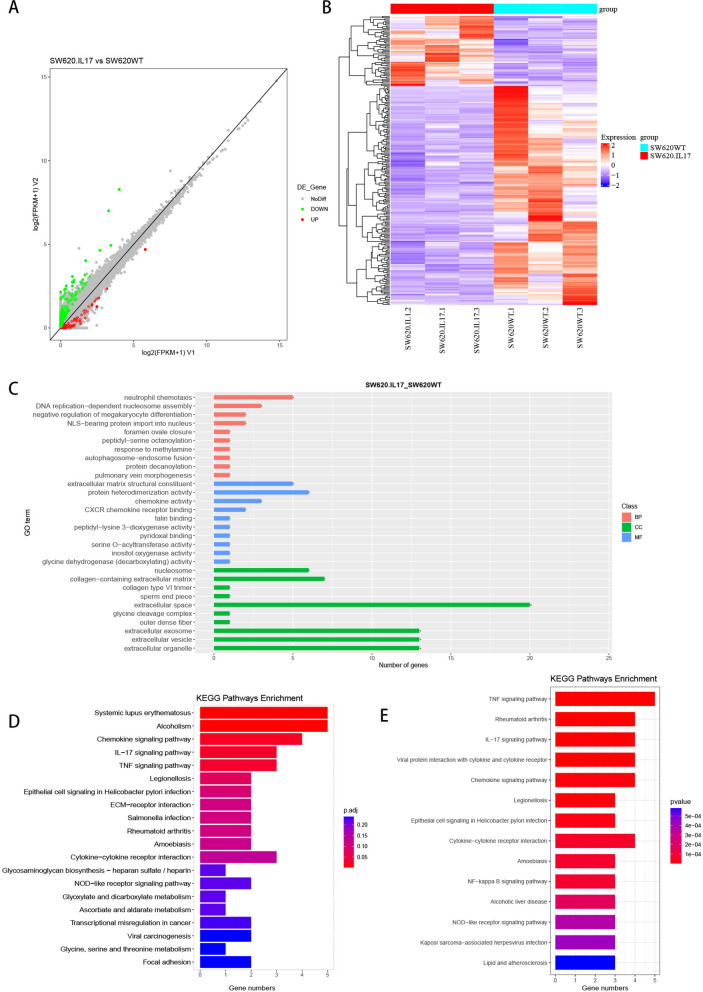
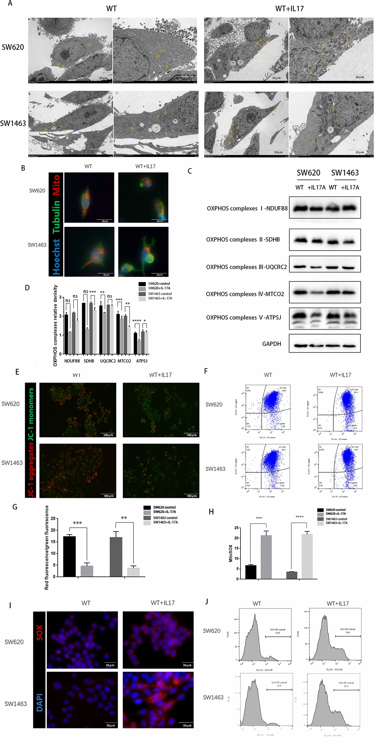
Method: The records of 78 patients diagnosed with CRC were reviewed via the public database to evaluate clinicopathological parameters and prognosis associations of IL-17A expression. The colorectal cancer cells were treated with IL-17A, and the morphological characteristics of those cells were indicated by scanning electron microscope and transmission electron microscope. After IL-17A treatment, mitochondrial dysfunction was tested by mitochondrial membrane potential (MMP) and reactive oxygen species (ROS). The expression of pyroptosis associated proteins including cleaved caspase-4, cleaved gasdermin-D (GSDMD), IL-1β, receptor activator of nuclear NOD-like receptor family pyrin domain containing 3 (NLRP3), apoptosis-associated speck like protein containing a card (ASC), and factor-kappa B was measured through western blotting.

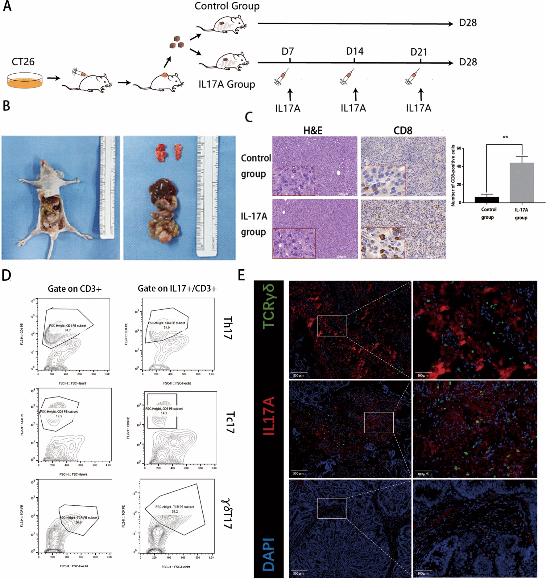
Results: Positive IL-17A protein expression was observed in CRC compared to the non-tumour tissue. IL-17A expression indicates a better differentiation, earlier stage, and better overall survival in CRC. IL-17A treatment could induce mitochondrial dysfunction and stimulate intracellular reactive oxygen species (ROS) production. Furthermore, IL-17A could promote pyroptosis of colorectal cancer cells and significantly increase the secretion of inflammatory factors. Nevertheless, the pyroptosis induced by IL-17A could be inhibited through the pre-treatment with Mito-TEMPO (a mitochondria-targeted superoxide dismutase mimetic with superoxide and alkyl radical scavenging properties) or Z-LEVD-FMK (caspase-4 inhibitor, fluoromethylketone). Additionally, after being treated with IL-17A, an increasing number of CD8 + T cells showed in mouse-derived allograft colon cancer models.

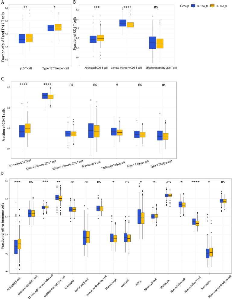
Conclusion: IL-17A, as a cytokine mainly secreted by γδT cells in the colorectal tumour immune microenvironment, can regulate the tumour microenvironment in multiple ways. IL-17A could induce mitochondrial dysfunction and pyroptosis through the ROS/NLRP3/caspase-4/GSDMD pathway, and promote intracellular ROS accumulation. In addition, IL-17A can promote the secretion of inflammatory factors such as IL-1β、IL-18 and immune antigens, and recruit CD8 + T cells to infiltrate tumours.

Keywords: CD8 + T; Colorectal cancer; IL-17A; Mitochondrial dysfunction; Pyroptosis.


Conflict of interest statement

He authors have declared that no competing interest exists.

Figures

Fig. 1
Fig. 1
Increased expression levels of IL-17A in patients with CRC. A Differentially expressed IL-17A between tumour and normal tissues; ACC: Adrenocortical carcinoma; BLCA: Bladder Urothelial Carcinoma; BRCA: Breast invasive carcinoma; CESC: Cervical squamous cell carcinoma and endocervical adenocarcinoma; CHOL: Cholangiocarcinoma; COAD: Colon adenocarcinoma; DLBC: Lymphoid Neoplasm Diffuse Large B-cell Lymphoma; ESCA: Esophageal carcinoma; GBM: Glioblastoma multiforme; HNSC: Head and Neck squamous cell carcinoma; KICH: Kidney Chromophobe; KIRC: Kidney renal clear cell carcinoma; KIRP: Kidney renal papillary cell carcinoma; LAML: Acute Myeloid Leukemia; LGG: Brain Lower Grade Glioma; LIHC: Liver hepatocellular carcinoma; LUAD: Lung adenocarcinoma; LUSC: Lung squamous cell carcinoma; OV: Ovarian serous cystadenocarcinoma; PAAD: Pancreatic adenocarcinoma; PCPG: Pheochromocytoma and Paraganglioma; PRAD: Prostate adenocarcinoma; READ: Rectum adenocarcinoma; SARC: Sarcomav; SKCM: Skin Cutaneous. Melanoma B Differentially expressed IL-17A between colorectal tumour and normal tissues; C Comparison of overall survival in IL-17A high and IL-17A low groups; D-E Representative western blot and quantification analysis of IL-17A expression in paired CRC samples. Data conforms to normal distribution, and tested by two independent samples t-test; F Immunohistochemical results showing expression of IL-17A in colorectal tumour and normal tissues; G Comparison of overall survival in IL-17A high and IL-17A low groups
Fig. 2
Fig. 2
Boxplots showing the relative abundances of immune cell types among the IL17A-hi group and IL17A-lo group. AThe relative abundances of γδ T cells among the IL17A-hi and IL17A-lo groups; B The relative abundances of CD8 + T cells and their subsets among the IL17A-hi and IL17A-lo groups; C The relative abundances of CD4 + T cells and their subsets among the IL17A-hi and IL17A-lo groups; D The relative proportions of other immune cells among the IL17A-hi and IL17A-lo groups. The difference between groups of normally distributed data was assessed by independent samples t-test, whereas the difference between two groups of non-normally distributed data was assessed by the Mann–Whitney U test. ns, not significant; *P < 0.05; **P < 0.01; ***P < 0.001; ****P < 0.0001
Fig. 3
Fig. 3
IL-17A activates NF-kB signaling pathway in CRC. A, B Differential genes expression of the IL-17A group versus that of the control group(pvalue < 0.05 and FoldChange (FC) ≥ 2); C Enrichment of the Gene Ontology by DEGs of the IL-17A group versus that in the control group; D Enrichment of the Kyoto Encyclopedia of Genes and Genomes pathway by DEGs of the IL-17A group versus that in the control group; E Enrichment of the Kyoto Encyclopedia of Genes and Genomes pathway by DEGs of the GSE127757
Fig. 4
Fig. 4
The morphologic and molecular characteristics of IL-17A–induced pyroptosis in CRC cells. A Representative microscopic images of CRC cells treated with IL-17A (100 ng/ml) for 72 h. Red arrows indicate the characteristic balloon in the cell membrane; B-C Expression of GSDME-N terminal by western blotting analysis after treated with IL-17A (100 ng/ml) for 72 h, data conforms to normal distribution, and tested by two independent samples t-test; D Representative scanning electron micrographs of CRC cells after treatment with IL-17A.Red arrows indicate formation of pyroptotic membrane pits and pores of varying size; E LDH release was detected in the supernates of CRC cells treated with IL-17A (100 ng/ml) for 72 h, data conforms to normal distribution, and tested by two independent samples t-test; F The release of IL-1β in the supernate was determined by ELISA, data conforms to normal distribution, and tested by two independent samples t-test; H-G Cell apoptosis rates were determined by using caspase-4 and propidium iodide co-dyeing staining assay after CRC cells treated with IL-17A (100 ng/ml) for 72 h, data conforms to normal distribution, and tested by two independent samples t-test; I Immunohistochemical results showing expression of IL-17A and IL-1β in colorectal tumour and normal tissues J Correlations between IL-17A and IL-1β in CRC
Fig. 5
Fig. 5
Mitochondrial dysfunction increases in CRC cells by treatment with IL-17A. A Transmission electron microscopy of CRC cells after treatment with IL-17A.Arrowheads indicate mitochondria; B CRC cells after treatment with IL-17A were immunostained with Mito-Tracker (red), anti-α-tubulin (green), and DAPI (nuclei, blue); C, D The expression of OxPhos complex subunits in CRC cells after treatment with IL-17A was analysed by western blotting, data conforms to normal distribution, and tested by two independent samples t-test. The mitochondrial membrane potential was measured using JC-1 dye. E representative microscopic and (F-G) flow cytometry analysis and quantitation, data conforms to normal distribution, and tested by two independent samples t-test. Immunostained for the Mitochondrial Superoxide Indicator with Mito-SOX. H representative microscopic and I-J flow cytometry analysis and quantitation, data conforms to normal distribution, and tested by two independent samples t-test
Fig. 6
Fig. 6
NF-κB activated pyroptosis-associated proteins treatment with IL-17A. A-B Protein levels of NF-κB p65, caspase 1, caspase 4, NLRP3, ASC, IL-1β, IL-18 and and GSDMD-N in CRC cells treated with IL-17A (100 ng/ml) for 72 h, data conforms to normal distribution, and tested by two independent samples t-test; C Representative IF staining of NLRP3 and ASC in CRC cells treated with IL-17A (100 ng/ml) for 72 h; D Representative microscopic images of CRC cells treated with IL-17A (100 ng/ml) and Z-YVAD(50 μM) or Z-LEVD(50 μM) or TEMPO(10 μM) for 72 h. Red arrows indicate the characteristic balloon in the cell membrane; E Protein levels of NF-κB p65, caspase1, caspase4, NLRP3, ASC, IL-1β, IL-18 and and GSDMD-N in CRC cells treated with IL-17A (100 ng/ml) and different types of inhibitors for 72 h
Fig. 7
Fig. 7
IL-17A are mainly producing by γδT cells and promotes CD8 + T cells infiltrating the tumour A-B The process of mouse-derived allograft colon cancer models; C H&E staining and immunostaining of CD8 from IL-17A or vehicle-treated colon tumour tissue. 3 fields under 40 × magnification were randomly selected, then counting and statistics CD8-positive cells, data conforms to normal distribution, and tested by two independent samples t-test; D Left panel representative flow cytometric analysis of all CD3 + cells from CD8 + T cells (Tc, top), CD4 + T cells (Th, middle), and γδTCR + T cells (γδT, bottom) and the right panel representative flow cytometric analysis of all CD3 + cells and IL-17A secretion from CD8 + T cells (Tc17, top), CD4 + T cells (Th17, middle), and γδTCR + T cells (γδT17, bottom) in tumour; E Paraffin sections from CRC patients were stained with anti-human pan-γδTCR (green) and anti-human IL-17A (red) for immunofluorescent (IF) staining

References

    1. Siegel RL, Miller KD, Fuchs HE, Jemal A. Cancer statistics, 2022. CA Cancer J Clin. 2022;72(1):7–33. doi: 10.3322/caac.21708. - DOI - PubMed
    1. Brahmer JR, Tykodi SS, Chow LQ, Hwu WJ, Topalian SL, Hwu P, Drake CG, Camacho LH, Kauh J, Odunsi K, et al. Safety and activity of anti-PD-L1 antibody in patients with advanced cancer. N Engl J Med. 2012;366:2455–2465. doi: 10.1056/NEJMoa1200694. - DOI - PMC - PubMed
    1. He R, Lao Y, Yu W, Zhang X, Jiang M, Zhu C. Progress in the application of immune checkpoint inhibitor-based immunotherapy for targeting different types of colorectal cancer. Front Oncol. 2021;23(11):764618. doi: 10.3389/fonc.2021.764618. - DOI - PMC - PubMed
    1. Cua DJ, Tato CM. Innate IL-17-producing cells: the sentinels of the immune system. Nat Rev Immunol. 2010;10:479–489. doi: 10.1038/nri2800. - DOI - PubMed
    1. Alshaker HA, Matalka KZ. IFN-γ, IL-17 and TGF-β involvement in shaping the tumor microenvironment: the significance of modulating such cytokines in treating malignant solid tumors[J] Cancer Cell Int. 2011;11(1):1–12. doi: 10.1186/1475-2867-11-33. - DOI - PMC - PubMed

Publication types

MeSH terms

Substances