

Evaluation of a photodynamic therapy agent using a canine prostate cancer model
- PMID: 37211857
- PMCID: PMC11135201
- DOI: 10.1002/pros.24560
Evaluation of a photodynamic therapy agent using a canine prostate cancer model
Abstract
Background: Male dogs can develop spontaneous prostate cancer, which is similar physiologically to human disease. Recently, Tweedle and coworkers have developed an orthotopic canine prostate model allowing implanted tumors and therapeutic agents to be tested in a more translational large animal model. We used the canine model to evaluate prostate-specific membrane antigen (PSMA)-targeted gold nanoparticles as a theranostic approach for fluorescence (FL) imaging and photodynamic therapy (PDT) of early stage prostate cancer.
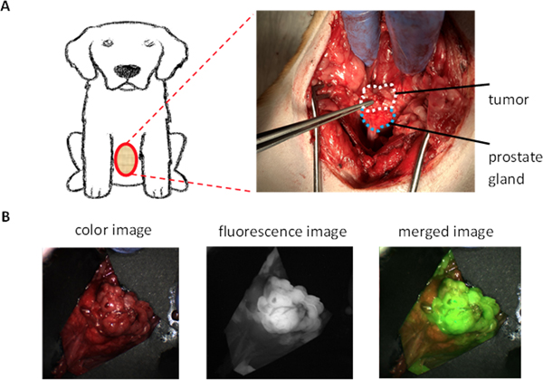
Methods: Dogs (four in total) were immunosuppressed with a cyclosporine-based immunosuppressant regimen and their prostate glands were injected with Ace-1-hPSMA cells using transabdominal ultrasound (US) guidance. Intraprostatic tumors grew in 4-5 weeks and were monitored by ultrasound (US). When tumors reached an appropriate size, dogs were injected intravenously (iv) with PSMA-targeted nano agents (AuNPs-Pc158) and underwent surgery 24 h later to expose the prostate tumors for FL imaging and PDT. Ex vivo FL imaging and histopathological studies were performed to confirm PDT efficacy.
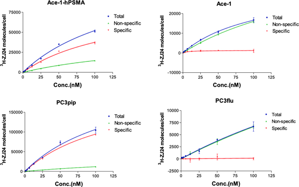
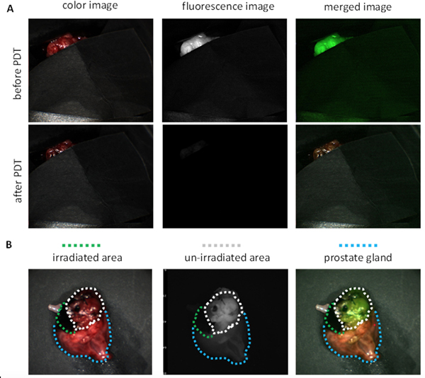
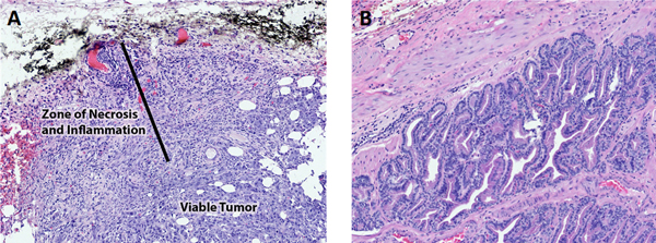
Results: All dogs had tumor growth in the prostate gland as revealed by US. Twenty-four hours after injection of PSMA-targeted nano agents (AuNPs-Pc158), the tumors were imaged using a Curadel FL imaging device. While normal prostate tissue had minimal fluorescent signal, the prostate tumors had significantly increased FL. PDT was activated by irradiating specific fluorescent tumor areas with laser light (672 nm). PDT bleached the FL signal, while fluorescent signals from the other unexposed tumor tissues were unaffected. Histological analysis of tumors and adjacent prostate revealed that PDT damaged the irradiated areas to a depth of 1-2 mms with the presence of necrosis, hemorrhage, secondary inflammation, and occasional focal thrombosis. The nonirradiated areas showed no visible damages by PDT.
Conclusion: We have successfully established a PSMA-expressing canine orthotopic prostate tumor model and used the model to evaluate the PSMA-targeted nano agents (AuNPs-Pc158) in the application of FL imaging and PDT. It was demonstrated that the nano agents allowed visualization of the cancer cells and enabled their destruction when they were irradiated with a specific wavelength of light.
Keywords: PDT; PSMA; canine prostate cancer; fluorescence imaging; nanoparticles.
© 2023 The Authors. The Prostate published by Wiley Periodicals LLC.
Conflict of interest statement
Figures
References
Publication types
MeSH terms
Substances
Grants and funding
LinkOut - more resources
Full Text Sources
Medical
Miscellaneous
