. 2023 Jul 1;325(1):R81-R95.
doi: 10.1152/ajpregu.00057.2023. Epub 2023 May 22.

Loss of hepatic PPARα in mice causes hypertension and cardiovascular disease

Affiliations

Loss of hepatic PPARα in mice causes hypertension and cardiovascular disease

Olufunto O Badmus et al. Am J Physiol Regul Integr Comp Physiol. .

Abstract

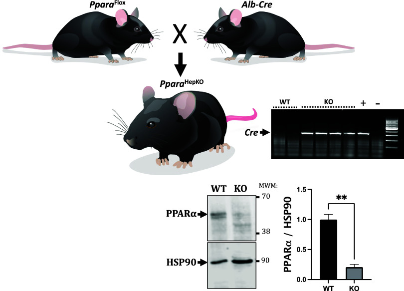
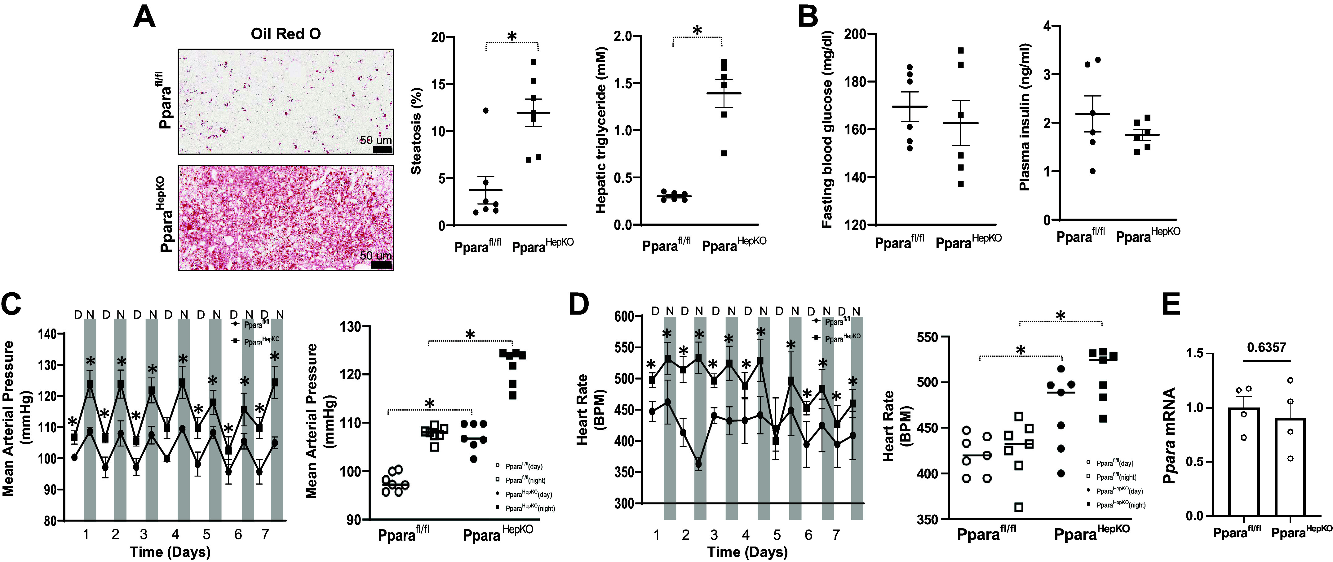
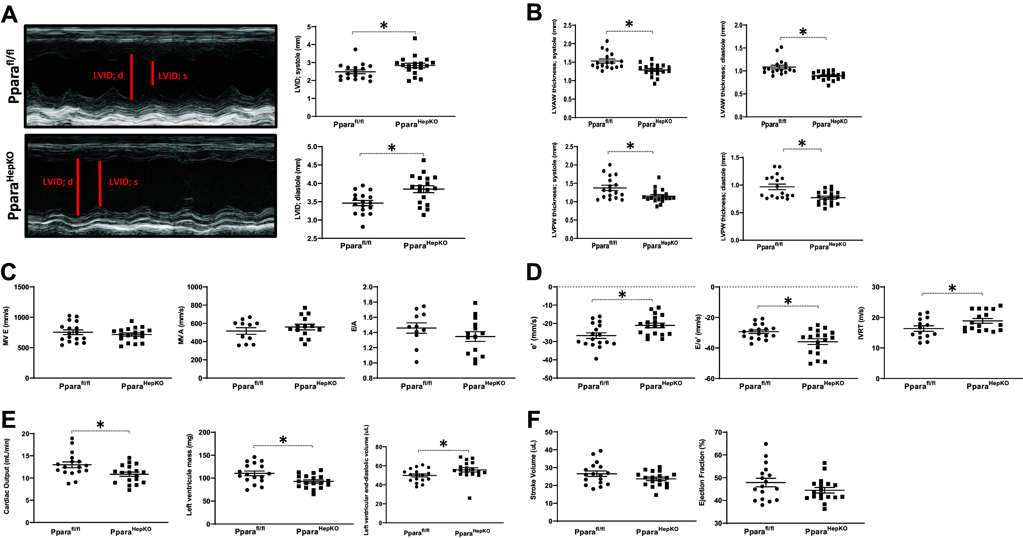
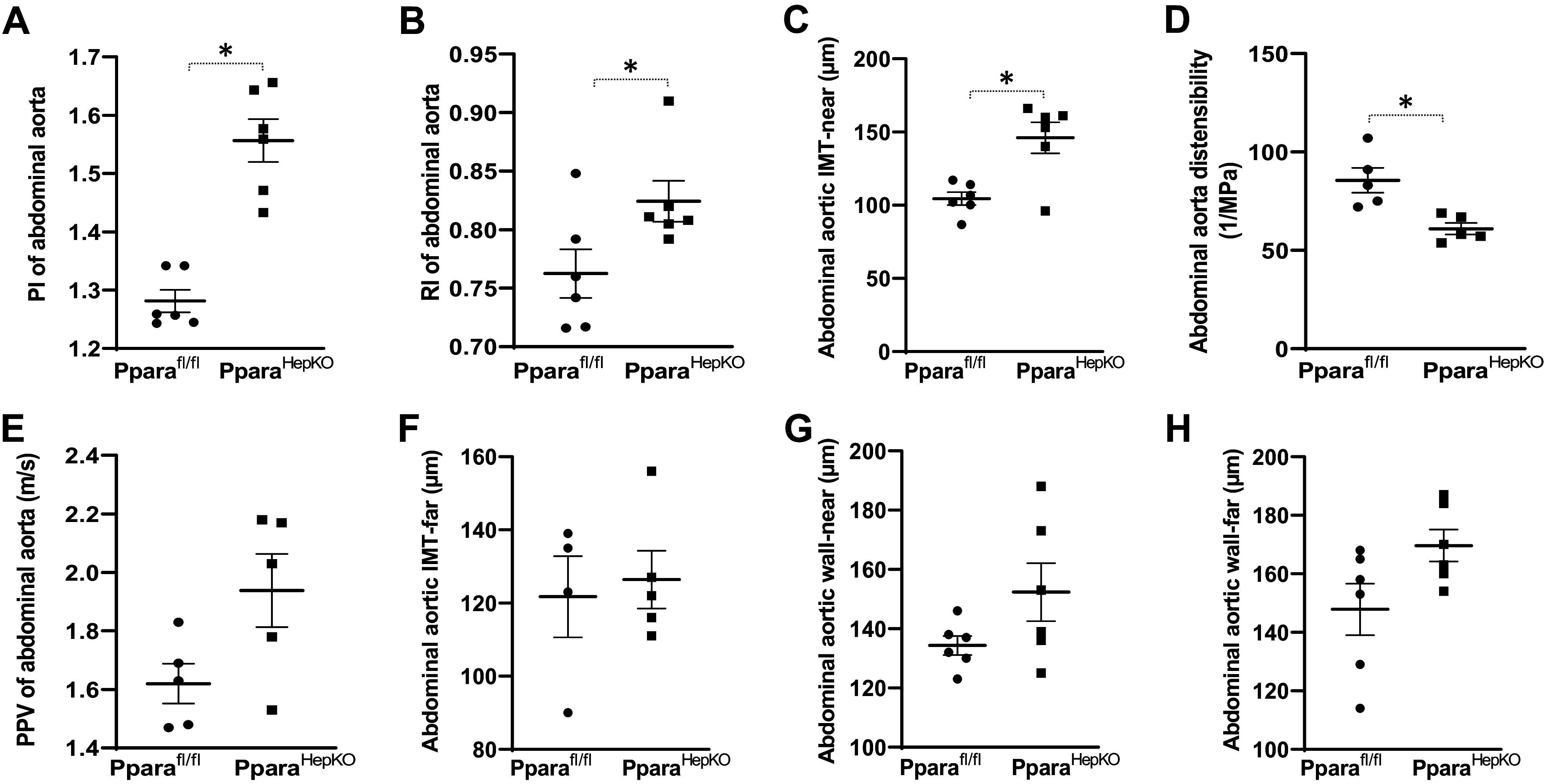
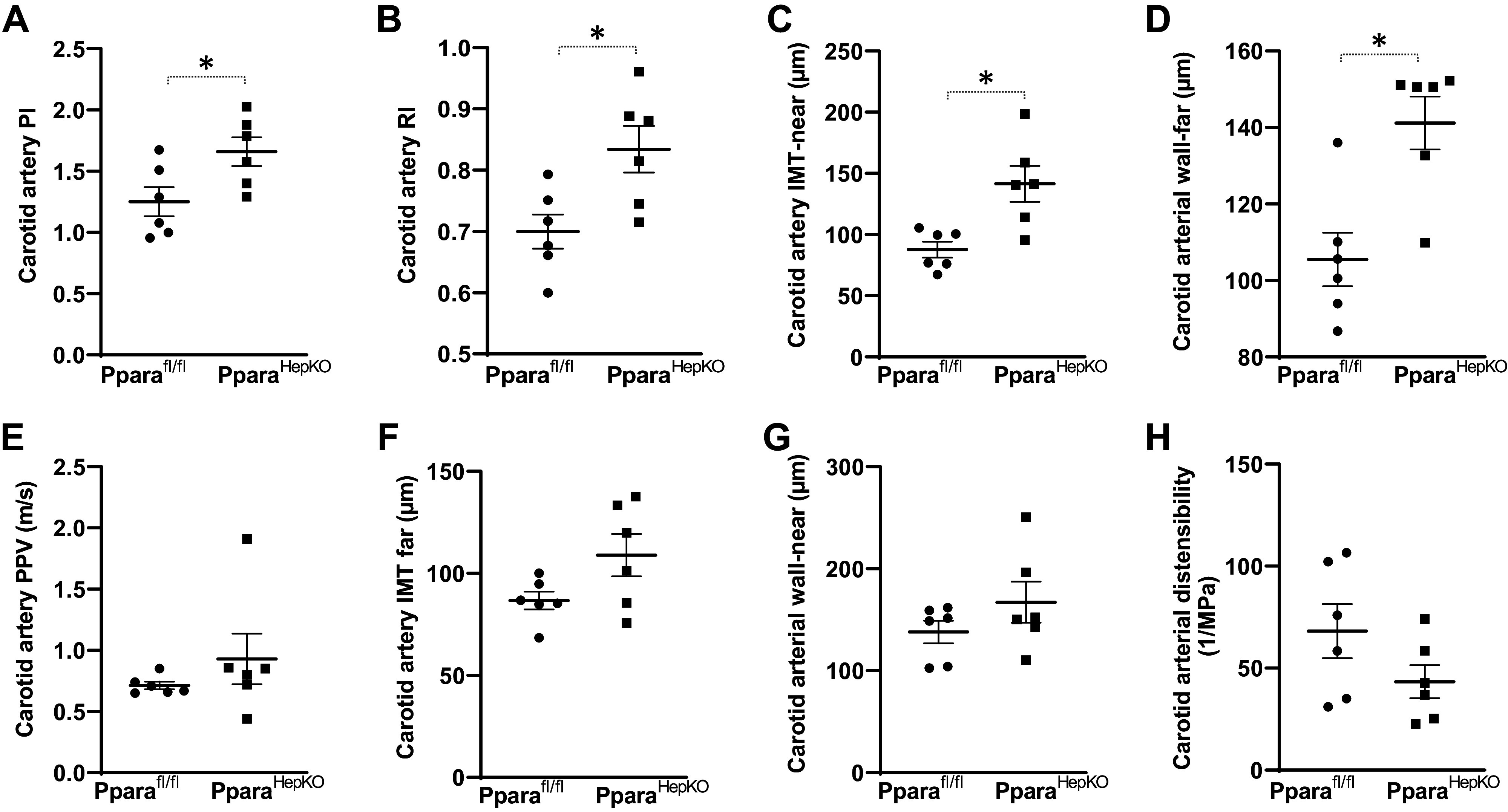
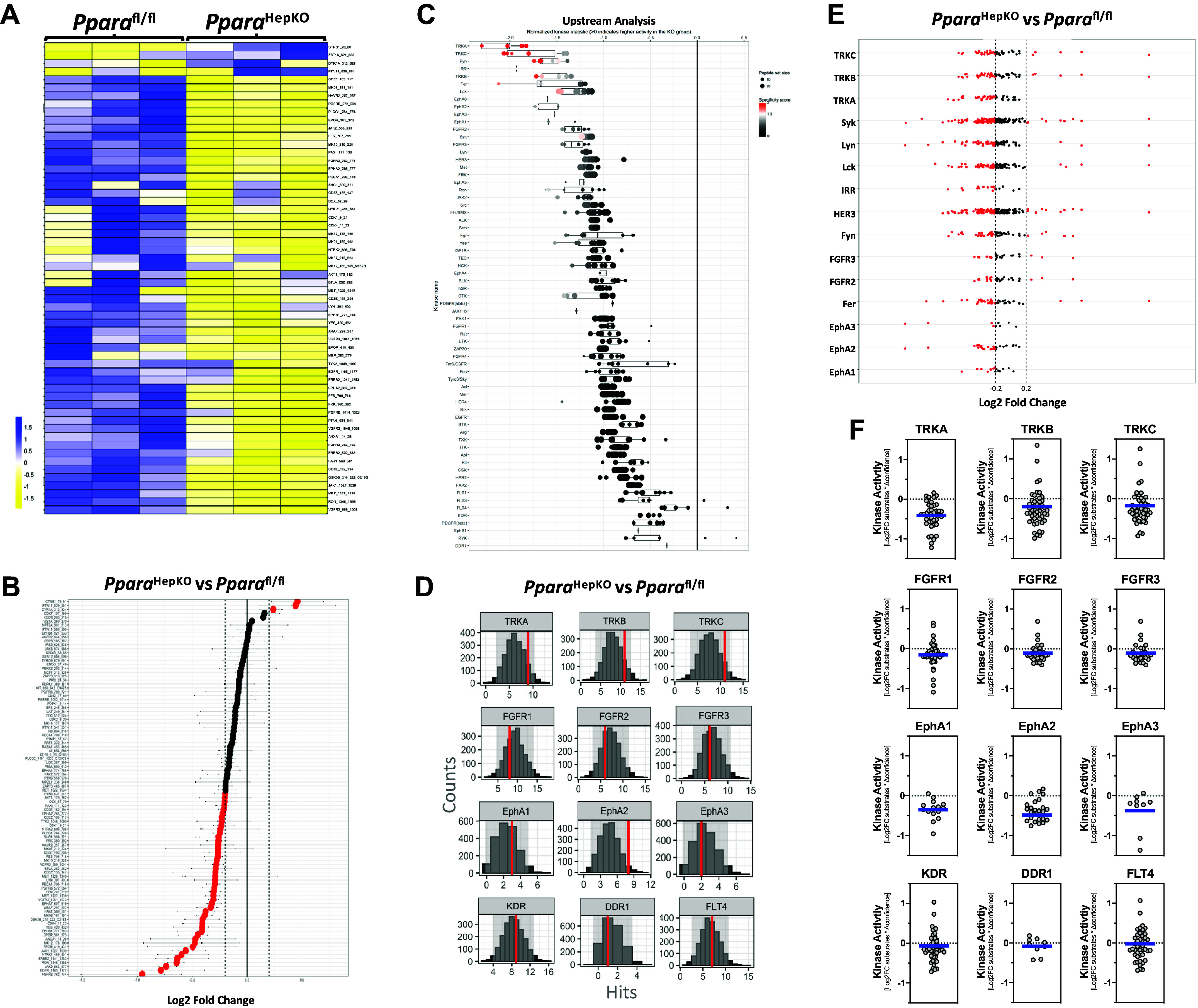
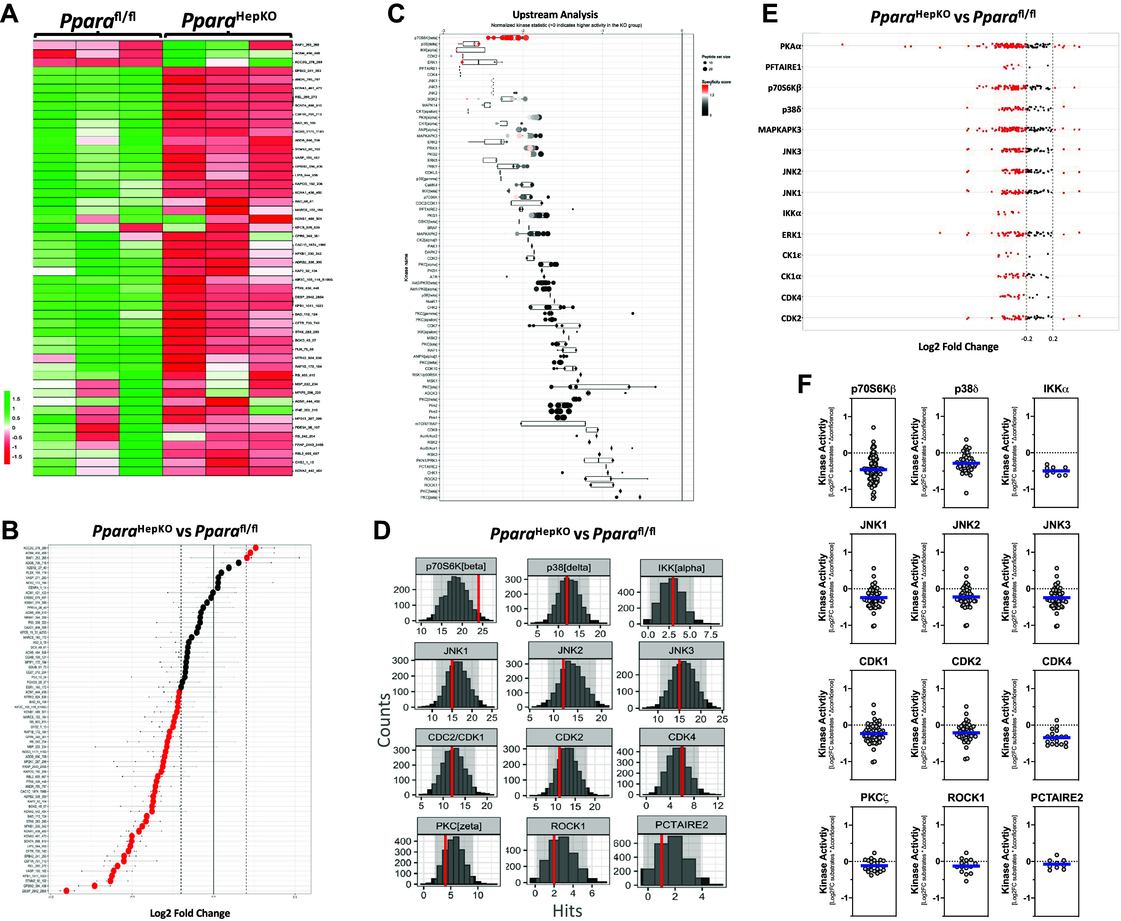
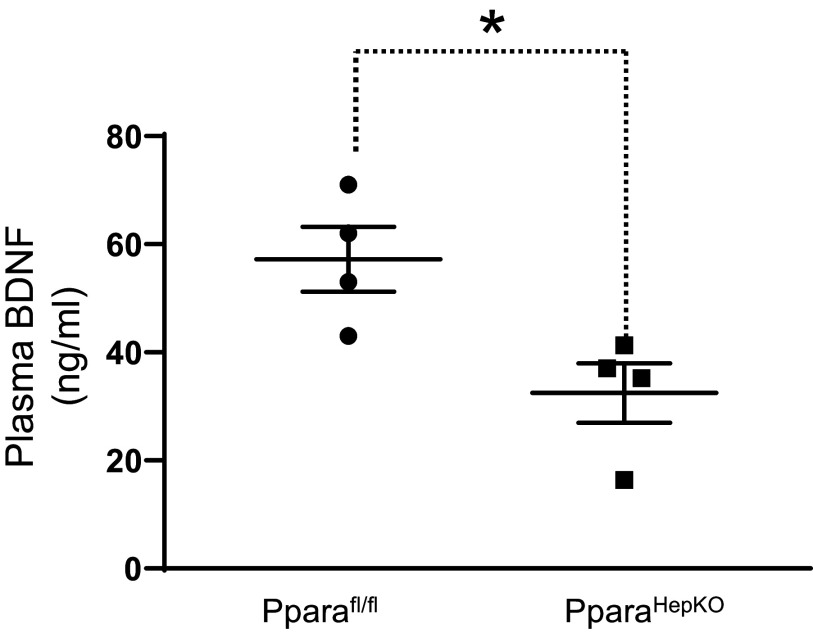
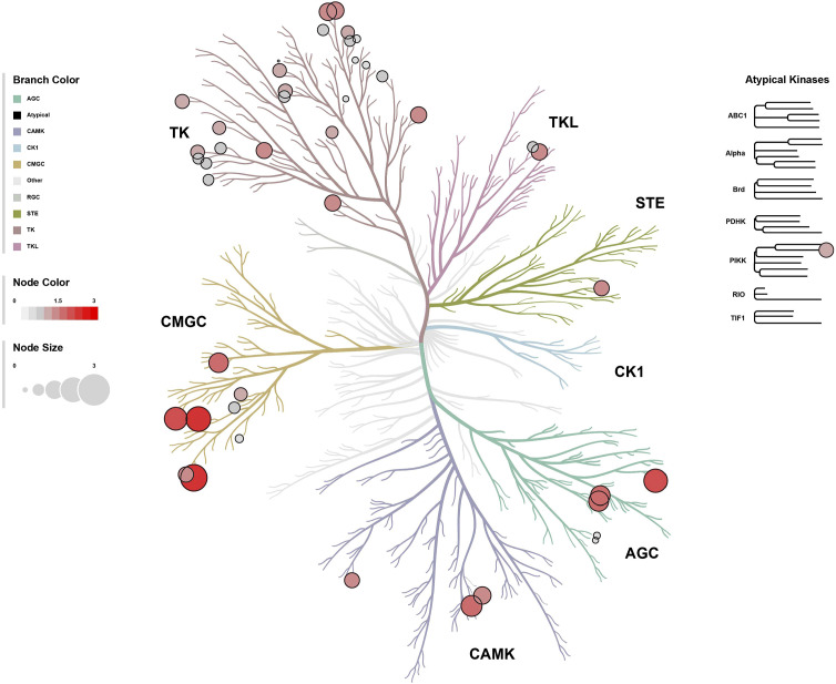
The leading cause of death in patients with nonalcoholic fatty liver disease (NAFLD) is cardiovascular disease (CVD). However, the mechanisms are unknown. Mice deficient in hepatocyte proliferator-activated receptor-α (PPARα) (PparaHepKO) exhibit hepatic steatosis on a regular chow diet, making them prone to manifesting NAFLD. We hypothesized that the PparaHepKO mice might be predisposed to poorer cardiovascular phenotypes due to increased liver fat content. Therefore, we used PparaHepKO and littermate control mice fed a regular chow diet to avoid complications with a high-fat diet, such as insulin resistance and increased adiposity. After 30 wk on a standard diet, male PparaHepKO mice exhibited elevated hepatic fat content compared with littermates as measured by Echo MRI (11.95 ± 1.4 vs. 3.74 ± 1.4%, P < 0.05), hepatic triglycerides (1.4 ± 0.10 vs. 0.3 ± 0.01 mM, P < 0.05), and Oil Red O staining, despite body weight, fasting blood glucose, and insulin levels being the same as controls. The PparaHepKO mice also displayed elevated mean arterial blood pressure (121 ± 4 vs. 108 ± 2 mmHg, P < 0.05), impaired diastolic function, cardiac remodeling, and enhanced vascular stiffness. To determine mechanisms controlling the increase in stiffness in the aorta, we used state-of-the-art PamGene technology to measure kinase activity in this tissue. Our data suggest that the loss of hepatic PPARα induces alterations in the aortas that reduce the kinase activity of tropomyosin receptor kinases and p70S6K kinase, which might contribute to the pathogenesis of NAFLD-induced CVD. These data indicate that hepatic PPARα protects the cardiovascular system through some as-of-yet undefined mechanism.

Keywords: cardiac dysfunction; hepatic steatosis; hypertension; lean NAFLD; nonalcoholic fatty liver disease.


Conflict of interest statement

No conflicts of interest, financial or otherwise, are declared by the authors.

Figures

Figure 1.
Figure 1.
Generation and characterization of PparaHepKO and Pparafl/fl mice. PparaHepKO mice were generated by crossing PparaHepKO mice heterozygous for Albumin-Cre with Pparafl/fl mice generating litters with Cre-positive and Cre-negative mice. Hepatic Ppara levels were determined by Western blot and normalized to the levels of HSP90. **P < 0.01 vs. Pparafl/fl; n = 3 mice per group. P < 0.01 by unpaired t test. KO, knockout, WT, wild-type.
Figure 2.
Figure 2.
Analysis of metabolic and cardiovascular parameters in PparaHepKO and Pparafl/fl mice. Representative Oil red O staining of livers from PparaHepKO and floxed littermate control (Pparafl/fl), echo MRI steatosis measurements, and hepatic triglycerides (A), and fasting blood glucose levels and fasting plasma insulin levels (B). Values are means ± SE; *P < 0.05 vs. Pparafl/ by uparied t test; A: n = 7 mice per group; B–D: n = 6 mice per group. Scale bar = 50 μm. C: day and night mean arterial pressure (MAP) over the 7-day recording period and 7-day average MAP. D: heart rates for day and night of the 7-day recording period and 7-day average heart rate. E: real-time PCR expression of PPARα mRNA in the ventricle. Values are means ± SE; *P < 0.05 vs. Pparafl/fl (during the day) by uparied t test; n = 4 mice per group. D = day, N = night. PPARα, proliferator-activated receptor-α.
Figure 3.
Figure 3.
Assessment of cardiac function and remodeling in PparaHepKO and Pparafl/fl mice. A: a representative image of the dilated left ventricular chamber, LVID during systole, LVID during diastole, and LVAW thickness during systole in PparaHepKO and Pparafl/fl mice. B: quantification of the LVAW thickness during diastole, LVPW thickness during systole, and LVPW thickness during diastole in PparaHepKO and Pparafl/fl mice. Values are means ± SE; *P < 0.05 vs. Pparafl/fl; n = 17 and n = 18 mice per group, Pparafl/fl, PparaHepKO. Measurements of MV E, MV A, and E/A ratio (C), and evaluation of e′, E/e′, and IVRT in PparaHepKO and Pparafl/fl mice (D). Values are means ± SE; *P < 0.05 vs. Pparafl/fl by unpaired t test; n = 17 and n = 18 mice per group, Pparafl/fl, PparaHepKO. Cardiac output, left ventricular mass, and left-ventricular end-diastolic volume (E), and stroke volume and ejection fraction in PparaHepKO and Pparafl/fl mice (F). Values are means ± SE; *P < 0.05 vs. PPARα floxed control (Pparafl/fl) by unpaired t test; n = 17 and n = 18 mice per group, Pparafl/fl, PparaHepKO. A, peak flow velocity in late diastole; e′, early diastolic mitral annular velocity; E, peak flow velocity in early diastole; IVRT, isovolumetric relaxation time; LVAW, left ventricular anterior wall; LVID, left ventricular internal diameter; LVPW, left ventricular posterior wall; MV, mitral valve; PPARα, proliferator-activated receptor-α.
Figure 4.
Figure 4.
Phenotypes of the abdominal aortic in PparaHepKO and Pparafl/fl mice. Measurements of PI (A), RI (B), IMT-near (C), distensibility (D), PPV (E), IMT-far (F), wall-near thickness (G), and wall-far thickness (H) in PparaHepKO and Pparafl/fl mice. Values are means ± SE; *P < 0.05 vs. Pparafl/fl by unpaired t test; A–H; n = 6 mice per group. IMT, intima-media thickness; PI, pulsatility index; PPV, pulse propagation velocity; RI, resistive index.
Figure 5.
Figure 5.
Common carotid artery phenotype in PparaHepKO and Pparafl/fl mice. Measurements of PI (A), RI (B), IMT-near (C), wall-far thickness (D), PPV (E), IMT-far (F), wall-near thickness (G), and distensibility (H) in PparaHepKO and Pparafl/fl mice. Values are means ± SE; *P < 0.05 vs. Pparafl/fl by unpaired t test; A–H; n = 6 mice per group. IMT, intima-media thickness; PI, pulsatility index; PPV, pulse propagative velocity; RI, resistive index.
Figure 6.
Figure 6.
Tyrosine kinase signaling pathways in the aortas of PparaHepKO and Pparafl/fl mice. A and B: heatmap of substrate phosphorylation levels for tyrosine kinases and waterfall plot for comparison of kinase activity in PparaHepKO and Pparafl/fl mice. C: upstream tyrosine kinase analysis is represented by a waterfall plot. D: quantification of tyrosine kinases by histogram peacock plots comparing PparaHepKO vs. Pparafl/fl mice. E: tyrosine kinase substrate activity for each subfamily of tyrosine kinases. The red dots of the Log2 fold change images indicate increased or decreased activity for each individual kinase in the aortas of the PparaHepKO and Pparafl/fl mice. Red coloring indicates higher specificity. F: differentially active individual tyrosine kinases in the aortas of the PparaHepKO and Pparafl/fl mice. The blue line represents the average of the activity for each subfamily of kinases.
Figure 7.
Figure 7.
Serine/threonine kinase signaling pathways in the aortas of PparaHepKO and Pparafl/fl mice. A and B: heatmap of substrate phosphorylation levels for serine/threonine kinases and waterfall plot for comparison of kinase activity in PparaHepKO and Pparafl/fl mice. C: upstream serine/threonine kinase analysis represented by waterfall plot. D: quantification of serine/threonine kinases by histogram peacock plots comparing PparaHepKO vs. Pparafl/fl mice. E: serine/threonine kinase substrate activity for each subfamily of kinases. Red dots of the Log2 fold change images indicate increased or decreased activity for each individual kinase in the aortas of the PparaHepKO and Pparafl/fl mice. Red coloring indicates higher specificity. F: differentially active individual serine/threonine kinases in the aortas of the PparaHepKO and Pparafl/fl mice. Blue line represents the average of the activity for each subfamily of kinases.
Figure 8.
Figure 8.
Plasma brain-derived neurotrophic factor (BDNF) in PparaHEPKO and Pparafl/fl mice. Plasma BDNF levels were measured in 30-wk-old male PparaHEPKO and Pparafl/fl mice. *P < 0.05 vs. Pparafl/fl by unpaired t test. n = 4 mice per group.
Figure 9.
Figure 9.
Kinase phyla tree analysis in the aortas of PparaHepKO and Pparafl/fl mice. The paralogous phylogenic relationships between differentially altered kinases in aortas of PparaHepKO and Pparafl/fl mice. Node color and size of the bubble plot on the paralogous phylogenic tree correspond to the final kinase score from the PTK and STK upstream kinase analysis. PTK, protein tyrosine kinase; STK, serine-threonine kinase.

References

    1. Muzurović E, Peng CC, Belanger MJ, Sanoudou D, Mikhailidis DP, Mantzoros CS. Nonalcoholic fatty liver disease and cardiovascular disease: a review of shared cardiometabolic risk factors. Hypertension 79: 1319–1326, 2022. doi:10.1161/HYPERTENSIONAHA.122.17982. - DOI - PubMed
    1. Ismaiel A, Dumitraşcu DL. Cardiovascular risk in fatty liver disease: the liver-heart axis-literature review. Front Med (Lausanne) 6: 202, 2019. doi:10.3389/fmed.2019.00202. - DOI - PMC - PubMed
    1. Hinds TD Jr, Stec DE. Bilirubin safeguards cardiorenal and metabolic diseases: a protective role in health. Curr Hypertens Rep 21: 87, 2019. doi:10.1007/s11906-019-0994-z. - DOI - PMC - PubMed
    1. Weaver L, Hamoud AR, Stec DE, Hinds TD Jr. Biliverdin reductase and bilirubin in hepatic disease. Am J Physiol Gastrointest Liver Physiol 314: G668–G676, 2018. doi:10.1152/ajpgi.00026.2018. - DOI - PMC - PubMed
    1. Hinds TD Jr, Stec DE. Bilirubin, a cardiometabolic signaling molecule. Hypertension 72: 788–795, 2018. [Erratum in Hypertension 72: e95, 2018]. doi:10.1161/HYPERTENSIONAHA.118.11130. - DOI - PMC - PubMed

Publication types

LinkOut - more resources