. 2023 Jun 15;207(12):1565-1575.
doi: 10.1164/rccm.202209-1707OC.

Distinct Epithelial-Innate Immune Cell Transcriptional Circuits Underlie Airway Hyperresponsiveness in Asthma

Affiliations

Distinct Epithelial-Innate Immune Cell Transcriptional Circuits Underlie Airway Hyperresponsiveness in Asthma

Ryan C Murphy et al. Am J Respir Crit Care Med. .

Abstract

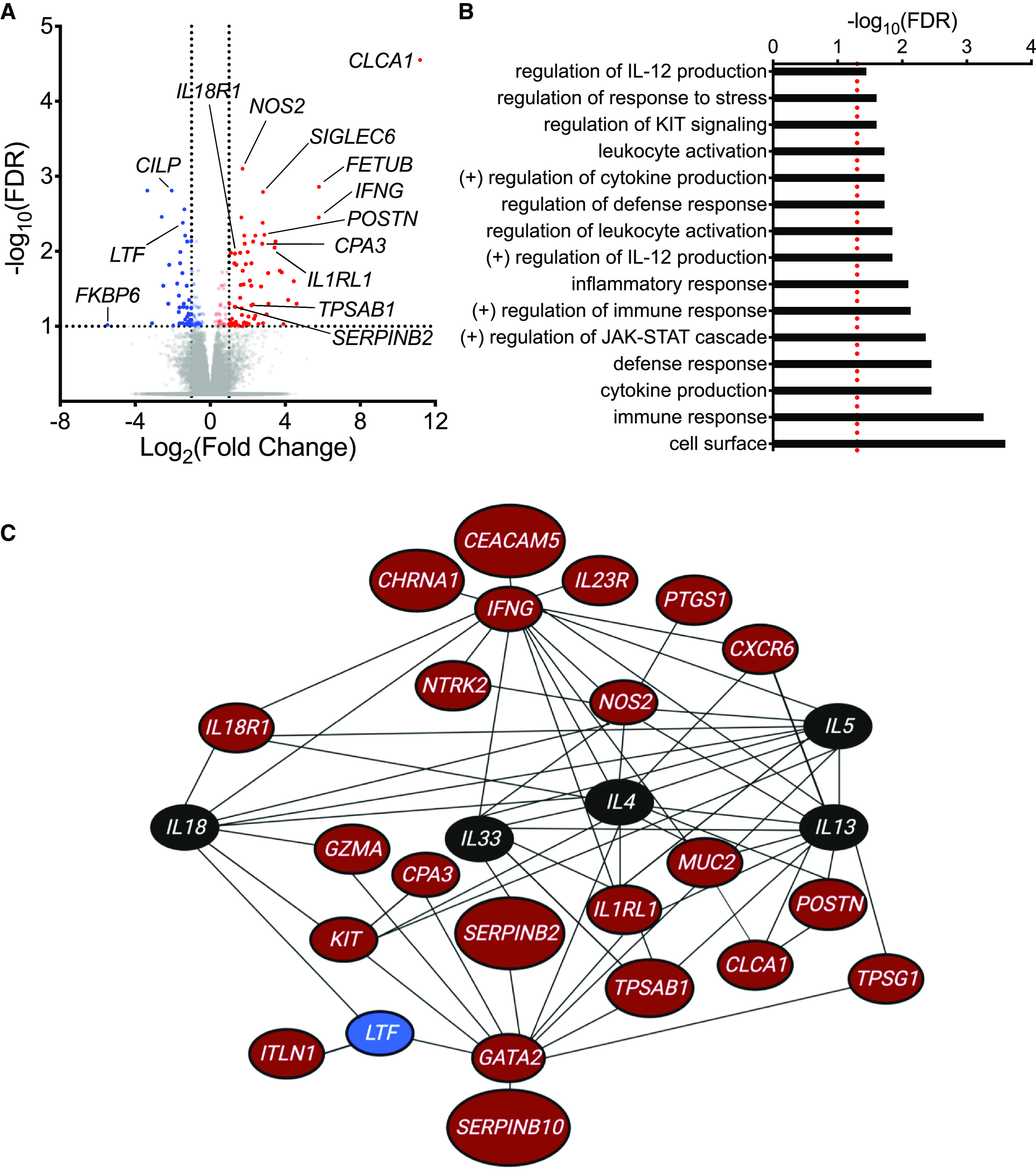
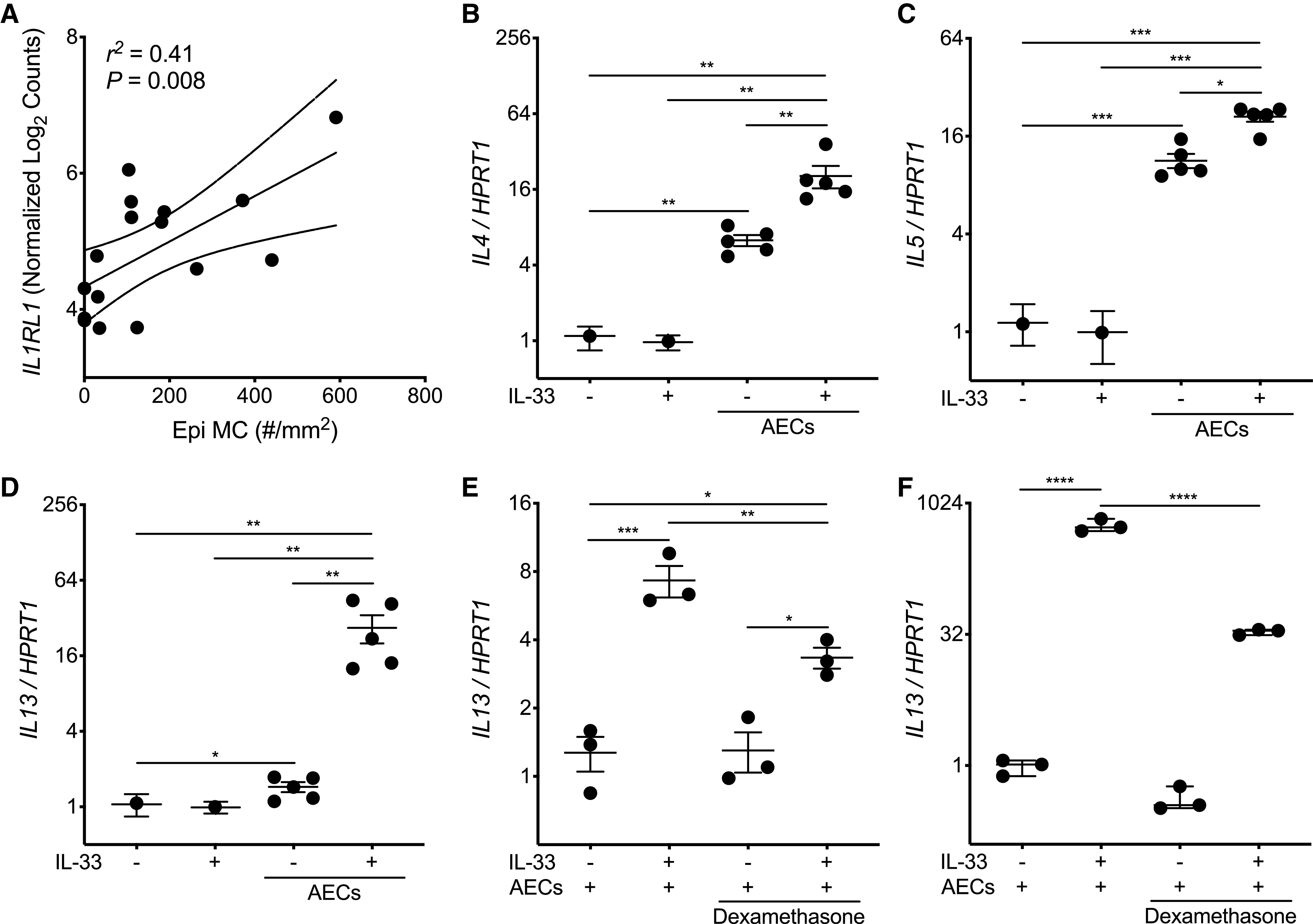
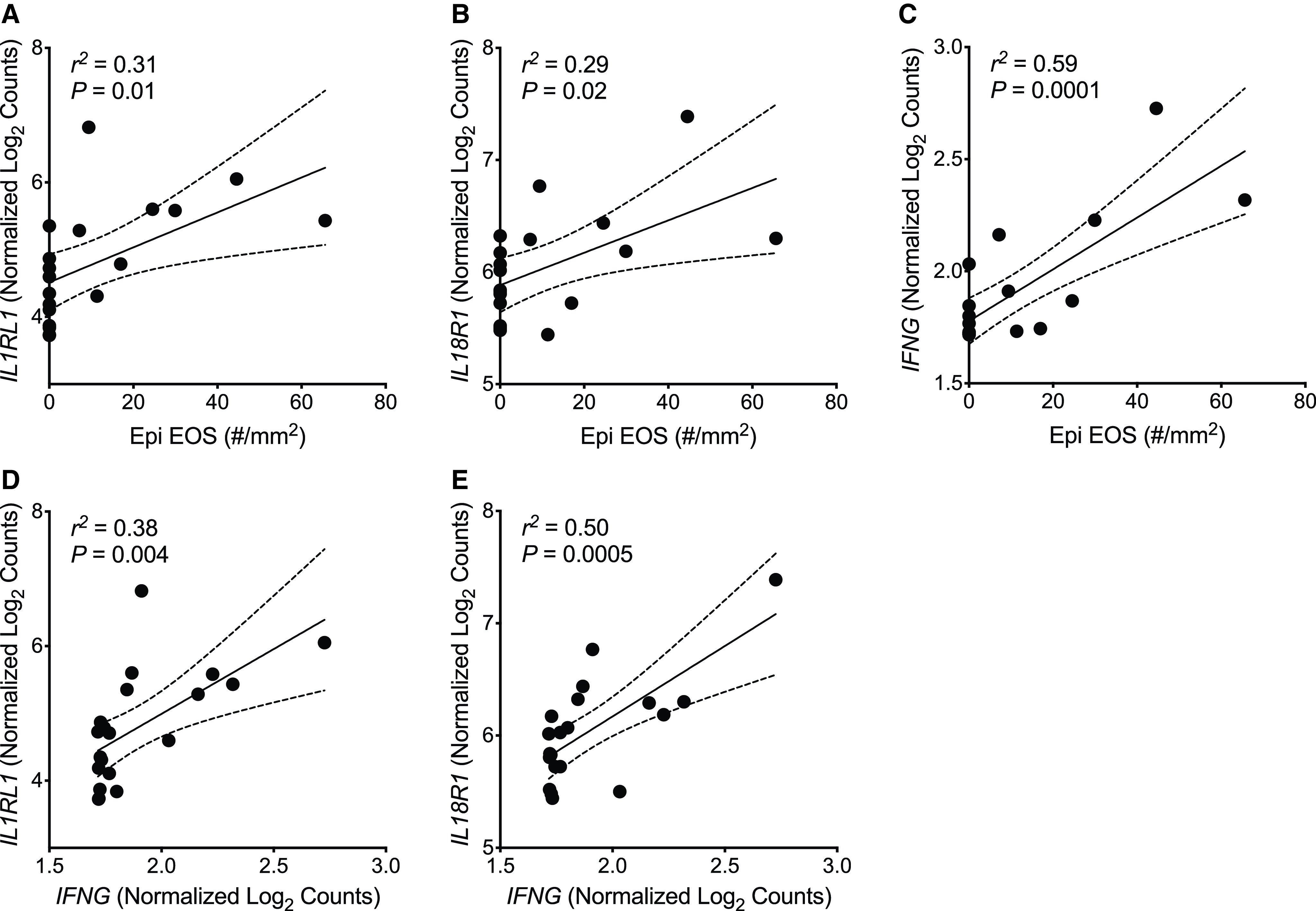
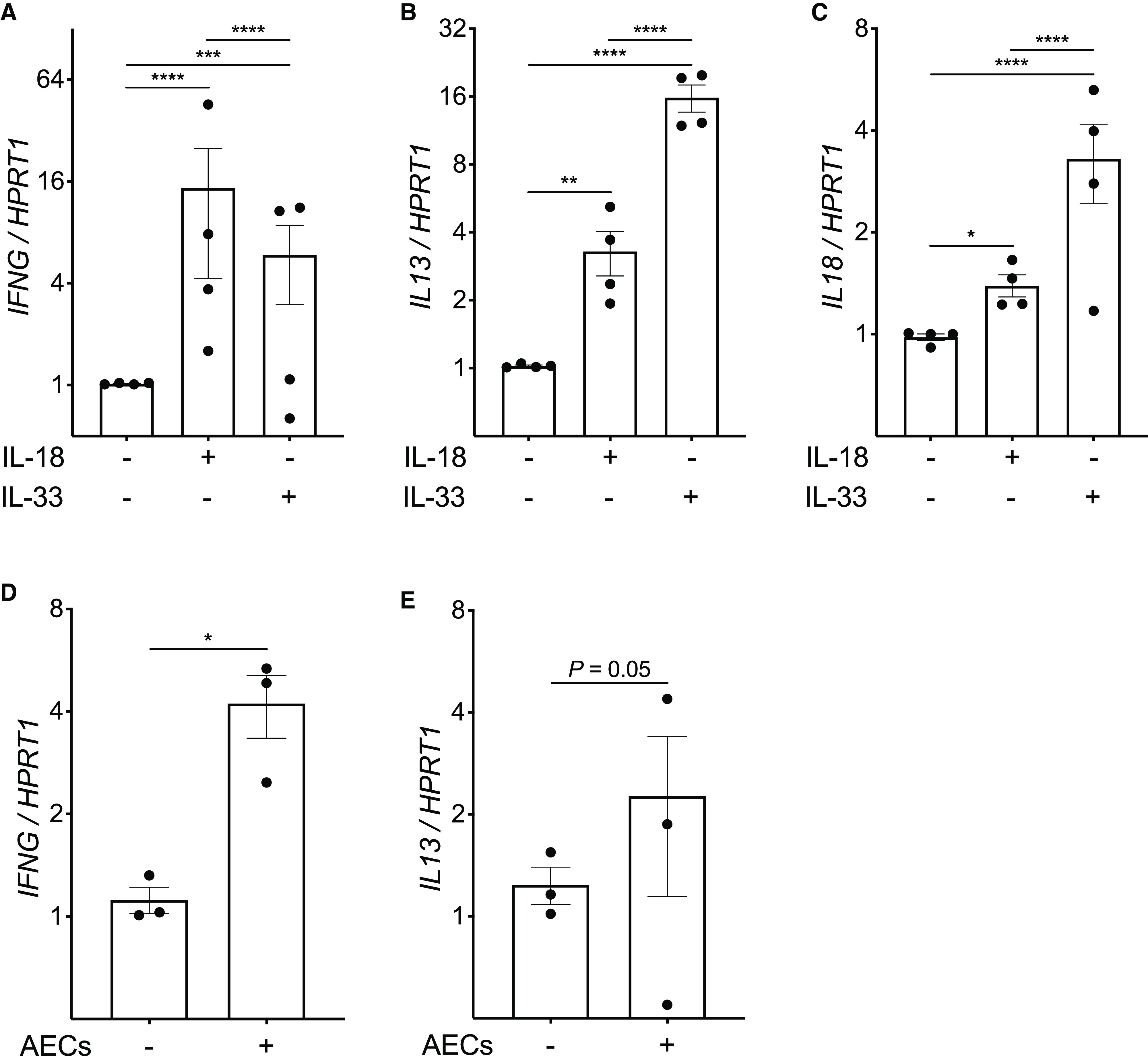
Rationale: Indirect airway hyperresponsiveness (AHR) is a highly specific feature of asthma, but the underlying mechanisms responsible for driving indirect AHR remain incompletely understood. Objectives: To identify differences in gene expression in epithelial brushings obtained from individuals with asthma who were characterized for indirect AHR in the form of exercise-induced bronchoconstriction (EIB). Methods: RNA-sequencing analysis was performed on epithelial brushings obtained from individuals with asthma with EIB (n = 11) and without EIB (n = 9). Differentially expressed genes (DEGs) between the groups were correlated with measures of airway physiology, sputum inflammatory markers, and airway wall immunopathology. On the basis of these relationships, we examined the effects of primary airway epithelial cells (AECs) and specific epithelial cell-derived cytokines on both mast cells (MCs) and eosinophils (EOS). Measurements and Main Results: We identified 120 DEGs in individuals with and without EIB. Network analyses suggested critical roles for IL-33-, IL-18-, and IFN-γ-related signaling among these DEGs. IL1RL1 expression was positively correlated with the density of MCs in the epithelial compartment, and IL1RL1, IL18R1, and IFNG were positively correlated with the density of intraepithelial EOS. Subsequent ex vivo modeling demonstrated that AECs promote sustained type 2 (T2) inflammation in MCs and enhance IL-33-induced T2 gene expression. Furthermore, EOS increase the expression of IFNG and IL13 in response to both IL-18 and IL-33 as well as exposure to AECs. Conclusions: Circuits involving epithelial interactions with MCs and EOS are closely associated with indirect AHR. Ex vivo modeling indicates that epithelial-dependent regulation of these innate cells may be critical in indirect AHR and modulating T2 and non-T2 inflammation in asthma.

Keywords: airway epithelium; airway hyperresponsiveness; asthma; eosinophil; mast cell.


Figures

Figure 1.
Figure 1.
RNA-sequencing analysis of epithelial brushings obtained from individuals with and without exercise-induced bronchoconstriction (EIB). (A) A volcano plot showing the significance and fold-change differences in gene expression between individuals with and without EIB. Red indicates genes that have significantly higher expression, and blue indicates genes that have significantly lower expression in individuals with EIB. False discovery rate (FDR), <0.1; log2(fold change), ⩾1.0 or ⩽−1.0. (B) Gene Ontology biologic processes, cellular components, and molecular functions as well as pathways from the Kyoto Encyclopedia of Genes and Genomes and Reactome databases that were overrepresented among the 120 differentially expressed genes (DEGs) between asthmatic individuals with and without EIB using WebGestalt (FDR <0.05, represented by a red dashed line). (C) Network analysis of DEGs using Ingenuity Pathway Analysis. Genes indicated in red have significantly higher expression, and genes indicated in blue have significantly lower expression in individuals with EIB relative to individuals without EIB. Genes indicated in black are not DEGs identified in our RNA-sequencing analysis but were added to the network to demonstrate interactions between key cytokines of interest (IL4, IL5, IL13, IL18, and IL33) and DEGs.
Figure 2.
Figure 2.
Airway epithelial cells (AECs) promote mast cell (MC) type-2 (T2) gene expression at baseline and enhance sustained T2 gene expression in response to IL-33. (A) Simple linear regression analysis of the density of MCs in the epithelial compartment (Epi MC) versus IL1RL1 expression identified from the epithelial brushings of matched study participants. A solid linear regression line is shown, with the 95% confidence interval indicated with dotted lines. (B–D) Quantitative PCR analysis for expression of IL4 (B), IL5 (C), and IL13 (D) (in comparison with the housekeeping gene HPRT1) in the Laboratory of Allergic Diseases-2 (LAD2) MCs cultured for 48 hours in the presence or absence of AECs obtained from five healthy adult AEC donors and/or IL-33 (10 ng/ml). Individual data points represent the mean value of four LAD2 MC replicates, and each replicate was the result of three PCR reactions. Thus, data columns with AECs have five individual data points, each representing four LAD2 MC replicates cocultured with a single AEC donor. Mean values are indicated, with error bars representing the SEM. P values represent the result of one-way ANOVAs with repeated measures. (E) Quantitative PCR of LAD2 MCs (n = 4; each replicate was the result of three PCR reactions) cocultured with a single healthy adult AEC donor in the presence of IL-33 (10 ng/ml) or dexamethasone (1 μM). Mean values are indicated, with error bars representing the SEM. P values represent the result of a one-way ANOVA. (F) Human MCs derived from CD34+ cells isolated from the peripheral blood of a single donor were cocultured with primary AECs from a single healthy adult donor for 48 hours in the presence or absence of IL-33 (10 ng/ml) and/or dexamethasone (1 μM). n = 3 per condition, and each data point is the result of three PCR reactions. Mean values are indicated, with error bars representing the SEM. P values represent the result of a one-way ANOVA. *P < 0.05, **P < 0.01, ***P < 0.001, and ****P < 0.0001.
Figure 3.
Figure 3.
Eosinophils (EOS) within the epithelial compartment are positively correlated with expression of IFN-γ and the receptors for IL-33 and IL-18. (A–C) Simple linear regression analyses of the density of EOS within the epithelial compartment (Epi EOS) and expression of IL1RL1 (A), IL18R1 (B), and IFNG (C) identified from the epithelial brushings of matched study participants. (D and E) Simple linear regression analyses of IFNG expression and expression of IL1RL1 (D) and IL18R1 (E) identified from the epithelial brushings of matched study participants. A solid linear regression line is shown, with the 95% confidence interval indicated with dotted lines.
Figure 4.
Figure 4.
IL-18, IL-33, and airway epithelial cells (AECs) induce IFN-γ and type-2 (T2) gene expression in eosinophils (EOS). (A–C) Quantitative PCR analysis for expression of IFNG (A), IL13 (B), and IL18 (C) (in comparison with the housekeeping gene HPRT1) in EOS isolated from the peripheral blood of four human donors cultured for 2 hours in the presence or absence of IL-18 (10 ng/ml) or IL-33 (10 ng/ml). Individual data points represent the mean value of four EOS replicates per EOS donor; each replicate was the result of three PCR reactions. Mean values are indicated, with error bars representing the SEM. (D and E) Quantitative PCR analysis for expression of IFNG (D) and IL13 (E) in EOS isolated from the peripheral blood of three human donors cultured for 8 hours in the presence or absence of primary AECs from a single healthy adult donor. Individual data points represent the mean value of three EOS replicates per EOS donor; each replicate was the result of three PCR reactions. Mean values are indicated, with error bars representing the SEM. P values represent the result of two-way ANOVAs. *P < 0.05, **P < 0.01, ***P < 0.001, and ****P < 0.0001.

Comment in

Similar articles

Cited by

References

    1. Hallstrand TS, Leuppi JD, Joos G, Hall GL, Carlsen KH, Kaminsky DA, et al. American Thoracic Society (ATS)/European Respiratory Society (ERS) Bronchoprovocation Testing Task Force ERS technical standard on bronchial challenge testing: pathophysiology and methodology of indirect airway challenge testing. Eur Respir J . 2018;52:1801033. - PubMed
    1. Coates AL, Wanger J, Cockcroft DW, Culver BH, Carlsen K-H, Diamant Z, et al. Bronchoprovocation Testing Task Force ERS technical standard on bronchial challenge testing: general considerations and performance of methacholine challenge tests. Eur Respir J . 2017;49:1601526. - PubMed
    1. Stern DA, Morgan WJ, Halonen M, Wright AL, Martinez FD. Wheezing and bronchial hyper-responsiveness in early childhood as predictors of newly diagnosed asthma in early adulthood: a longitudinal birth-cohort study. Lancet . 2008;372:1058–1064. - PMC - PubMed
    1. Cabral AL, Conceição GM, Fonseca-Guedes CH, Martins MA. Exercise-induced bronchospasm in children: effects of asthma severity. Am J Respir Crit Care Med . 1999;159:1819–1823. - PubMed
    1. Altman MC, Lai Y, Nolin JD, Long S, Chen CC, Piliponsky AM, et al. Airway epithelium-shifted mast cell infiltration regulates asthmatic inflammation via IL-33 signaling. J Clin Invest . 2019;129:4979–4991. - PMC - PubMed

Publication types