. 2023 Oct;60(10):e14336.
doi: 10.1111/psyp.14336. Epub 2023 May 22.

Multimodal study of the neural sources of error monitoring in adolescents and adults

Affiliations

Multimodal study of the neural sources of error monitoring in adolescents and adults

Stefania Conte et al. Psychophysiology. 2023 Oct.

Erratum in

Abstract

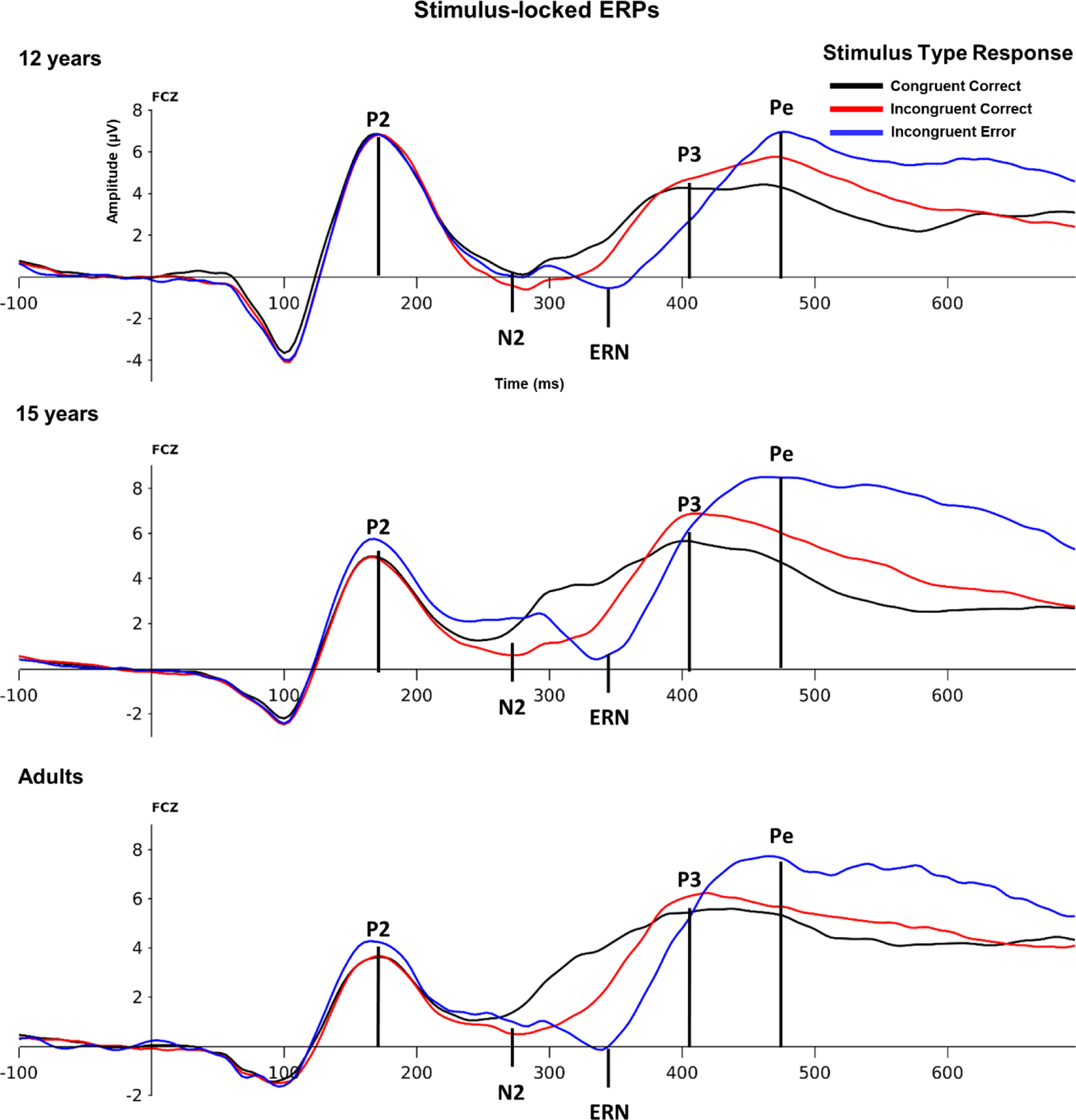
The ability to monitor performance during a goal-directed behavior differs among children and adults in ways that can be measured with several tasks and techniques. As well, recent work has shown that individual differences in error monitoring moderate temperamental risk for anxiety and that this moderation changes with age. We investigated age differences in neural responses linked to performance monitoring using a multimodal approach. The approach combined functional MRI and source localization of event-related potentials (ERPs) in 12-year-old, 15-year-old, and adult participants. Neural generators of two components related to performance and error monitoring, the N2 and ERN, lay within specific areas of fMRI clusters. Whereas correlates of the N2 component appeared similar across age groups, age-related differences manifested in the location of the generators of the ERN component. The dorsal anterior cingulate cortex (dACC) was the predominant source location for the 12-year-old group; this area manifested posteriorly for the 15-year-old and adult groups. A fMRI-based ROI analysis confirmed this pattern of activity. These results suggest that changes in the underlying neural mechanisms are related to developmental changes in performance monitoring.

Keywords: ERN; ERP; adolescents; decision-making; fMRI; flanker task.


Figures

Figure 1.
Figure 1.
Neurosynth ROIs overlaid on the rendered adult brain template.
Figure 2.
Figure 2.
Stimulus-locked ERPs for the congruent correct (dotted lines), incongruent error (solid lines), and incongruent correct (dashed lines) conditions on FCz, separately for the three testing ages.
Figure 3.
Figure 3.
a) Response-locked ERPs for the congruent correct and incorrect conditions over fronto- and parietal-central channels. b) algebraic difference of the ERN activity between error and correct trials (i.e., ΔERN) over five midline channels from frontal to parietal areas. In all panels the black lines represent the activity of 12-year-old participants, the red lines are for the 15-year-old group, and the blue lines are for adult participants.
Figure 4.
Figure 4.
Top panels depict the scalp activity at the ΔERN peak for the three age groups. A white circle is placed to mark the location of the reference channels (Cz) to show how the negative central activity moves posteriorly in adult participants versus adolescents. Mean activity around the ERN peak is depicted in then bar graphs as a function of participant age for the midline channels. A raincloud plot is reported for the CPz channel. Full dots represent individual data points. Error bars represent the standard error from the mean.
Figure 5.
Figure 5.
Sagittal view of the CDR source activity for each ERP component plotted on the age-specific templates. Note that P1, N1, and N2 are stimulus-locked activities, whereas ERN and Pe are response-locked activities.
Figure 6.
Figure 6.
The pattern of ΔCDR activity at the latency of the ΔERN component is depicted in the line plots for each considered ROI as a function of participant’s age. Bar plots report the average ΔCDR at the peak of the ERN activity. The largest ΔCDR activity was localized in the dACC for the adolescent groups and more posteriorly (PCC) in adults. Raincloud plots depict the ΔCDR activity in PCC as a function of age. Dots represent individual data points. Error bars indicate the SE form the mean.
Figure 7.
Figure 7.
T-values of the PALM clustering procedure along with the Gamma and Gaussian-Gamma distributions plotted separately for the three testing ages and the Neurosynth data.
Figure 8.
Figure 8.
Sagittal view of the group ΔCDR activity at the peak of the response-locked ERN component (second row) overlaid on the age-specific average template, and the fMRI-conditioned ΔCDR (third row). The cross is centered to the anterior commissure of each template. The top row displays the BOLD activity for the error>correct contrast. The reconstructed source activates of both ERN and N2 components are depicted in the bottom row. Overlapping sources are evident for the 12-year-old group, whereas more posterior ERN activity occurred in adults.
Figure 9.
Figure 9.
The pattern of fMRI-conditioned ΔCDR activity at the latency of the ΔERN component is depicted in the line plots for each considered ROI as a function of participant’s age. Bar plots report the average fMRI-conditioned ΔCDR at the peak of the ERN activity. The largest fMRI-conditioned ΔCDR activity was localized in the dACC for the adolescent groups and more posteriorly (PCC) in adults. Raincloud plots depict the fMRI-conditioned ΔCDR activity in PCC as a function of age. Dots represent individual data points. Error bars indicate the SE form the mean.
Figure 10.
Figure 10.
The pattern of ΔCDR and fMRI-conditioned ΔCDR activity at the latency of the ΔERN component for the two Neurosynth ROIs showing age effects, PCC and superior parietal cortex, along with the dACC.

References

    1. Aarts K, & Pourtois G (2010). Anxiety not only increases, but also alters early error-monitoring functions. Cognitive, Affective, & Behavioral Neuroscience, 10(4), 479–492. 10.3758/CABN.10.4.479 - DOI - PubMed
    1. Agam Y, Hamalainen MS, Lee AK, Dyckman KA, Friedman JS, Isom M, Makris N, & Manoach DS (2011). Multimodal neuroimaging dissociates hemodynamic and electrophysiological correlates of error processing. Proc Natl Acad Sci U S A, 108(42), 17556–17561. 10.1073/pnas.1103475108 - DOI - PMC - PubMed
    1. Alain C, McNeely HE, He Y, Christensen BK, & West R (2002). Neurophysiological Evidence of Error-monitoring Deficits in Patients with Schizophrenia. Cerebral cortex, 12(8), 840–846. 10.1093/cercor/12.8.840 - DOI - PubMed
    1. Araki T, Niznikiewicz M, Kawashima T, Nestor PG, Shenton ME, McCarley RW. (2013). Disruption of function-structure coupling in brain regions sub-serving self monitoring in schizophrenia. Schizophr Res 146(1–3):336–43. 10.1016/j.schres.2013.02.028 - DOI - PMC - PubMed
    1. Bartholow BD, Pearson MA, Dickter CL, Sher KJ, Fabiani M, & Gratton G (2005). Strategic control and medial frontal negativity: Beyond errors and response conflict. Psychophysiology, 42(1), 33–42. 10.1111/j.1469-8986.2005.00258.x - DOI - PubMed

Publication types