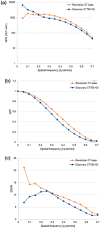
Comparison of image quality, arterial depiction, and radiation dose between two rapid kVp-switching dual-energy CT scanners in CT angiography at 40-keV
- PMID: 37212946
- PMCID: PMC10613589
- DOI: 10.1007/s11604-023-01448-5
Comparison of image quality, arterial depiction, and radiation dose between two rapid kVp-switching dual-energy CT scanners in CT angiography at 40-keV
Abstract
Purpose: To compare the quantitative parameters and qualitative image quality of dual-energy CT angiography (CTA) between two rapid kVp-switching dual-energy CT scanners.
Materials and methods: Between May 2021 and March 2022, 79 participants underwent whole-body CTA using either Discovery CT750 HD (Group A, n = 38) or Revolution CT Apex (Group B, n = 41). All data were reconstructed at 40-keV and with adaptive statistical iterative reconstruction-Veo of 40%. The two groups were compared in terms of CT numbers of the thoracic and abdominal aorta, and the iliac artery, background noise, signal-to-noise ratio (SNR) of the artery, CT dose-index volume (CTDIvol), and qualitative scores for image noise, sharpness, diagnostic acceptability, and arterial depictions.
Results: The median CT number of the abdominal aorta (p = 0.04) and SNR of the thoracic aorta (p = 0.02) were higher in Group B than in Group A, while no difference was observed in the other CT numbers and SNRs of the artery (p = 0.09-0.23). The background noises at the thoracic (p = 0.11), abdominal (p = 0.85), and pelvic (p = 0.85) regions were comparable between the two groups. CTDIvol was lower in Group B than in Group A (p = 0.006). All qualitative scores were higher in Group B than in Group A (p < 0.001-0.04). The arterial depictions were nearly identical in both two groups (p = 0.005-1.0).
Conclusion: In dual-energy CTA at 40-keV, Revolution CT Apex improved qualitative image quality and reduced radiation dose.
Keywords: CT angiography; Dual-energy CT; Modulation transfer function; Noise power spectrum; Rapid kVp-switching.
© 2023. The Author(s).
Figures


References
-
- Achenbach S, Delgado V, Hausleiter J, Schoenhagen P, Min JK, Leipsic JA. SCCT expert consensus document on computed tomography imaging before transcatheter aortic valve implantation (TAVI)/transcatheter aortic valve replacement (TAVR) J Cardiovasc Comput Tomogr. 2012;6(6):366–380. doi: 10.1016/j.jcct.2012.11.002. - DOI - PubMed
-
- Arbab-Zadeh A, Di Carli MF, Cerci R, George RT, Chen MY, Dewey M, et al. Accuracy of computed tomographic angiography and single-photon emission computed tomography-acquired myocardial perfusion imaging for the diagnosis of coronary artery disease. Circ Cardiovasc Imaging. 2015;8(10):e003533. doi: 10.1161/CIRCIMAGING.115.003533. - DOI - PMC - PubMed
-
- Oikonomou EK, Marwan M, Desai MY, Mancio J, Alashi A, Hutt Centeno E, et al. Non-invasive detection of coronary inflammation using computed tomography and prediction of residual cardiovascular risk (the CRISP CT study): a post-hoc analysis of prospective outcome data. Lancet. 2018;392(10151):929–939. doi: 10.1016/S0140-6736(18)31114-0. - DOI - PMC - PubMed
MeSH terms
Substances
LinkOut - more resources
Full Text Sources
Medical
Miscellaneous

