

Comparing confocal and two-photon Ca2+ imaging of thin low-scattering preparations
- PMID: 37213258
- PMCID: PMC10192416
- DOI: 10.1016/j.bpr.2023.100109
Comparing confocal and two-photon Ca2+ imaging of thin low-scattering preparations
Abstract
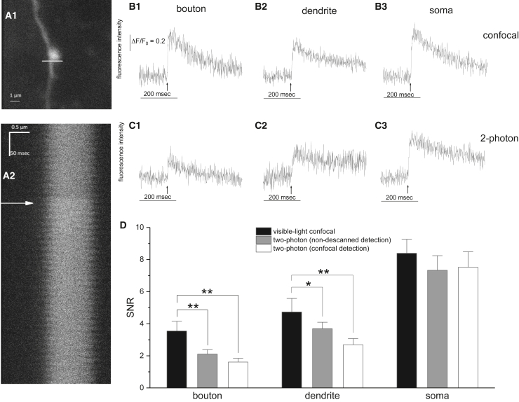
Ca2+ imaging provides insight into biological processes ranging from subcellular dynamics to neural network activity. Two-photon microscopy has assumed a dominant role in Ca2+ imaging. The longer wavelength infra-red illumination undergoes less scattering, and absorption is confined to the focal plane. Two-photon imaging can thus penetrate thick tissue ∼10-fold more deeply than single-photon visible imaging to make two-photon microscopy an exceptionally powerful method for probing function in intact brain. However, two-photon excitation produces photobleaching and photodamage that increase very steeply with light intensity, limiting how strongly one can illuminate. In thin samples, illumination intensity can assume a dominant role in determining signal quality, raising the possibility that single-photon microscopy may be preferable. We therefore tested laser scanning single-photon and two-photon microscopy side by side with Ca2+ imaging in neuronal compartments at the surface of a brain slice. We optimized illumination intensity for each light source to obtain the brightest signal without photobleaching. Intracellular Ca2+ rises elicited by one action potential had twice the signal/noise ratio with confocal as with two-photon imaging in axons, were 31% higher in dendrites, and about the same in cell bodies. The superior performance of confocal imaging in finer neuronal processes likely reflects the dominance of shot noise when fluorescence is dim. Thus, when out-of-focus absorption and scattering are not issues, single-photon confocal imaging can yield better quality signals than two-photon microscopy.
© 2023 The Author(s).
Conflict of interest statement
The authors declare no competing interests.
Figures

References
-
- Jia H., Rochefort N.L., et al. Konnerth A. In vivo two-photon imaging of sensory-evoked dendritic calcium signals in cortical neurons. Nat. Protoc. 2011;6:28–35. - PubMed
-
- Ohki K., Chung S., et al. Reid R.C. Functional imaging with cellular resolution reveals precise micro-architecture in visual cortex. Nature. 2005;433:597–603. - PubMed
-
- Denk W., Strickler J.H., Webb W.W. Two-photon laser scanning fluorescence microscopy. Science. 1990;248:73–76. - PubMed
Publication types
Grants and funding
LinkOut - more resources
Full Text Sources
Miscellaneous
