. 2022;2(1):9.
doi: 10.1038/s43586-022-00098-7. Epub 2022 Feb 10.

High-content CRISPR screening

Affiliations

High-content CRISPR screening

Christoph Bock et al. Nat Rev Methods Primers. 2022.

Abstract

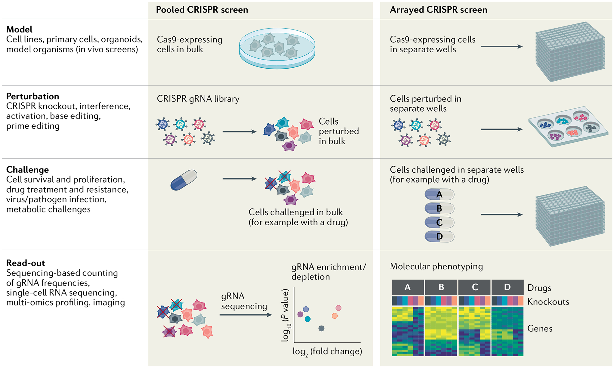
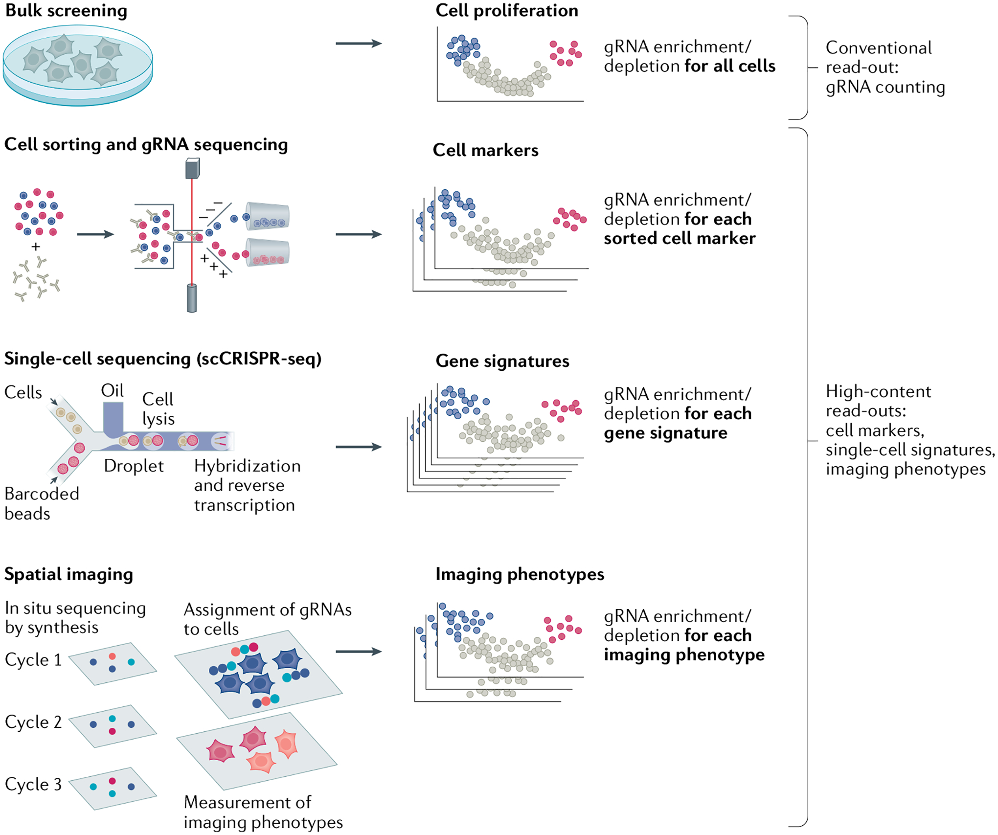
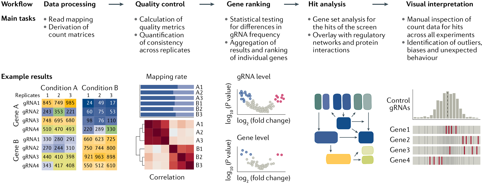
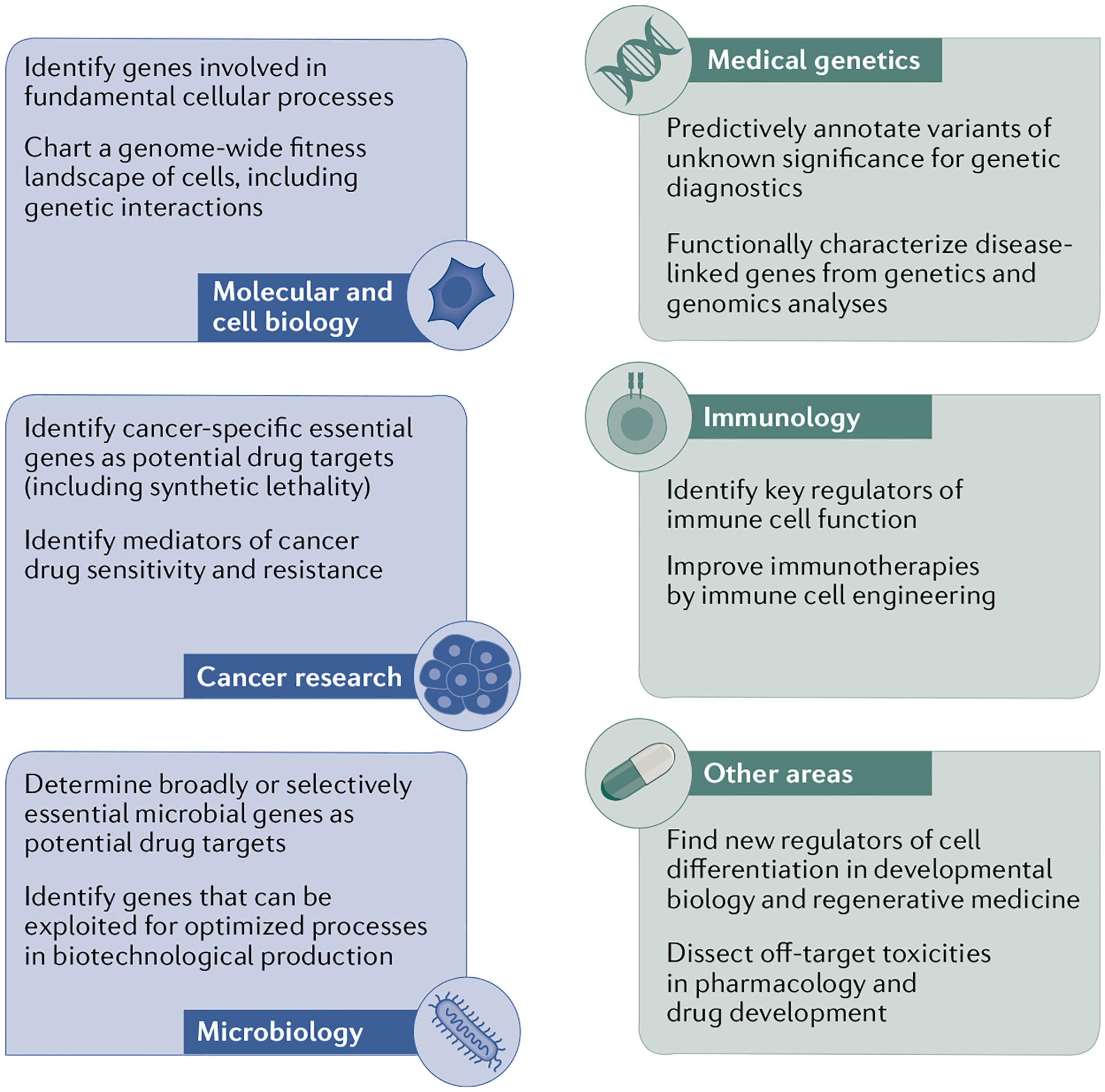
CRISPR screens are a powerful source of biological discovery, enabling the unbiased interrogation of gene function in a wide range of applications and species. In pooled CRISPR screens, various genetically encoded perturbations are introduced into pools of cells. The targeted cells proliferate under a biological challenge such as cell competition, drug treatment or viral infection. Subsequently, the perturbation-induced effects are evaluated by sequencing-based counting of the guide RNAs that specify each perturbation. The typical results of such screens are ranked lists of genes that confer sensitivity or resistance to the biological challenge of interest. Contributing to the broad utility of CRISPR screens, adaptations of the core CRISPR technology make it possible to activate, silence or otherwise manipulate the target genes. Moreover, high-content read-outs such as single-cell RNA sequencing and spatial imaging help characterize screened cells with unprecedented detail. Dedicated software tools facilitate bioinformatic analysis and enhance reproducibility. CRISPR screening has unravelled various molecular mechanisms in basic biology, medical genetics, cancer research, immunology, infectious diseases, microbiology and other fields. This Primer describes the basic and advanced concepts of CRISPR screening and its application as a flexible and reliable method for biological discovery, biomedical research and drug development - with a special emphasis on high-content methods that make it possible to obtain detailed biological insights directly as part of the screen.


Conflict of interest statement

C.B. is a co-founder and scientific advisor of Aelian Biotechnology and Neurolentech. S.C. is a co-founder of EvolveImmune Therapeutics and Cellinfinity Bio. M.G. has performed consultancy for Sanofi, receives research funding from AstraZeneca and GlaxoSmithKline, and is a co-founder of Mosaic Therapeutics. J.M. is a shareholder of Northern Biologics and Pionyr Immunotherapeutics, and a scientific advisor and shareholder of Century Therapeutics and Aelian Biotechnology. L.S.Q. is a co-founder and scientific advisor of Epicrispr Biotechnologies and Refuge Biotechnologies. J.S. is a scientific advisor of Maze Therapeutics, Camp4 Therapeutics, Cajal Biosciences, Adaptive Biotechnologies and Guardant Health, and a co-founder of Scale Bio and Phase Genomics. J.S.W. consults for and holds equity in KSQ Therapeutics, Maze Therapeutics and Tenaya Therapeutics, is a venture partner at 5AM Ventures and is a member of the Amgen Scientific Advisory Board. X.Z. is a co-founder and consultant of Vizgen. The other authors declare no competing interests.

Figures

Fig. 1 |
Fig. 1 |. Experimental design for CRISPR screening.
CRISPR screens can be described along four dimensions: the biological model in which the screen is conducted; perturbations introduced using CRISPR technology; challenges to which the perturbed cells are exposed; and a read-out that measures the induced molecular or cellular effects. In pooled CRISPR screens, perturbations are introduced in bulk. They are genetically encoded and typically read out by guide RNA (gRNA) sequencing. In arrayed CRISPR screens, different perturbations are introduced separately — for example, in different wells of a 96-well plate. As each reaction compartment is subjected to a defined perturbation, the read-out does not need to include gRNA sequencing.
Fig. 2 |
Fig. 2 |. Preparation of CRISPR gRNA libraries.
The guide RNA (gRNA) library defines which genes are probed in a CRISPR screen. Application-specific libraries are designed using bioinformatic tools (detailed in TABLE 1), synthesized as oligonucleotide pools, cloned in bulk into the plasmid vector and packaged into a lentivirus for delivery into cells.
Fig. 3 |
Fig. 3 |. CRISPR-mediated perturbation of cells.
CRISPR technology provides many options to perturb cells. a | Genome editing. Directed by a guide RNA (gRNA), Cas9 nucleases introduce double-strand breaks into the target site; subsequent DNA repair results in compromised gene function (CRISPR knockout (CRISPRko)). CRISPR base editors induce specific mutations, by combining a base modification enzyme, a uracil DNA glycosylase inhibitor (UGI) domain that inhibits base excision repair and a Cas nickase that nicks the non-edited strand of DNA to favour repair with the edited base. A cytosine base editor is shown; adenosine base editors are also available. CRISPR prime editing can introduce new sequence information into the genome; it uses a Cas9 nickase fused to a reverse transcriptase and a prime editing gRNA (pegRNA) corresponding to the target locus, which also provides new genetic information. b | Epigenome editing. Cas9 endonuclease dead (dCas9) can be combined with epigenetic writer and eraser enzymes such as the demethylase Tet1 (not pictured), the methyltransferase DNMT3A or the H3K27 acetyltransferase p300 to induce changes in DNA methylation or histone marks. c | Transcriptional control. CRISPR interference (CRISPRi) uses dCas9 fused to transcriptional repressors such as Krüppel associated box (KRAB), causing repression of genes close to the gRNA target site. CRISPR activation (CRISPRa) uses dCas9 with transcriptional activators such as the VP64 domain and MCP–p65–HSF1 fusion proteins recruited via an MS2 stem–loop sequence. d | RNA modulation. RNA base editors induce specific mutations into RNA molecules using an adenosine deaminase (ADAR) targeted to RNA; it converts adenosine into inosine, which acts as guanine during translation. RNA splicing can be altered at the RNA level by substituting the RNA-binding domain of the RBFOX1 protein with dCas9. Finally, RNA interference (RNAi) can be achieved by a ribonuclease (CasRx) that binds to RNA and cleaves it. PAM, protospacer adjacent motif; RT, reverse transcription.
Fig. 4 |
Fig. 4 |. CRISPR screening with high-content read-out.
Sequencing-based counting of guide RNAs (gRNAs) is a straight-forward and widely used read-out of pooled CRISPR screens, especially for phenotypes that affect cell proliferation and survival. To broaden CRISPR screening to additional cellular phenotypes, several high-content read-outs have been introduced. Cell sorting prior to gRNA sequencing makes it possible to identify gRNAs and genes that affect predefined, sortable cellular phenotypes — such as expression of a fluorescent reporter. Single-cell sequencing of transcriptomes and matched gRNAs can identify regulators of gene expression and transcriptome-linked cell states. Spatial imaging combined with methods to distinguish gRNAs in individual cells makes it possible to identify genes that affect imaging-based cellular phenotypes, for example inducing changes in cell shape.
Fig. 5 |
Fig. 5 |. Bioinformatic analysis of CRISPR screening data.
Starting from sequencing data, typical steps in the analysis of a pooled CRISPR screen comprise data processing, quality control, gene ranking, hit analysis and visual interpretation. This workflow is depicted with a description of tasks and an illustration of typical results. gRNA, guide RNA.
Fig. 6 |
Fig. 6 |. Applications of CRISPR screening.
CRISPR screens are broadly contributing to our understanding of biology. Carefully designed screens can help address a wide range of research topics, some of which are outlined here.
Fig. 7 |
Fig. 7 |. Applications of CRISPR screening in diverse microorganisms.
a | Gene repression by CRISPR interference (CRISPRi) facilitated genome-wide screening for essential genes in the model organism Escherichia coli. In the depicted system, the guide RNA (gRNA) sequence is constitutively expressed from a replicating plasmid, while the Cas9 endonuclease dead (dCas9) gene is integrated in the bacterial genome under an inducible Ptet promoter. Gene repression is induced by addition of anhydrotetracycline (aTc), an antibiotic derivative of tetracycline. b | A similar inducible CRISPRi approach was used in the bacterial pathogen Streptococcus pneumoniae to study population bottlenecks during in vivo infection of a murine host. c | CRISPR knockout (CRISPRko) enabled the large-scale construction of double mutants to map genetic interactions in the yeast pathogen Candida albicans. d | CRISPRko screening and barcode tagging of clones in the parasite Leishmania mexicana uncovered genes involved in stress adaptation during sandfly infection.

References

    1. Grimm S The art and design of genetic screens: mammalian culture cells. Nat. Rev. Genet 5, 179–189 (2004). - PubMed
    1. St Johnston D The art and design of genetic screens: Drosophila melanogaster. Nat. Rev. Genet 3, 176–188 (2002). - PubMed
    1. Wieschaus E & Nüsslein-Volhard C The Heidelberg screen for pattern mutants of Drosophila: a personal account. Annu. Rev. Cell Dev. Biol 32, 1–46 (2016). - PubMed
    1. Jorgensen EM & Mango SE The art and design of genetic screens: Caenorhabditis elegans. Nat. Rev. Genet 3, 356–369 (2002). - PubMed
    1. Forsburg SL The art and design of genetic screens: yeast. Nat. Rev. Genet 2, 659–668 (2001). - PubMed

RELAtEd Links

    1. Addgene: https://www.addgene.org/
    1. Depmap: https://depmap.org/
    1. EBI Arrayexpress: https://www.ebi.ac.uk/arrayexpress/
    1. EBI European Genome-phenome Archive (eGA): https://ega-archive.org/
    1. International Nucleotide Sequence Database Collaboration: https://www.insdc.org/

LinkOut - more resources