Review
. 2023 Apr 25;13(2):51-63.
eCollection 2023.

Stroke and molecular imaging: a focus on FDG-PET

Affiliations
Review

Stroke and molecular imaging: a focus on FDG-PET

Cyrus Ayubcha et al. Am J Nucl Med Mol Imaging. .

Abstract

Stroke is the leading cause of disability worldwide, the second most common cause of dementia and the third leading cause of death. Though the etiology of stroke has been explored extensively, there remains open questions in the scientific and clinical study of stroke. Traditional imaging techniques, such as magnetic resonance imaging and computed tomography, have been applied extensively and remain mainstays in clinical practice. Nevertheless, positron emission tomography has proven to be a powerful molecular imaging tool in exploring the scientific aspects of neurological disease, and stroke remains an area of great interest. This review article examines the role of positron emission tomography in the study of stroke including its contributions to elaborating related pathophysiology and delving into possible clinical applications.

Keywords: PET; infarction; ischemic stroke; molecular imaging; stroke.


Conflict of interest statement

None.

Figures

Figure 1
Figure 1
The diagram illustrates the pathophysiology of the two main types of strokes: ischemic stroke (left) and hemorrhagic (right). Reproduced with permission from Peng et al. [7].
Figure 2
Figure 2
The left carotid contains a suspected culprit plaque in a symptomatic patient and illustrates the high FDG uptake in (A) CT angiography, (B) fused PET/CT, and (C) PET alone. Reproduced with permission from Græbe et al. [26].
Figure 3
Figure 3
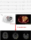
A 75-year-old woman with persistent atrial fibrillation for 8 years underwent positron emission tomography (PET)/computer tomography (CT) due to elevated CEA marker (final diagnosis of gastric cancer) and developed stroke after 13 months of the follow-up. (B) Shows the red oval circle outlines the RA FDG uptake (SUVmax = 4.76). (C) Shows acute infarcts in the left thalamus, hippocampus, and occipital lobe (yellow arrow) (A. Electrocardiogram; B1. PET image; B2. PET/CT image; C. DWI image). Reproduced with permission from B. Wang et al. [34].
Figure 4
Figure 4
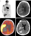
64-year-old male presented with an acute hemorrhagic stroke on 18F-FDG PET/CT. Maximum intensity projection (A), non-contrast CT axial image (B), fused PET/CT axial (C), and a short-term follow-up enhanced head CT (D). Reproduced with permission from Dundar et al. [11].

References

    1. Woodruff TM, Thundyil J, Tang SC, Sobey CG, Taylor SM, Arumugam TV. Pathophysiology, treatment, and animal and cellular models of human ischemic stroke. Mol Neurodegener. 2011;6:11. - PMC - PubMed
    1. Tadi P, Lui F. StatPearls. Treasure Island (FL): StatPearls Publishing Copyright © 2022, StatPearls Publishing LLC.; 2022. Acute Stroke.
    1. Xing C, Arai K, Lo EH, Hommel M. Pathophysiologic cascades in ischemic stroke. Int J Stroke. 2012;7:378–385. - PMC - PubMed
    1. Carmichael ST. The 3 Rs of stroke biology: radial, relayed, and regenerative. Neurotherapeutics. 2016;13:348–359. - PMC - PubMed
    1. Lovblad KO, Altrichter S, Mendes Pereira V, Vargas M, Marcos Gonzalez A, Haller S, Sztajzel R. Imaging of acute stroke: CT and/or MRI. J Neuroradiol. 2015;42:55–64. - PubMed

LinkOut - more resources