. 2023 May 4:11:1168136.
doi: 10.3389/fbioe.2023.1168136. eCollection 2023.

Effect of stem cell conditional medium-loading adhesive hydrogel on TGF-β1-induced endometrial stromal cell fibrosis

Affiliations

Effect of stem cell conditional medium-loading adhesive hydrogel on TGF-β1-induced endometrial stromal cell fibrosis

Yuan Zhu et al. Front Bioeng Biotechnol. .

Abstract

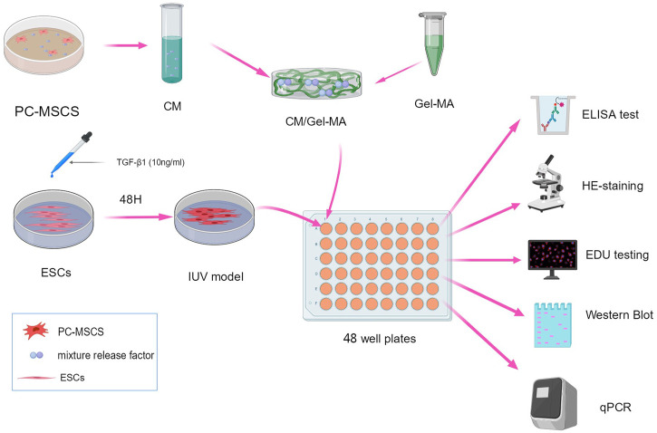
Introduction: Uterine adhesion (IUA) is a severe complication that results from uterine operations or uterine infections. Hysteroscopy is considered the gold standard for the diagnosis and treatment of uterine adhesions. Yet, this invasive procedure leads to re-adhesions after hysteroscopic treatment. Hydrogels loading functional additives (e.g., placental mesenchymal stem cells (PC-MSCs)) that can act as physical barriers and promote endometrium regeneration are a good solution. However, traditional hydrogels lack tissue adhesion which makes them unstable under a rapid turnover of the uterus, and PC-MSCs have biosafety risks when used as functional additives. Methods: In this study, we coupled an adhesive hydrogel with a PC-MSCs conditioned medium (CM) to form a hybrid of gel and functional additives (CM/Gel-MA). Results and Discussion: Our experiments show that CM/Gel-MA enhances the activity of endometrial stromal cells (ESCs), promotes cell proliferation, and reduces the expression of α-SMA, collagen I, CTGF, E-cadherin, and IL-6, which helps to reduce the inflammatory response and inhibit fibrosis. We conclude that CM/Gel-MA can more potentially prevent IUA by combining the physical barriers from adhesive hydrogel and functional promotion from CM.

Keywords: adhesive hydrogel; endometrial stromal cell; fibrosis; stem cell conditional medium; uterine adhesion.


Conflict of interest statement

The authors declare that the research was conducted in the absence of any commercial or financial relationships that could be construed as a potential conflict of interest.

Figures

FIGURE 1
FIGURE 1
Scheme of the experimental procedure. All schematics are drawn using MedPeer (www.medpeer.com).
FIGURE 2
FIGURE 2
PC-MSC immunophenotype identification. (A) Schematic diagram of flow analysis. (B) Test for CD73 and CD105 expressions. (C) Test for CD14 and CD45 expressions.
FIGURE 3
FIGURE 3
Evaluation of gel properties. (A and D) Schemes showing the lap shear test (A) and wound closure test of Gel-MA and CM/Gel-MA. (B and C) The shear strength of Gel-MA and CM/Gel-MA (n = 3, mean ± SD, ns: not significant). (E and F) The wound closure strength of Gel-MA and CM/Gel-MA (n = 3, mean ± SD, ns: not significant). Statistical significance calculated using Student’s t-test is given in C and F. (G) The tensile resistance of HA. (H) Adhesion test. (I) The results of the ELISA. CM/Gel-MA group: Gel containing 75% CM (100 μL) + PBS(800 μL); CM group: 75% CM solution (100 μL) + PBS (800 μL).
FIGURE 4
FIGURE 4
CCK-8 assays for cell proliferation. (A) 75% CM promotes cell proliferation. Control group: Complete medium (800 μL) + serum-free medium (100 μL); 75% CM group: 75% fresh CM (800 μL) +75% serum-free CM medium (100 μL); (B) Proliferation ability of ESCs in different media. All subgroups contain 800 μL of 75% fresh CM as the basic medium. Control group: serum-free medium (100 μL); Gel-MA group: Gel containing serum-free medium (100 μL); CM group: 75% serum-free CM medium (100 μL); CM/Gel-MA group: Gel containing 75% serum-free CM (100 μL). (***: p < 0.001; **: p < 0.01; *: p < 0.01; ns: >0.05).
FIGURE 5
FIGURE 5
CM/Gel-MA enhances cell viability and reduces fibrosis. (A) H&E staining (100×). The nucleus is stained blue and the cytoplasm is stained pink. Blue arrows indicate normal ESC and red arrows indicate post-fibrotic ESC. (B) Percentage of normal ESCs. (C) EDU assay for cell viability, cells in the proliferative phase show strong red fluorescence after EDU staining and blue fluorescence reaction of the nucleus after Hoechst 33324 staining. (D) Statistical analysis table of EDU assay for cell viability. (***: p < 0.001; **: p < 0.01; *: p < 0.01; ns:>0.05).
FIGURE 6
FIGURE 6
Expression level of some bio-factors. (A) Relative mRNA level of α-SMA. (B) Relative mRNA level of collagen I. (C) Relative mRNA level of CTGF. (D) Relative mRNA level of E-cadherin. (E) Relative mRNA level of IL-6. (F) The results of WB.
FIGURE 7
FIGURE 7
Effect of CM/Gel-MA on IUA cell model. The IUA model was induced by adding TGF-β1 into endometrial stromal cells. After treating the model with CM/Gel-MA, the fibrosis was inhibited with decreased the expression of α-SMA, collagen I, CTGF, E-cadherin, and IL-6. At the same time, the cell vitality and proliferation ability were enhanced after treatment.

References

    1. AAGL Elevating Gynecologic Surgery (2017). AAGL practice report: Practice guidelines on intrauterine adhesions developed in collaboration with the European society of gynaecological endoscopy (ESGE). Gynecol. Surg. 14 (1), 6. 10.1186/s10397-017-1007-3 - DOI - PMC - PubMed
    1. Ai Y., Chen M., Liu J., Ren L., Yan X., Feng Y. (2020). lncRNA TUG1 promotes endometrial fibrosis and inflammation by sponging miR-590-5p to regulate Fasl in intrauterine adhesions. Int. Immunopharmacol. 86, 106703. 10.1016/j.intimp.2020.106703 - DOI - PubMed
    1. Annabi N., Zhang Y. N., Assmann A., Sani E. S., Cheng G., Lassaletta A. D., et al. (2017). Engineering a highly elastic human protein–based sealant for surgical applications. Sci. Transl. Med. 9 (410), eaai7466. 10.1126/scitranslmed.aai7466 - DOI - PMC - PubMed
    1. Benor A., Gay S., DeCherney A. (2020). An update on stem cell therapy for Asherman syndrome. J. Assist. Reprod. Genet. 37 (7), 1511–1529. 10.1007/s10815-020-01801-x - DOI - PMC - PubMed
    1. Bonafe F., Govoni M., Giordano E., Caldarera C. M., Guarnieri C., Muscari C. (2014). Hyaluronan and cardiac regeneration. J. Biomed. Sci. 21, 100. 10.1186/s12929-014-0100-4 - DOI - PMC - PubMed