Review
. 2023 May 6:20:100660.
doi: 10.1016/j.mtbio.2023.100660. eCollection 2023 Jun.

Functionalization of biomimetic mineralized collagen for bone tissue engineering

Affiliations
Review

Functionalization of biomimetic mineralized collagen for bone tissue engineering

Xiujie Zhu et al. Mater Today Bio. .

Abstract

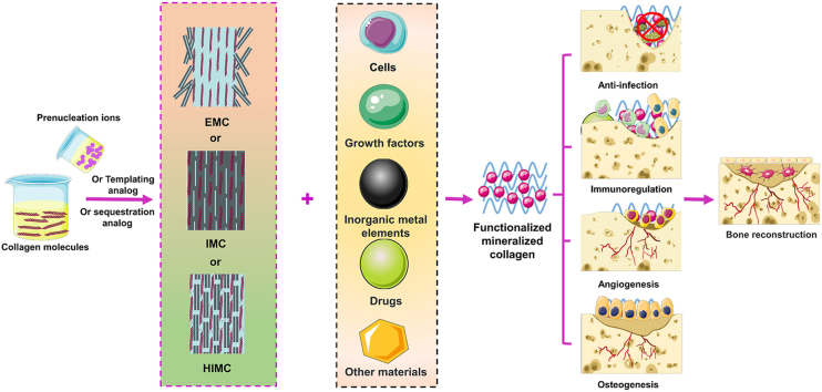
Mineralized collagen (MC) is the basic unit of bone structure and function and is the main component of the extracellular matrix (ECM) in bone tissue. In the biomimetic method, MC with different nanostructures of neo-bone have been constructed. Among these, extra-fibrous MC has been approved by regulatory agencies and applied in clinical practice to play an active role in bone defect repair. However, in the complex microenvironment of bone defects, such as in blood supply disorders and infections, MC is unable to effectively perform its pro-osteogenic activities and needs to be functionalized to include osteogenesis and the enhancement of angiogenesis, anti-infection, and immunomodulation. This article aimed to discuss the preparation and biological performance of MC with different nanostructures in detail, and summarize its functionalization strategy. Then we describe the recent advances in the osteo-inductive properties and multifunctional coordination of MC. Finally, the latest research progress of functionalized biomimetic MC, along with the development challenges and future trends, are discussed. This paper provides a theoretical basis and advanced design philosophy for bone tissue engineering in different bone microenvironments.

Keywords: Bioactive factors; Biomimetic; Bone tissue repair; Functionalization; Mineralized collagen; Osteogenesis.


Conflict of interest statement

The authors declare that they have no known competing financial interests or personal relationships that could have appeared to influence the work reported in this paper.

Figures

Image 1
Graphical abstract
Fig. 1
Fig. 1
The multiscale structure of bone [1]. Reproduced with permission [1]. Copyright 2019, Elsevier.
Scheme 1
Scheme 1
Schematic illustration of functional MC synthesis strategies, modification, and some of the active factors and cellular aspects that take part in the bone repair processes in each stage.
Fig. 2
Fig. 2
Common methods used to prepare MC with different nanostructures and the corresponding transmission electron microscopy (TEM) images. (A) The preparation process of EMC by classical ion-mediated crystallization strategy. (B) The preparation of IMC by procollagen 1 intact N-terminal (PINP) pathway. (C) The preparation process of hierarchical, intrafibrillarly MC (HIMC) by dual biomimetic analog-based bottom-up strategy.
Fig. 3
Fig. 3
Comparison of MC with different nanostructures. (A) Nanotopography (a–c) and nanomechanical (d–f) properties of MC with different nanostructures. Scanning electron microscope (SEM) image of HIMC (a), IMC (b) and EMC (c). Corresponding atomic force microscopy property maps and section analyses of Young's modulus of parts a–c, respectively. (B) rBMSC morphology (a′-c') after 1 ​d of culturing on the a) HIMC, b) IMC, and c) EMC. Cell morphology quantified for d′) the number of branch points and e′) the cell area in each group. f′) Cell viability and g′) quantitative results of ALP. (C) Representative HE staining images of mandibular defect areas in each group. (C) Micro-CT images of mandibular defect areas in each group. ∗α ​< ​0.05 versus HIMC; #α ​< ​0.05 versus IMC. Reproduced with permission [90]. Copyright 2016, John Wiley & Sons.
Fig. 4
Fig. 4
Loading strategies of active factors on MC. (A) Highly efficient loading of active factor (AF) into MC by immersing in AF solution. (B) Heparin-modified surface of MC develops an affinity for AF. (C) AF was loaded onto MC scaffolds, and polymer was introduced into the scaffolds by injection or mixing. (D) AF was mixed with the raw materials of MC to form functionalized mineralized collagen (FMC). FMCS, functionalized MC scaffolds; FMC, functionalized MC; AF, active factors.
Fig. 5
Fig. 5
(A) (a) Saddle-type bone defect with dental implant insertion. (b) Lateral view following the placement of HAp/TCP/Col composite and cover screw. (B) Representative 3D CT reconstruction. (C) Merged confocal microscope images of the two fluorochromes. Dotted line: original bone level [160]. Reproduced under the Creative Commons Attribution 4.0 International License [160]. Copyright 2021, John Wiley & Sons.
Fig. 6
Fig. 6
(A) Representative images of the fluorescently labeled BP (Alexa Pam 647 – red color) within both scaffold types. (B) Quantification of 14C-ZA elution from porous collagen and carbonated hydroxyapatite (CHA) scaffolds post-washing. ∗p ​< ​0.01 in comparison to CHA elution of both 1 ​μg and 2 ​μg 14C-ZA. (C) (a, b) Representative 3D CT reconstruction; (c, d) corresponding transaxial slices (stack of 50 slices) of μCT images of bone nodules resulting from each group. (D) Representative TRAP-stained histological sections of osteoclasts (stained in red) in trabecular-like structure of ectopic bone formed following 4 weeks of intramuscular implantation. The arrows indicate the stained osteoclasts. Scale bars ​= ​500 ​μm [110]. Reproduced under the terms of the Creative Commons Attribution 4.0 International License [110]. Copyright 2014, Elsevier. (For interpretation of the references to colour in this figure legend, the reader is referred to the Web version of this article.)
Fig. 7
Fig. 7
(A) Experimental design in vivo. SEM image of a strontium-containing MC type I scaffold (a). The scaffolds were functionalized with and without rhBMP-2 and implanted into 2 ​mm bone defects of nude mice (b–d). The defects were stabilized by an external fixator (c). (B) μCT evaluation of the bone volume at the defect site. (C) The result of three-point binding stiffness. (D) Histological staining of femurs at 6 weeks after surgery. (E) Morphological scoring of the HE stained defect areas [154]. Reproduced under the Creative Commons Attribution License [154]. Copyright 2020, John Wiley & Sons.
Fig. 8
Fig. 8
(A) Simple method to prepare HA/COL composite. (B) Composites are implanted into animals for four weeks after removal and observed. (C) Masson staining images of each group, four weeks after implantation. NB, deposition of new bone; M, bone-filling material; arrows refer to the new blood vessels. Reproduced with permission [208]. Copyright 2019, Elsevier.
Fig. 9
Fig. 9
Potential molecular mechanism of how macrophage polarization activated by MC with different nanostructures affects the process of endogenous bone regeneration.
Fig. 10
Fig. 10
(A) The amount of adsorbed antibiotic (mg) per 1 ​g of HAp/Col. N/A, not available. (B) Representative photographs of culture dishes. The translucent circles are inhibitory zones. ∗Effective inhibitory zone. (C) 3D CT reconstruction of bone holes at 4 weeks after implantation. (D) Hematoxylin and eosin staining images of the implant site at different times after implantation (scale bar: 1000 ​μm) [107]. Reproduced with permission [107]. Copyright 2019, the Authors. Published by Wiley Periodicals.
Fig. 11
Fig. 11
(A) Schematic illustration of the MC coating on titanium with the aid of metal-organic framework nanocrystals to control the release of naringin, which could enhance osseointegration and simultaneously inhibit microbial cell growth. (B) Morphological observation of MSCs on various substrates. Filopodia are indicated by white arrows [226]. Reproduced under the terms of the Creative Commons Attribution License [226]. Copyright 2017, American Chemical Society.

Similar articles

Cited by

References

    1. Shah F.A., Thomsen P., Palmquist A. Osseointegration and current interpretations of the bone-implant interface. Acta Biomater. 2019;84:1–15. doi: 10.1016/j.actbio.2018.11.018. - DOI - PubMed
    1. De Mori A., Hafidh M., Mele N., Yusuf R., Cerri G., Gavini E., Tozzi G., Barbu E., Conconi M., Draheim R.R., Roldo M. Sustained release from injectable composite gels loaded with silver nanowires designed to combat bacterial resistance in bone regeneration applications. Pharmaceutics. 2019:11. doi: 10.3390/pharmaceutics11030116. - DOI - PMC - PubMed
    1. Tampieri A., Iafisco M., Sandri M., Panseri S., Cunha C., Sprio S., Savini E., Uhlarz M., Herrmannsdoerfer T. Magnetic bioinspired hybrid nanostructured collagen-hydroxyapatite scaffolds supporting cell proliferation and tuning regenerative process. ACS Appl. Mater. Interfaces. 2014;6:15697–15707. doi: 10.1021/am5050967. - DOI - PubMed
    1. Tampieri A., D'Alessandro T., Sandri M., Sprio S., Landi E., Bertinetti L., Panseri S., Pepponi G., Goettlicher J., Bañobre-López M., Rivas J. Intrinsic magnetism and hyperthermia in bioactive Fe-doped hydroxyapatite. Acta Biomater. 2012;8:843–851. doi: 10.1016/j.actbio.2011.09.032. - DOI - PubMed
    1. Qi Y., Mai S., Ye Z., Aparicio C. Biomimetic fabrication and characterization of collagen/strontium hydroxyapatite nanocomposite. Mater. Lett. 2020;274 doi: 10.1016/j.matlet.2020.127982. - DOI

LinkOut - more resources