. 2023 Mar 1;4(1):26-34.
doi: 10.1089/phage.2022.0038. Epub 2023 Mar 17.

Cytotoxic Evaluation in HaCaT Cells of the Pa.7 Bacteriophage from Cutibacterium (Propionibacterium) acnes, Free and Encapsulated Within Liposomes

Affiliations

Cytotoxic Evaluation in HaCaT Cells of the Pa.7 Bacteriophage from Cutibacterium (Propionibacterium) acnes, Free and Encapsulated Within Liposomes

Daniela Torres Di Bello et al. Phage (New Rochelle). .

Abstract

Introduction: Acne is a multifactorial disease involving the colonization of skin follicles by Cutibacterium (formerly Propionibacterium) acnes. A combination of different retinoid-derived products, antibiotics, and hormonal antiandrogens are used to treat the disease, but these treatments require extended periods, may have secondary effects, are expensive, and not always effective. Owing to antibiotic resistance, the use of bacteriophages has been proposed as an alternative treatment. However, if they are intended for a cosmetic or pharmaceutical use, it is necessary to evaluate the safety of the phages and the preparations containing them.

Materials and methods: In this study, the cytotoxicity of Pa.7 bacteriophage was evaluated in HaCaT cells, along with a liposome suitable for their encapsulation, using 3-(4,5-dimethylthiazol-2-yl)-2,5-diphenyltetrazolium bromide and trypan blue assays.

Results: We found that Pa.7 was not cytotoxic for HaCaT cells. Also, 30 mM of liposomes, or below are considered noncytotoxic concentrations.

Conclusion: Phages encapsulated in the liposomes presented in this study can be used safely for skin treatments.

Keywords: Cutibacterium acnes; acne; cytotoxicity; liposomes; phage therapy.


Conflict of interest statement

M.J.V. declares she was in the past a member of the spin-off company SciPhage S.A.S., which works for the development of phage therapy in Colombia. She is also among the inventors of the patent no. NC2018/0008178, entitled “Composicionestópicas que comprendenbacteriófagos que se encapsulanenliposomas” (Topical compositions comprising bacteriophages encapsulated in liposomes). Other authors declare no competing interests.

Figures
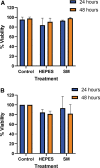
FIG. 1.
FIG. 1.
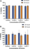
HaCaT cell viability after exposure to the two buffers HEPES and SM buffer evaluated through the (A) trypan blue exclusion assay and (B) MTT assay. The data shown in each bar represents three biological replicates, repeated three times. The comparisons were made between the control versus other concentrations and were evaluated by a two-way ANOVA and Dunnett's multiple comparisons tests. No significant differences were found. ANOVA, analysis of variance; HEPES, 4-(2-hydroxyethyl)-1-piperazineethanesulfonic acid; MTT, 3-(4,5-dimethylthiazol-2-yl)-2,5-diphenyltetrazolium bromide.
FIG. 2.
FIG. 2.
HaCaT cell viability after exposure to the Pa.7 phage suspended in SM buffer evaluated through the (A) trypan blue assay and (B) MTT assay. The data shown in each bar represents three biological replicates, repeated three times. The comparisons were made between the control versus other concentrations and were evaluated by a two-way ANOVA and Dunnett's multiple comparisons tests. No significant differences were found.
FIG. 3.
FIG. 3.
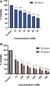
HaCaT cell viability after exposure to the different concentrations of liposomes per well through the MTT assay being (A) 10–60 mM for 24 h, (B) 75–300 mM for 24 and 48 h. The data in this figure represent the three biological replicates of each treatment, performed three times. The comparisons were made between the control versus other concentrations and were evaluated by a one-way ANOVA (A) and two-way ANOVA (B) and Dunnett's multiple comparisons tests. Significant differences: *p < 0.05, **p < 0.01, and ***p < 0.001.
FIG. 4.
FIG. 4.
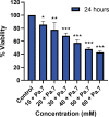
HaCaT cell viability after exposure to Pa.7 bacteriophage-containing liposomes, according to the MTT assay. Concentration of liposomes varied between 0 and 60 mM per microplate well. Data shown in this figure represents the three biological replicate of each treatment, performed three times. The comparisons were made between the control versus other concentrations and were evaluated by a one-way ANOVA and Dunnett's multiple comparisons tests. Significant differences: *p < 0.05, **p < 0.01, and ***p < 0.001.
FIG. 5.
FIG. 5.
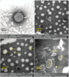
Liposomes morphology and size characterization by focused ion beam/scanning electron microscopy.

References

    1. Ashkenazi H, Malik Z, Harth Y, et al. . Eradication of Propionibacterium acnes by its endogenic porphyrins after illumination with high intensity blue light. FEMS Immunol Med Microbiol 2003;35(1):17–24; doi: 10.1111/j.1574-695x.2003.tb00644.x - DOI - PubMed
    1. Toyoda M, Morohashi M. Pathogenesis of acne. Med Electron Microsc 2001;34:29–40; doi: 10.1007/s007950100002 - DOI - PubMed
    1. Shaw JC. Acne: Effect of hormones on pathogenesis and management. Am J Clin Dermatol 2002;3:571–578; doi: 10.2165/00128071-200203080-00007 - DOI - PubMed
    1. Pochi PE, Strauss JS, Downing DT. Skin surface lipid composition, acne, pubertal development, and urinary excretion of testosterone and 17 ketosteroids in children. J Invest Dermatol 1977;69(5):485–489; doi: 10.1111/1523-1747.ep12511753 - DOI - PubMed
    1. Titus S, Hodge J. Diagnosis and treatment of acne. Am Fam Physician 2012;86(8):734–740. - PubMed

LinkOut - more resources