[Preprint]. 2023 May 8:rs.3.rs-2883247.
doi: 10.21203/rs.3.rs-2883247/v1.

Disease-Modifying Effects of a Glial-targeted Inducible Nitric Oxide Synthase Inhibitor (1400W) in Mixed-sex Cohorts of a Rat Soman (GD) Model of Epilepsy

Affiliations

Disease-Modifying Effects of a Glial-targeted Inducible Nitric Oxide Synthase Inhibitor (1400W) in Mixed-sex Cohorts of a Rat Soman (GD) Model of Epilepsy

Suraj S Vasanthi et al. Res Sq. .

Update in

Abstract

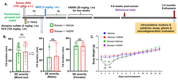
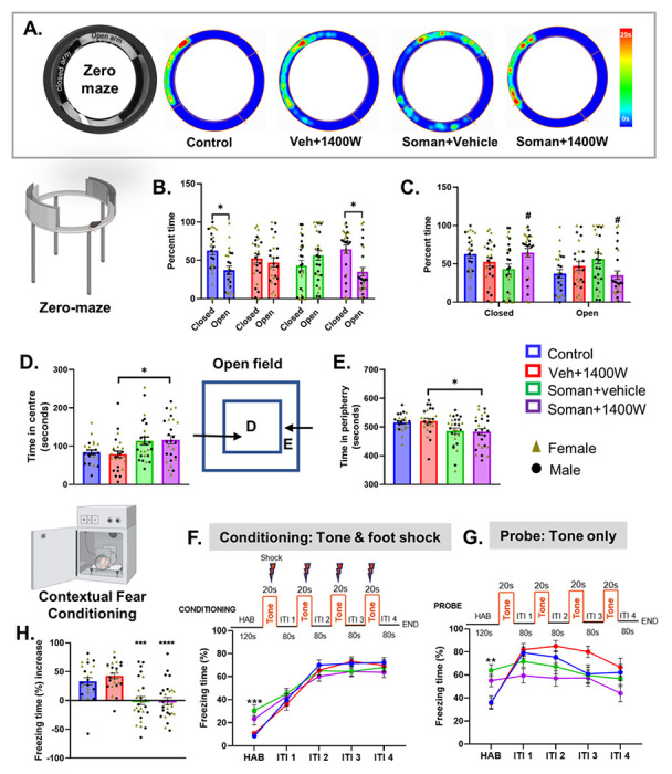
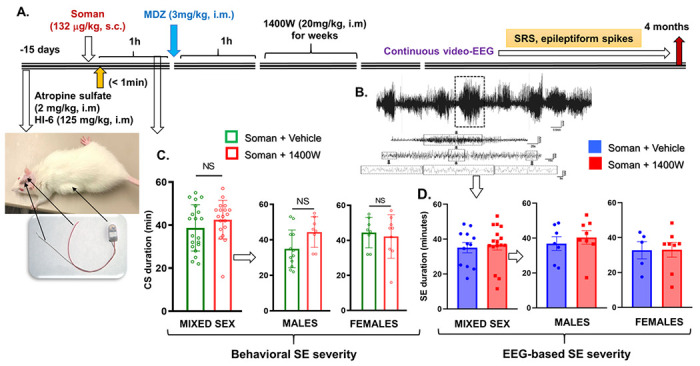
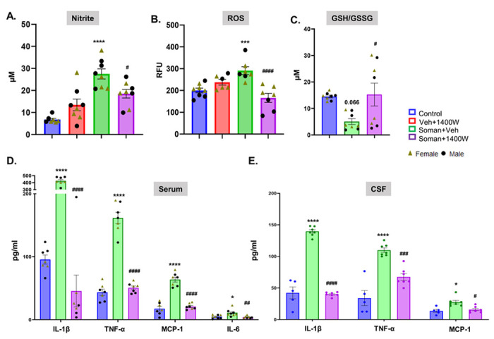
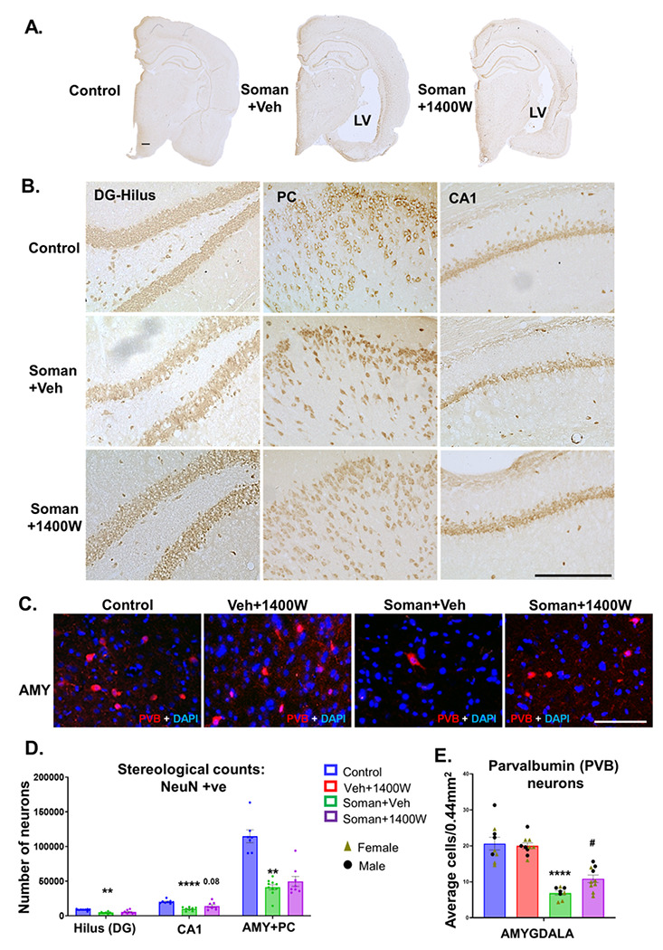
Background Acute exposure to seizurogenic organophosphate (OP) nerve agents (OPNA) such as diisopropylfluorophosphate (DFP) or soman (GD), at high concentrations, induce immediate status epilepticus (SE), reactive gliosis, neurodegeneration, and epileptogenesis as a consequence. Medical countermeasures (MCMs- atropine, oximes, benzodiazepines), if administered in < 20 minutes of OPNA exposure, can control acute symptoms and mortality. However, MCMs alone are inadequate to prevent OPNA-induced brain injury and behavioral dysfunction in survivors. We have previously shown that OPNA exposure-induced SE increases the production of inducible nitric oxide synthase (iNOS) in glial cells in both short- and long- terms. Treating with a water soluble and highly selective iNOS inhibitor, 1400W, for three days significantly reduced OPNA-induced brain changes in those animals that had mild-moderate SE in the rat DFP model. However, such mitigating effects and the mechanisms of 1400W are unknown in a highly volatile nerve agent GD exposure. Methods Mixed-sex cohort of adult Sprague Dawley rats were exposed to GD (132µg/kg, s.c.) and immediately treated with atropine (2mg/kg, i.m) and HI-6 (125mg/kg, i.m.). Severity of seizures were quantified for an hour and treated with midazolam (3mg/kg, i.m.). An hour post-midazolam, 1400W (20mg/kg, i.m.) or vehicle was administered daily for two weeks. After behavioral testing and EEG acquisition, animals were euthanized at 3.5 months post-GD. Brains were processed for neuroinflammatory and neurodegeneration markers. Serum and CSF were used for nitrooxidative and proinflammatory cytokines assays. Results We demonstrate a significant long-term (3.5 months post-soman) disease-modifying effect of 1400W in animals that had severe SE for > 20min of continuous convulsive seizures. 1400W significantly reduced GD-induced motor and cognitive dysfunction; nitrooxidative stress (nitrite, ROS; increased GSH: GSSG); proinflammatory cytokines in the serum and some in the cerebrospinal fluid (CSF); epileptiform spikes and spontaneously recurring seizures (SRS) in males; reactive gliosis (GFAP + C3 and IBA1 + CD68 positive glia) as a measure of neuroinflammation, and neurodegeneration (including parvalbumin positive neurons) in some brain regions. Conclusion These findings demonstrate the long-term disease-modifying effects of a glial-targeted iNOS inhibitor, 1400W, in a rat GD model by modulating reactive gliosis, neurodegeneration, and neuronal hyperexcitability.


Conflict of interest statement

Competing Interests

None

Figures

Figure 1
Figure 1
(A) Experimental design, animals grouping based on the initial SE severity (B), and bodyweight comparison post-exposure/treatment (C). There were no significant differences in SE severity between the vehicle and 1400W treated groups in either mixed sex cohort or males or females (B). There was a significant weight loss in soman exposed animals, irrespective of sex, during the first 3 days of postsoman exposure (C). (B. Mixed-sex) Mann-Whitney test, n= 27-33; (B. Males) Mann-Whitney test, n=12-13; (B. Females) Unpaired t-test, n=12-13. (C) Repeated measures two-way ANOVA (Šídák’s multiple comparison), n= 24-26 for mixed-sex cohort (12-13/sex). **p<0.01, ***,xxx p<0.001 vs control. CS, convulsive seizures; NS, non-significant.
Figure 2
Figure 2
1400W significantly reduced soman-induced motor deficit, measured by rotarod, in mixed sex cohort (A). The impact of mild and severe SE (B). Experimental design for the Novel Object Recognition (NOR) test (C) and representative heatmap from each group during 24 hours probe testing (D). 1400W significantly rescued soman-induced long-term memory deficits at 24h post-familiarization (F) but not at 3h (E). The effects of soman /vehicle or 1400W in mild and severe SE groups were compared (G). No sex interaction was detected in either rotarod or NOR test either at 3hrs or 24hrs. (A, E, F) Two-way ANOVA (Tukey’s multiple comparisons test), n= 16-26 for mixed-sex cohort (8-13/sex); (B, G) Two-way ANOVA Sidak’s multiple comparisons test, n= 4-21 (4-12/sex). * p< 0.05, ** p< 0.01 vs control; # p< 0.05, ## p< 0.01 vs Soman+vehicle.
Figure 3
Figure 3
The Elevated Zero/Circular Maze (A-C) and Open Field tests (D, E). Representative heatmaps from each group is shown (A). Control and soman+1400W groups significantly spent more time in closed arms (B, C). Soman exposed 1400W treated group spent significantly more time at the center (D) or less time in the periphery (E) in an open field (illustrated in the schematic diagram) than the vehicle 1400W group. Contextual Fear Conditioning Test (F-H). During habituation before conditioning (F) and probing (G), soman exposed groups froze longer than the controls. There were no significant differences in freezing time during the inter-trial intervals (ITI) between any groups during the conditioning phase (F). Differences in freezing time emerged during the ITI during probing (G). Significant differences in percent freezing time increase were observed in soman exposed animals (H). (B) Paired t-test (Control, VEH 1400W) and Wilcoxon matched-pairs signed rank test (Soman VEH, Soman 1400W). (C-E, H) Two-way ANOVA (Tukey’s multiple comparisons test), n= 11-15/sex; (F, G) Two-way repeated measures ANOVA (Tukey’s multiple comparisons test), n= 24-27 for mixed-sex cohort, n= 11-15/sex. *p< 0.05, **p< 0.01, ***p< 0.001 vs respective control; #p<0.05 vs soman+vehicle.
Figure 4
Figure 4
(A) Telemetry experimental design, initial SE severity quantification and grouping (C, D). A telemetry device was implanted two weeks before exposure to soman. (B) A representative EEG trace during SE is shown. (C) Animals were grouped based on the initial behavioral SE severity. There were no significant differences in SE severity between the vehicle and 1400W treated groups in either mixed sex cohort or males or females (C, D). (C) Unpaired t-test, n=19-20 for mixed-sex cohort (n=8-12/sex); (D. Mixed-sex) Mann-Whitney test, n=12-16; (D. Males-Females) Unpaired t-test, n= 5-8. NS, non-significant.
Figure 5
Figure 5
1400W treatment significantly reduced soman-induced epileptiform spikes and spontaneously recurring seizures (SRS) in males but not in females. (A) Representative EEG traces showing spontaneous recurrent seizures (SRS) in a male and a female rat and the corresponding gamma powerband elevation. (B) Heatmap of SRS episodes during the entire 4-month study period. (C-E) Epileptiform spike rate comparison between soman+vehicle and soman+1400W (mixed sex cohort-C; males-D, females-E). (F, G) SRS frequency in males and females compared between vehicle and 1400W treated soman exposed animals; (C. Males, D) Unpaired t-test; (C. Females, E) Mann-Whitney test; (D) Mixed-effects analysis (Šídák’s multiple comparisons test); for all, n=12-13 for mixed sex cohort or 5-8/sex; *p<0.05, **p<0.01.
Figure 6
Figure 6
1400W significantly reduced the soman-induced nitrite (A) and ROS (B) production in the serum, and mitigated soman-induced glutathione oxidation (C). 1400W significantly reduced soman-induced proinflammatory cytokines (IL1β, TNFα) and a chemokine, MCP1 in both serum and CSF (D, E). Serum IL-6 was also affected by the treatment (D). Assays were performed at 3.5 months post-treatment. (A-D) Two Way ANOVA (Tukey’s multiple comparisons test); (E) One Way ANOVA (Tukey’s multiple comparison test for TNF-α and IL1-β) and One Way ANOVA (Dunn’s multiple comparison test for MCP-1); for all, n=6-8 for serum; 5-7 for CSF; *p<0.05, ***p<0.001, ****p<0.0001 vs Soman+vehicle; #p<0.05, ##p<0.01, ###p<0.001, ####p<0.0001, vs Soman+vehicle
Figure 7
Figure 7
(A) Representative photomicrographs of the brain sections stained for IBA1 (microglia), and DAPI (nuclear stain); Scale bar 100μm. (B) Representative higher magnification images of reactive/homeostatic microglia. Microgliosis (C-F) and reactive microgliosis (G-J) quantification. Overall effect (C, G) and regional differences (D, H) in mixed sex, and differences in males (E, I) and females (F, J) are shown for microgliosis and reactive microgliosis. Differences in CD68+IBA1 colocalization in AMY from different groups is shown (K, L). The brain regions where significant increase in CD68+IBA1 colocalization were observed in soman exposed animals were quantified and compared with other groups (M). (C, G) Two-way ANOVA (Tukey’s multiple comparisons test); (D-F, H-J, M) Region-wise two-way ANOVA (Tukey’s multiple comparisons test). For all, n=8-10 for mixed sex cohort or 3-5/sex. *p<0.05, **p<0.01, ***p<0.001, ****p<0.0001 vs control; #p<0.05, ##p<0.01, ###p<0.001, ####p<0.0001 vs soman+vehicle.
Figure 8
Figure 8
(A) Representative photomicrographs of the brain sections stained for GFAP (astrocytes), and DAPI (nuclear stain); Scale bar 100μm. (B) Representative higher magnification images of reactive/homeostatic astrocytes (green labeled cells). Astrogliosis (C, D) and reactive astrogliosis (E, F) quantification. Overall effect (C, E) and regional differences (D, F) in mixed sex are presented. Sex differences were observed only in the centromedial thalamic nuclei (CMT) (G) for reactive astrogliosis. Representative images of GFAP and C3 positive cells in AMY from each group is shown (H, I); Scalebar 100μm. Quantification of C3 containing GFAP positive cells in other brain regions (J). (C, E) Two-way ANOVA (Tukey’s multiple comparisons test; (D, F-G, J) Region-wise two-way ANOVA (Tukey’s multiple comparisons test). For all, n=8-10 for mixed sex cohort or 3-5/sex. **p<0.01, ***p<0.001 ****p<0.0001 vs control; #p<0.05, ##p<0.01, ###p<0.001, ####p<0.0001 vs soman+vehicle.
Figure 9
Figure 9
(A) Representative photomicrographs of the brain sections stained for NeuN (red) and FJB (green, co-label appears yellow). Amygdala (AMY) and mediodorsal thalamus (MDT); Scalebar 100μm. (B-E) cell quantification. Overall effect (B) and regional differences (C) in mixed sex are presented. Sex differences were observed in subiculum (SUB), amygdala (AMY), mediodorsal and centromedial thalamic nuclei (MDT, CMT). (B) Two-way ANOVA (Tukey’s multiple comparisons test); (C-E) Region-wise Two-Way ANOVA (Tukey’s multiple comparisons test). For all, n=9-10 for mixed sex cohort or 3-6/sex. *p<0.05, **p<0.01, ***p<0.001, ****p<0.0001 vs control; #p<0.05 vs soman+vehicle.
Figure 10
Figure 10
Representative brain sections used for stereological NeuN (DAB stained; A, B) and parvalbumin (PVB) immunostained images (C) are shown. Neuronal cell quantification, by stereology, from the hilus of the dentate gyrus, CA1 region of the hippocampus, amygdala and piriform cortex (D). PVB positive neurons from AMY (E). (D) One-way ANOVA with multiple comparisons test, n= 7-10; (E) Two-way ANOVA (Tukey’s multiple comparisons test), n= 8-10 (4-7 per sex). **p<0.01, ****p<0.0001 vs control; #p<0.05 vs soman+vehicle. Scalebar 500μm (A), 50μm (B), 100μm (C).

Similar articles

References

    1. Gage M, Rao NS, Samidurai M, Putra M, Vasanthi SS, Meyer C, Wang C, Thippeswamy T (2022) Soman (GD) Rat Model to Mimic Civilian Exposure to Nerve Agent: Mortality, Video-EEG Based Status Epilepticus Severity, Sex Differences, Spontaneously Recurring Seizures, and Brain Pathology. Front Cell Neurosci. 10.3389/fncel.2021.798247 - DOI - PMC - PubMed
    1. Rao NS, Meyer C, Vasanthi SS, Massey N, Samidurai M, Gage M, Putra M, Almanza AN, Wachter L, Thippeswamy T (2022) DFP-Induced Status Epilepticus Severity in Mixed-Sex Cohorts of Adult Rats Housed in the Same Room: Behavioral and EEG Comparisons. Front Cell Dev Biol 10:895092. - PMC - PubMed
    1. Hulse EJ, Haslam JD, Emmett SR, Woolley T (2019) Organophosphorus nerve agent poisoning: managing the poisoned patient. Br J Anaesth 123:457–463 - PubMed
    1. Nallapaneni A, Liu J, Karanth S, Pope C (2006) Modulation of paraoxon toxicity by the cannabinoid receptor agonist WIN 55,212-2. Toxicology 227:173–183 - PubMed
    1. Jackson C, Ardinger C, Winter KM, McDonough JH, McCarren HS (2019) Validating a model of benzodiazepine refractory nerve agent-induced status epilepticus by evaluating the anticonvulsant and neuroprotective effects of scopolamine, memantine, and phenobarbital. J Pharmacol Toxicol Methods 97:1–12 - PMC - PubMed

Publication types

LinkOut - more resources