[Preprint]. 2023 May 9:2023.05.09.540025.
doi: 10.1101/2023.05.09.540025.

The Regulator FleQ Post-Transcriptionally Regulates the Production of RTX Adhesins by Pseudomonas fluorescens

Affiliations

The Regulator FleQ Post-Transcriptionally Regulates the Production of RTX Adhesins by Pseudomonas fluorescens

Alexander B Pastora et al. bioRxiv. .

Update in

Abstract

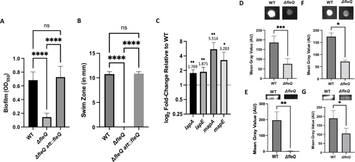
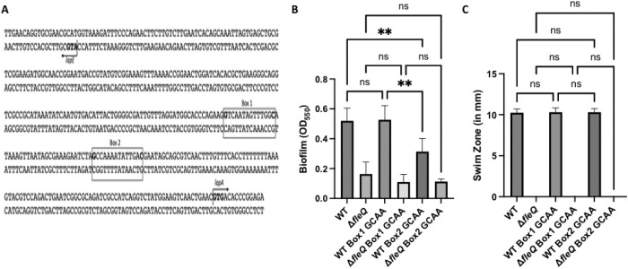
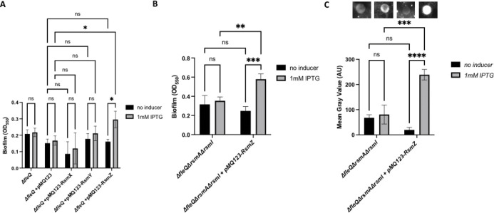
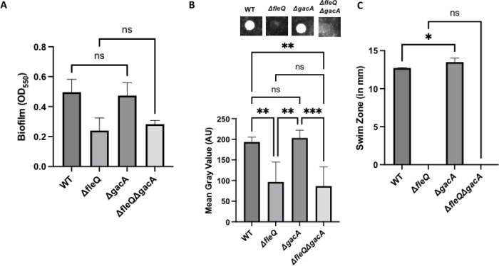
Biofilm formation by the Gram-negative gammaproteobacterium Pseudomonas fluorescens relies on the production of the repeat-in-toxin (RTX) adhesins LapA and MapA in the cytoplasm, secretion of these adhesins through their respective type 1 secretion systems, and retention at the cell surface. Published work has shown that retention of the adhesins occurs via a post-translational mechanism involving the cyclic-di-GMP receptor LapD and the protease LapG. However, little is known about the underlying mechanisms that regulate the production of these adhesins. Here, we demonstrate that the master regulator FleQ modulates biofilm formation by post-transcriptionally regulating the production of LapA and MapA. We find that a Δ fleQ mutant has a biofilm formation defect compared to the WT strain, which is attributed in part to a decrease in LapA and MapA production, despite the Δ fleQ mutant having increased levels of lapA and mapA transcripts compared to the WT strain. Through transposon mutagenesis and subsequent genetic analysis, we found that over-stimulation of the Gac/Rsm pathway partially rescues biofilm formation in the Δ fleQ mutant background. Collectively, these findings provide evidence that FleQ regulates biofilm formation by post-transcriptionally regulating the production of LapA and MapA, and that activation of the Gac/Rsm pathway can enhance biofilm formation by P. fluorescens .

Importance: Biofilm formation is a highly coordinated process that bacteria undergo to colonize a variety of surfaces. For Pseudomonas fluorescens , biofilm formation requires the production and localization of RTX adhesins to the cell surface. To date, little is known about the underlying mechanisms that regulate biofilm formation by P. fluorescens . Here, we identify FleQ as a key regulator of biofilm formation that modulates the production of LapA and MapA through a post-transcriptional mechanism. We provide further evidence implicating activation of the Gac/Rsm system in FleQ-dependent regulation of biofilm formation. Together, our findings uncover evidence for a mechanism of post-transcriptional regulation of the LapA/MapA adhesins.


Figures

FIG 1
FIG 1. A FleQ-deficient strain has a biofilm formation defect due to a decrease in adhesin production.
(A) Quantification of the biofilm formed by the WT strain, ΔfleQ mutant, and a ΔfleQ mutant complemented with the wild-type fleQ gene at the att site measured at OD550 after 16h of growth in KA minimal medium. Statistical significance was determined using an unpaired t-test. ****, P<0.0001. (B) Swim zone (in millimeters) of the WT strain, ΔfleQ mutant, and a ΔfleQ mutant complemented with the wild-type fleQ gene at the att site after toothpick inoculation on KA medium supplemented with 0.3% agar after 24h of growth at 30°C. Statistical significance was determined using an unpaired t-test. ****, P<0.0001. (C) Expression of lapA, lapE, mapA, and mapE genes in the ΔfleQ mutant relative to the WT strain, after 16h of growth on KA medium supplemented with 1.5% agar (KA agar), using the 2−ΔΔCt (Livak) method. For statistical significance, paired t-tests were conducted for each gene between the WT strain and ΔfleQ mutant. *, P<0.05; **, P<0.01. (D,F) Quantification of cell surface-associated LapA after 16h of growth on KA agar (D) or MapA after 24h of growth on KA agar (F) for the WT strain and ΔfleQ mutant using ImageJ by measuring the mean gray value of each spot using a predefined region of interest (ROI) and subtracting the background, which was determined by measuring the mean gray value of a section of the blot without any sample. Representative images are included above each graph. Statistical significance was determined using unpaired t-tests. *, P<0.05; ***, P< 0.001. (E,G) Quantification of LapA after 16h of growth on KA agar (E) or MapA after 24h of growth on KA medium (G) from whole cell lysates that were prepared from 25 ml of cultures, concentrated to 100 μl in 3mg/mL lysozyme with sonication and quantified for total protein using the BCA assay. 25μg (E) or 50μg (G) of total protein was resolved on a 7.5% TGX Gel and then blotted for LapA or MapA, respectively. Representative images are included above each graph. Statistical significance was determined using unpaired t-tests. *, P<0.05; **, P<0.01. All error bars represent standard deviation.
FIG 2
FIG 2. A FleQ-deficient strain produces adhesin sufficient to restore biofilm formation in a lapG mutant.
(A) Quantification of the biofilm formed for the WT and the ΔfleQ, ΔlapG, and ΔfleQΔlapG mutant strains measured at OD550 after 24h of growth in KA minimal medium. This timepoint was chosen because both LapA and MapA are detected at the cell surface at this time point. (B-C) Quantification of cell surface-associated LapA after 16h of growth on KA agar (B) or MapA after 24h of growth on KA agar (C) in the WT, ΔfleQ, ΔlapG, and ΔfleQΔlapG strains as described in Figure 1. Representative images are included above each graph. (D) Swim Zone (in millimeters) for the WT, ΔfleQ, ΔlapG, and ΔfleQΔlapG strains after toothpick inoculation on KA supplemented with 0.3% agar and 24h of growth at 30°C. Statistical significance for this figure was determined using one-way ANOVAs with Tukey’s multiple comparisons tests. **, P<0.01; ***, P<0.001. All error bars represent standard deviation.
FIG 3
FIG 3. Mutagenizing the FleQ consensus sequences in the lapA promoter has a transcriptional effect that is masked in the ΔfleQ mutant.
(A) Schematic showing the divergent lapA/lapE promoter. FleQ binding Box 1 and Box 2 are illustrated, and the respective sites of the point mutations are highlighted in bold. The lapA and lapE start codons are highlighted in bold and direction of translation is indicated by an arrow. (B) Quantification of the biofilm formed for the WT and the ΔfleQ mutant with a mutated FleQ binding Box 1 or Box 2, or the WT sequence at OD550 after 24h of growth in KA minimal medium. (C) Swim Zone (in millimeters) of WT and ΔfleQ strains with a mutated FleQ binding Box 1 or Box 2, or the WT sequence after toothpick inoculation on KA medium supplemented with 0.3% agar and 24h of growth at 30°C. Statistical significance for this figure was determined using one-way ANOVAs with Tukey’s multiple comparisons tests. **, P<0.01. All error bars represent standard deviation.
FIG 4
FIG 4. Identifying other factors that contribute to FleQ-dependent regulation of adhesins.
(A) Schematic showing the location of FleQ-deficient transposon mutants that restore biofilm formation. Triangles indicate insertion into the genome and the arrow above each triangle indicates directionality of the Ptac promoter. Genomic coordinates are included for reference. (B) Quantification of the biofilm formed by the WT strain, the ΔfleQ mutant and the indicated ΔfleQ derivatives at OD550 after 24h of growth in KA minimal medium. (C) Swim Zone (in millimeters) measurements of the WT strain, the ΔfleQ mutant, and ΔfleQ derivatives after toothpick inoculation on KA medium supplemented with 0.3% agar and 24h of growth at 30°C. Statistical significance for this figure was determined using one-way ANOVAs with Tukey’s multiple comparisons tests. ****, P<0.0001. All error bars represent standard deviation.
FIG 5
FIG 5. Modulation of the Gac/Rsm system increases biofilm formation in a WT and FleQ-deficient strain.
(A) Quantification of the biofilm formed by the WT, ΔfleQ, ΔrsmAΔrsmEΔrsmI, and ΔfleQ ΔrsmAΔrsmEΔrsmI strains measured at OD550 after 24h of growth in KA minimal medium. (B) Swim Zone (in millimeters) of the WT, ΔfleQ, ΔrsmAΔrsmEΔrsmI, and ΔfleQ ΔrsmAΔrsmEΔrsmI strains after toothpick inoculation on KA medium supplemented with 0.3% agar and 24h of growth at 30°C. (C) Quantification of cell surface-associated LapA after 16h of growth on KA agar as described in Figure 1. Representative images are included above each graph. Statistical significance for this figure was determined using one-way ANOVAs with Tukey’s multiple comparisons tests. *, P<0.05; **, P<0.01; ****, P<0.0001. All error bars represent standard deviation.
FIG 6
FIG 6. Overexpressing the small regulatory RNA RsmZ in a FleQ-deficient increases biofilm formation.
(A) Quantification of the biofilm formed by the ΔfleQ strain with and without pMQ123, pMQ123-RsmX, pMQ123-RsmY, or pMQ123-RsmZ measured at OD550 after 24h of growth in KA minimal medium with or without 1mM IPTG for induction. (B) Quantification of the biofilm formed by the ΔfleQΔrsmAΔrsmI strain with and without the pMQ123-RsmZ construct measured at OD550 after 24h of growth in KA minimal medium with or without 1mM IPTG for induction. (C) Quantification of cell surface-associated LapA after 16h of growth on KA agar with or without 1mM IPTG for induction as described in Figure 1. Statistical significance for this figure was determined using two-way ANOVAs with Tukey’s multiple comparisons tests. *, P<0.05; **, P<0.01; ***, P<0.001; ****, P<0.0001. All error bars represent standard deviation.
FIG 7
FIG 7. The Gac/Rsm system contributes to LapA production.
(A) Quantification of the biofilm formed by the WT, ΔfleQ, ΔgacA, and ΔfleQΔgacA strains measured at OD550 after 24h of growth in KA minimal medium. (B) Quantification of cell surface-associated LapA after 16h of growth on KA agar as described in Figure 1. Statistical significance for this figure was determined using one-way ANOVAs with Tukey’s multiple comparisons tests. *, P<0.05; **, P<0.01; ***, P<0.001. All error bars represent standard deviation. (C) Swim Zone (in millimeters) of the WT, ΔfleQ, ΔgacA, and ΔfleQΔgacA strains after toothpick inoculation on KA supplemented with 0.3% agar and 24h of growth at 30°C.
FIG 8
FIG 8. Replacing the native lapA promoter with a non-native FleQ-independent promoter partially restores biofilm formation in a FleQ-deficient strain.
(A) Quantification of the biofilm formed by the WT and ΔfleQ strains with the native lapA or Plac promoter measured at OD550 after 24h of growth in KA minimal medium. (B) Quantification of cell surface-associated LapA after 16h of growth on KA agar as described in Figure 1. (C) Swim Zone (in millimeters) of the WT and ΔfleQ strains with the native lapA or Plac promoter strains after toothpick inoculation on KA supplemented with 0.3% agar and 24h of growth at 30°C. Statistical significance for this figure was determined using one-way ANOVAs with Tukey’s multiple comparisons tests. **, P<0.01; ***, P<0.001; ****, P<0.0001. All error bars represent standard deviation.

References

    1. Paulsen IT, Press CM, Ravel J, Kobayashi DY, Myers GSA, Mavrodi DV, DeBoy RT, Seshadri R, Ren Q, Madupu R, Dodson RJ, Durkin AS, Brinkac LM, Daugherty SC, Sullivan SA, Rosovitz MJ, Gwinn ML, Zhou L, Schneider DJ, Cartinhour SW, Nelson WC, Weidman J, Watkins K, Tran K, Khouri H, Pierson EA, Pierson LS, Thomashow LS, Loper JE. 2005. Complete genome sequence of the plant commensal Pseudomonas fluorescens Pf-5. Nat Biotechnol 23:873–878. - PMC - PubMed
    1. Howell CR. 1979. Control of Rhizoctonia solani on cotton seedlings with Pseudomonas fluorescens and with an antibiotic produced by the bacterium. J Phytopathol 69:480.
    1. Keel C. 1992. Suppression of root diseases by Pseudomonas fluorescens CHA0: importance of the bacterial secondary metabolite 2,4-diacetylphloroglucinol. MPMI 5:4.
    1. Haas D, Défago G. 2005. Biological control of soil-borne pathogens by fluorescent pseudomonads. Nat Rev Microbiol 3:307–319. - PubMed
    1. Khabbaz RF, Arnow PM, Highsmith AK, Herwaldt LA, Chou T, Jarvis WR, Lerche NW, Allen JR. 1984. Pseudomonas fluorescens bacteremia from blood transfusion. Am J Med 76:62–68. - PubMed

Publication types