[Preprint]. 2023 May 12:2023.05.11.540291.
doi: 10.1101/2023.05.11.540291.

Elevated nuclear TDP-43 induces constitutive exon skipping

Affiliations

Elevated nuclear TDP-43 induces constitutive exon skipping

Rogger P Carmen-Orozco et al. bioRxiv. .

Update in

  • Elevated nuclear TDP-43 induces constitutive exon skipping.
    Carmen-Orozco RP, Tsao W, Ye Y, Sinha IR, Chang K, Trinh VT, Chung W, Bowden K, Troncoso JC, Blackshaw S, Hayes LR, Sun S, Wong PC, Ling JP. Carmen-Orozco RP, et al. Mol Neurodegener. 2024 Jun 9;19(1):45. doi: 10.1186/s13024-024-00732-w. Mol Neurodegener. 2024. PMID: 38853250 Free PMC article.

Abstract

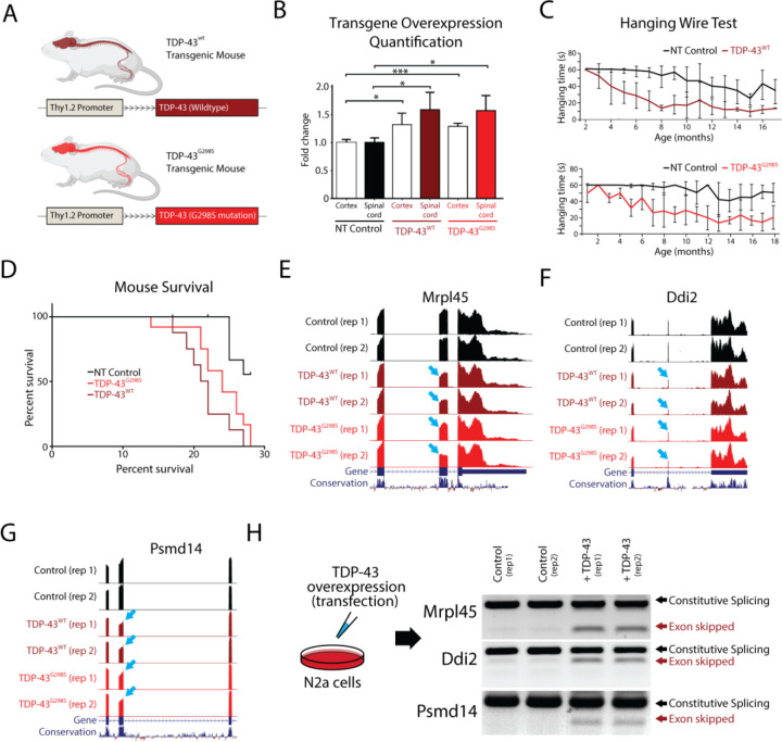
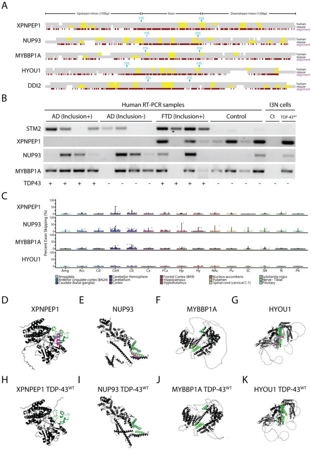
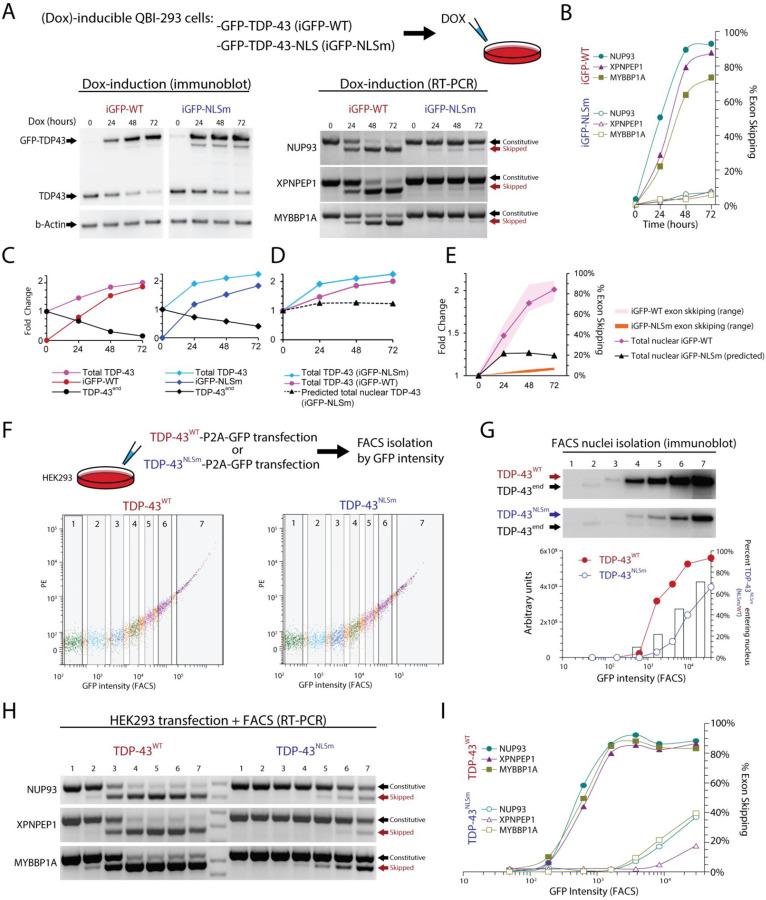
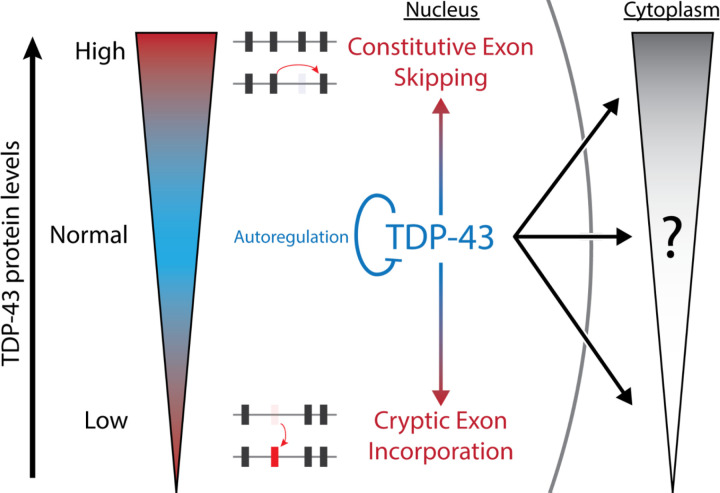
Cytoplasmic inclusions and loss of nuclear TDP-43 are key pathological features found in several neurodegenerative disorders, suggesting both gain- and loss-of-function mechanisms of disease. To study gain-of-function, TDP-43 overexpression has been used to generate in vitro and in vivo model systems. Our study shows that excessive levels of nuclear TDP-43 protein lead to constitutive exon skipping that is largely species-specific. Furthermore, while aberrant exon skipping is detected in some human brains, it is not correlated with disease, unlike the incorporation of cryptic exons that occurs after loss of TDP-43. Our findings emphasize the need for caution in interpreting TDP-43 overexpression data, and stress the importance of controlling for exon skipping when generating models of TDP-43 proteinopathy. Understanding the subtle aspects of TDP-43 toxicity within different subcellular locations is essential for the development of therapies targeting neurodegenerative disease.


Figures

Figure 1.
Figure 1.. TDP-43 overexpression in mice leads to skipping of constitutive exons.
Overexpression of TDP-43 has been shown to be toxic when under the control of different promoters, leading to premature death in animals. (A) In this study, we investigated the effects of overexpressing both human TDP-43 (TDP-43WT) and TDP-43 carrying a G298S mutation (TDP-43G298S), under the control of the weak Thy1.2 promoter in mice. (B) We compared TDP-43 levels in the spinal cord and cortex of transgenic mice to controls using immunoblotting. We observed approximately 1.5x and 1.3x overexpression of both transgenes in the spinal cord and cortex, respectively (* p<0,05, ***p<0.001). (C) We measured the hanging time of transgenic mice compared to their littermate controls and found a reduction associated with age, indicative of a motor neuron deficit. (D) Both transgenic lines had shorter survival times compared to nontransgenic (NT) controls (NT vs WT: p=0.0005, NT vs G298S: p=0.0035), but there were no differences between the two transgenic lines (WT vs G298S: p=0.1260). (E-G) To investigate the underlying mechanism of TDP-43 toxicity, we performed RNA-Seq analysis on isolated mouse spinal cords and found several instances of exon skipping (arrows) in both transgenic lines. (H) To confirm that these skipping events were directly caused by TDP-43 overexpression and not an indirect effect of disease progression, we transfected mouse N2a cells with human TDP-43 and evaluated different targets using double band RT-PCR. We observed exon skipping events only in transfected cells.
Figure 2.
Figure 2.. TDP-43 overexpression induces exon repression in humans.
(A) To investigate whether exon skipping events also occur in humans, we infected human iPS cells with a lentivirus expressing human TDP-43 and analyzed the resulting RNA-Seq dataset. (B) Our analysis revealed several genes with skipping events, including genes involved in several molecular pathways. (C) Network analysis identified genes related to intellectual disability, synaptic activity, and mitochondrial proteins. We identified genes that had exons with particularly high levels of exon skipping, including HYOU1, NUP93, and XPNPEP1 (arrows). (D-F) When we cross-referenced these results with samples from transgenic mice, we found that exons repressed in humans were not repressed in mice. We validated these findings using RT-PCR in i3Neurons. We used double band RT-PCR (G) with primers located in the adjacent exon to the repressed exon or single band RT-PCR (H) with primers spanning the skipped junction. (I) Analysis of UG repeats in 25 targets revealed that these TDP-43 recognition motives are found in both repressed exons and intronic sequences. UG motifs appear slightly more frequently around the downstream 5’ splice site, but with far shorter UG repeat lengths than those found adjacent to cryptic exons (73).
Figure 3.
Figure 3.. TDP-43 exon skipping events are found in aging human brains but do not correlate with disease.
(A) Alignment of syntenic mouse (mm10) and human (hg38) genomic sequences surrounding exons repressed by TDP-43 overexpression in human cells. Constitutively spliced exons in the genes XPNPEP1, NUP93, MYBBP1A, and HYOU1 are skipped in human cells but not mouse cells when TDP-43 is overexpressed. By contrast, the exon in DDI2 is repressed in both mouse and human cells. An analysis of UG motifs (yellow highlights) reveals that slightly higher frequencies in UG repeat frequency surrounding the 3’ and 5’ splice sites may account for species-specific exon skipping. (B) We performed single-band RT-PCR to amplify cryptic junctions or exon-repressed junctions in human brain samples from patients with AD pathology with or without TDP-43 inclusions, frontotemporal dementia with inclusions, and control patients who did not have TDP-43 inclusions. As previously reported, the cryptic exon in the gene STMN2 is highly correlated with TDP-43 proteinopathy (16, 17). RT-PCR analysis showed that exon skipping occurred in both control and disease samples, indicating that aberrant exon skipping does not correlate with TDP-43 proteinopathy. (C) Since aging is a primary factor for developing neurodegenerative disease, we wanted to explore whether skipping events appear normally in the CNS and whether skipping events correlated with aging. We analyzed publicly available human RNA-Seq datasets (GTEx) and measured the percent spliced-in values of skipping events in patients aged between 60 to 69 years old. Exon skipping was found at low levels in most of the different brain areas analyzed, with slightly higher levels in the cerebellum. Using AlphaFold 2, we modeled protein structures with (D-G) and without (H-K) exons repressed by TDP-43 overexpression. Purple highlights indicate the repressed exons while green highlights indicate flanking amino acid sequences. TDP-43 induced exon skipping can dramatically influence protein structure and may lead to functional deficits.
Figure 4.
Figure 4.. Overexpression of the ΔNLS mutant TDP-43 (TDP-43NLSm) induces exon repression when expressed at higher levels than wildtype TDP-43.
To mimic mislocalization of TDP-43 into the cytoplasm, we used mutant forms of TDP-43. We wanted to explore to what extent this studied protein can passively migrate into the nucleus and repress exons. We used QBI-293 cells that have a stable cassette with TDP-43 tagged with GFP (iGFP-WT) and a TDP-43 mutated form in its nuclear localization signal (iGFP-NLSm), both lines under the control of the Doxycycline (Dox) inducible promoter. Cultures were exposed to Dox for different times (0, 24, 48, 72 hours). (A) Exon skipping events were seen when wild type or NLS TDP-43 forms were induced. (B) Quantification from RT-PCR results in iGFP-NLSm Dox induction shows that exon repression only reached ~9% exon skipping compared to ~95% in iGFP-WT. (C) Protein quantification shows TDP-43 endogenous self-regulation and total levels of TDP-43 for both WT and NLSm reached approximately ~2 times the normal levels of TDP-43. (D and E) Approximate levels of total nuclear TDP-43 based on estimates of TDP-43NLSm passive diffusion into the nucleus. (F) To evaluate to what extent TDP-43NLSm was passively diffusing in the nucleus, we transfected HEK-293 cells with TDP-43WT-P2A-GFP or TDP-43NLSm-P2A-GFP under the control of a constitutive promoter and FACS isolated seven fractions according to GFP intensity. (G) We isolated nuclei from all seven fractions and detected TDP-43 by immunoblotting, TDP-43 endogenous is reduced while TDP-43WT or TDP-43NLSm are increasing. Raw intensity value in arbitrary units is plotted and the bar graph shows the ratio between overexpressed TDP-43NLSm and TDP-43WT, which suggest that TDP-43NLSm passive diffusion rate to the nucleus increases as the expression of this protein also progressively increases. (H) Double band RT-PCR from whole cell fractions showed the progressive increase of exon skipping when either TDP-43WT or TDP-43NLSm are overexpressed. (I) TDP-43WT can almost completely repress exons evaluated (~95%), however, TDP-43NLSm only reached up to ~40% of exon repression. Using these data, we estimate the proportion of TDP-43NLSm in the Dox-inducible system, dotted line (D). Levels of total TDP-43WT and total predicted TDP-43NLSm in the nucleus are plotted together with their respective exon expression levels (E).
Fig 5.
Fig 5.. Summary Diagram.
TDP-43 is a highly autoregulated protein due to different forms of cellular toxicity when TDP-43 protein levels are either too low (cryptic exon incorporation) or too high (aberrant exon skipping). Our study has demonstrated that these splicing deficits are linked specifically to nuclear TDP-43, whereas toxicity due to cytoplasmic TDP-43 remains to be fully elucidated. Future studies that avoid constitutive exon skipping may identify biomarkers for cytoplasmic-specific TDP-43 toxicity.

Similar articles

Cited by

References

    1. Neumann M., Sampathu D. M., Kwong L. K., Truax A. C., Micsenyi M. C., Chou T. T., Bruce J., Schuck T., Grossman M., Clark C. M., McCluskey L. F., Miller B. L., Masliah E., Mackenzie I. R., Feldman H., Feiden W., Kretzschmar H. A., Trojanowski J. Q., Lee V. M.-Y., Ubiquitinated TDP-43 in frontotemporal lobar degeneration and amyotrophic lateral sclerosis. Science. 314, 130–133 (2006). - PubMed
    1. Arai T., Hasegawa M., Akiyama H., Ikeda K., Nonaka T., Mori H., Mann D., Tsuchiya K., Yoshida M., Hashizume Y., Oda T., TDP-43 is a component of ubiquitin-positive tau-negative inclusions in frontotemporal lobar degeneration and amyotrophic lateral sclerosis. Biochem. Biophys. Res. Commun. 351, 602–611 (2006). - PubMed
    1. Hayes L. R., Kalab P., Emerging Therapies and Novel Targets for TDP-43 Proteinopathy in ALS/FTD. Neurotherapeutics. 19, 1061–1084 (2022). - PMC - PubMed
    1. Meneses A., Koga S., O’Leary J., Dickson D. W., Bu G., Zhao N., TDP-43 Pathology in Alzheimer’s Disease. Molecular Neurodegeneration. 16 (2021), , doi:10.1186/s13024-021-00503-x. - DOI - PMC - PubMed
    1. Nelson P. T., Dickson D. W., Trojanowski J. Q., Jack C. R., Boyle P. A., Arfanakis K., Rademakers R., Alafuzoff I., Attems J., Brayne C., Coyle-Gilchrist I. T. S., Chui H. C., Fardo D. W., Flanagan M. E., Halliday G., Hokkanen S. R. K., Hunter S., Jicha G. A., Katsumata Y., Kawas C. H., Keene C. D., Kovacs G. G., Kukull W. A., Levey A. I., Makkinejad N., Montine T. J., Murayama S., Murray M. E., Nag S., Rissman R. A., Seeley W. W., Sperling R. A., White C. L. 3rd, Yu L., Schneider J. A., Limbic-predominant age-related TDP-43 encephalopathy (LATE): consensus working group report. Brain. 142, 1503–1527 (2019). - PMC - PubMed

Publication types