[Preprint]. 2023 Aug 9:2023.05.05.539606.
doi: 10.1101/2023.05.05.539606.

TGF-β Ligand Cross-Subfamily Interactions in the Response of Caenorhabditis elegans to Bacterial Pathogens

Affiliations

TGF-β Ligand Cross-Subfamily Interactions in the Response of Caenorhabditis elegans to Bacterial Pathogens

Emma Jo Ciccarelli et al. bioRxiv. .

Update in

Abstract

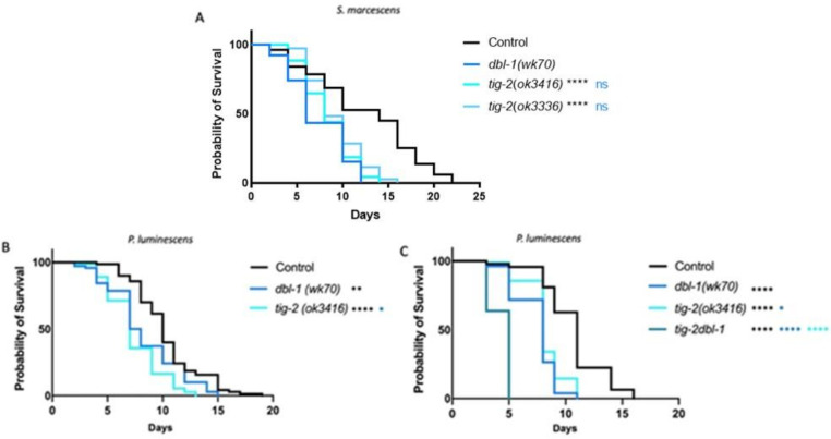
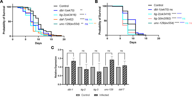
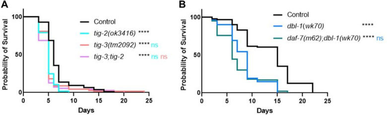
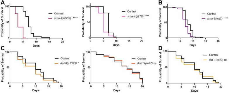
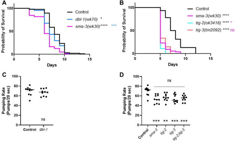
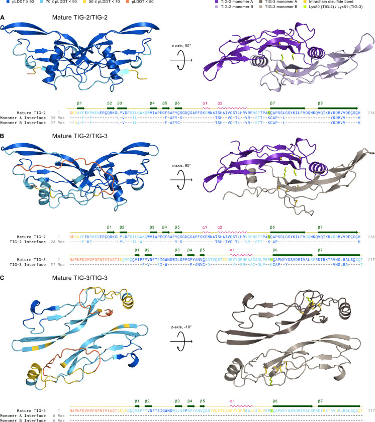
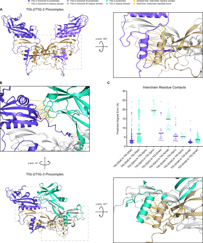
The Transforming Growth Factor beta (TGF-β) family consists of numerous secreted peptide growth factors that play significant roles in cell function, tissue patterning, and organismal homeostasis, including wound repair and immunity. Typically studied as homodimers, these ligands have the potential to diversify their functions through ligand interactions that are synergistic, cooperative, additive, and/or antagonistic. In the nematode Caenorhabditis elegans, there are only five TGF-β ligands, providing an opportunity to dissect ligand interactions in fewer combinations than in vertebrates. As in vertebrates, these ligands can be divided into bone morphogenetic protein (BMP) and TGF-β/Activin subfamilies that predominantly signal through discrete signaling pathways. The BMP subfamily ligand DBL-1 has been well studied for its role in the innate immune response in C. elegans. Here we show that all five TGF-β ligands play a role in the immune response. We also demonstrate that multiple TGF-β ligands act cooperatively as part of this response. We show that the two BMP-like ligands - DBL-1 and TIG-2 - function independently of each other in the immune response, while TIG-2/BMP and the TGF-β/Activin-like ligand TIG-3 function cooperatively. Structural modeling supports the potential for TIG-2 and TIG-3 to form heterodimers. Finally, we show that canonical DBL-1/BMP receptor and Smad signal transducers function in the response to bacterial pathogens, while components of the DAF-7 TGF-β/Activin signaling pathway do not play a role in survival. These results demonstrate a novel potential for BMP and TGF-β/Activin subfamily ligands to interact, and may provide a mechanism for distinguishing the developmental and homeostatic functions of these ligands from an acute response such as the innate immune response to bacterial pathogens.


Figures

Figure 1.
Figure 1.. The BMP-like Ligands DBL-1 and TIG-2 are Required for C. elegans Survival on Bacterial Pathogen.
A. Survival analysis of dbl-1(wk70) and two tig-2 mutants (tig-2(ok3416) and (tig-2(ok3336)) on S. marcescens bacteria. n values: Control (51), dbl-1 (39), tig-2 (43), tig-2 (35). B. Survival analysis of dbl-1(wk70) and tig-2(ok3416) on P. luminescens bacteria. n values: Control (70), dbl-1 (70), tig-2 (73). C. Survival analysis of tig-2dbl-1 double mutant on P. luminescens. n values: Control (94), dbl-1 (53), tig-2 (97), tig-2dbl-1 (55). Statistical analysis done using Log-rank (Mantel-Cox) Test. ns p > 0.05; * p ≤ 0.05; ** p ≤ 0.01; **** p < 0.0001. Black asterisks denote significance relative to control, blue is significance relative to dbl-1, and teal is significance relative to tig-2.
Figure 2.
Figure 2.. Five TGF-β Ligands Demonstrate Involvement in Survival Against Bacterial Infection.
A. Survival of TGF-β ligand mutants on P. luminescens bacteria. For this trial, strains were grown at 15°C to avoid dauer formation by daf-7 mutants and shifted to 20°C at L4 when exposed to pathogen. n values: Control (70), dbl-1 (70), tig-2 (73), daf-7 (84), unc-129 (82). B. Survival of TGF-β ligand mutants on P. luminescens bacteria. n values: Control (99), dbl-1 (99), tig-2 (108), tig-3 (62), unc-129 (73). C. qRT-PCR analysis showing relative expression of TGF-β ligand genes upon 24-hour exposure to P. luminescens in Control animals. qRT-PCR data represents repeated analyses of two biological replicates. Statistical analysis done using One-way ANOVA with multiple comparison test. For survivals, statistical analysis done using Log-rank (Mantel-Cox) Test. ns p > 0.05; * p ≤ 0.05; ** p ≤ 0.01; **** p < 0.0001. Black asterisks denote significance relative to control, blue is significance relative to dbl-1, teal is significance relative to tig-2, orange is significance relative to daf-7, and salmon is significance relative to tig-3.
Figure 3.
Figure 3.. Multiple TGF-β Ligands Demonstrate Cooperative Interaction in the C. elegans Response to Bacterial Infection.
A. Survival analysis of tig-3(tm2092);tig-2(ok3416) double mutants on P. luminescens bacteria. n values: Control (73), tig-2 (68), tig-3 (67), tig-3;tig-2 (71). B. Survival analysis of daf-7(m62);dbl-1(wk70) double mutants on P. luminescens bacteria. n values: Control (94), dbl-1 (94), daf-7 (83). For this trial, strains were grown at 15°C to avoid dauer formation by daf-7 mutants and shifted to 20°C at L4 when exposed to pathogen. Statistical analysis done using Log-rank (Mantel-Cox) Test. ns p > 0.05; **** p < 0.0001. Black asterisks show significance relative to control, blue is significance relative to dbl-1, teal is significance relative to tig-2, and salmon is significance relative to tig-3.
Figure 4.
Figure 4.. Canonical BMP Signaling Components are Involved in the C. elegans Immune Response.
A. Survival of DBL-1 R-Smad mutant sma-2(e502) and Co-Smad mutant sma-4(jj278) on P. luminescens bacteria. n values: Control (103), sma-2 (88), Control (78), sma-4 (72). B. Survival analysis of Type I receptor sma-6(wk7) on P. luminescens. n values: Control (88), sma-6 (100). C. Survival of DAF-7 pathway R-Smad mutants daf-8(e1393) and daf-14(m77) on P. luminescens bacteria. n values: Control (90), daf-8 (77), Control (81), daf-14 (78). D. Survival analysis of Type I receptor daf-1(m40) on P. luminescens. n values: Control (90), daf-1 (86). Statistical analysis done using Log-rank (Mantel-Cox) Test. ns p > 0.05; * p ≤ 0.05; **** p < 0.0001.
Figure 5.
Figure 5.. tig-2 and tig-3 Animals Share Reduced Survival and Pumping Rate Phenotypes with sma-3 Mutants.
A. Survival of dbl-1(wk70) and sma-3(wk30). n values: Control (88), dbl-1 (74), sma-3 (78). Black asterisks are compared to control. Blue asterisks are compared to dbl-1. B. Survival of sma-3(wk30) mutants compared to tig-2(ok3416) and tig-3(tm2092). Black asterisks are compared to control. Pink asterisks are compared to sma-3. n values: Control (92), sma-3 (74), tig-2 (55), tig-3 (54). Statistical analysis for all survivals done using Log-rank (Mantel-Cox) test. Survivals were repeated. C. Pumping rate per 20 seconds for dbl-1(wk70) compared to Control. Statistical analysis done using t test. D. Pumping rate per 20 seconds for sma-3(wk30), tig-2(ok3416), tig-3(tm2092), and tig-3;tig-2. Black asterisks are compared to Control. Pink asterisks are compared to sma-3. Statistical analysis done using One-way ANOVA with Multiple Comparison Test. n values for all pumping rate experiments: ten worms per strain. Pumping rate experiments were repeated on independent biological samples. ns p > 0.05; * p ≤ 0.05; ** p ≤ 0.01; *** p 0.001; **** p < 0.0001.
Figure 6.
Figure 6.. Structural Modeling of TIG-2 and TIG-3 Mature Ligand Homo- and Heterodimer Complexes Using AlphaFold2.
Left complex: 3D structures are colored by pLDDT confidence score with deep blue corresponding to regions of very high confidence and orange representing regions of very low confidence or disorder. Right complex: The mature dimer as in the left panel, but colored by monomer and following geometric rotation. All cysteine residues (underlined) in mature TIG-2 (with 15–81, 44–113, 48–115 disulfides) and TIG-3 (with 22–26, 25–82, 54–114, 59–116 disulfides) homo- and heterodimers are paired in intrachain disulfide bonds (gold) within the cystine knot domain. The cysteine residue that forms the interchain disulfide bond in the mature dimer is absent in TIG-2 and TIG-3, similar to GDF-3, GDF-9, and BMP-15 (Hinck et al., 2016). Interestingly, lysine (yellow-green), with its reactive ε-amino group upon deprotonation, is instead substituted for this cysteine residue in both TIG-2 (Lys80) and TIG-3 (Lys81). Amino Acid Sequence: Residues are colored by pLDDT score. The monomer interface tracks consist of confidently-predicted dimer-interface contacts (see Methods). A. Mature TIG-2(purple)/TIG-2(lavender) exhibits high confidence with respect to per-residue structural modeling (pLDDT: 92.7), pairwise residue alignment confidence (pTM: 0.868), and dimeric interaction confidence (ipTM: 0.875). B. Mature TIG-2(purple)/TIG-3(gray-beige) is confidently modeled regarding its structure and potential for dimerization (pLDDT: 84.9, pTM: 0.798, ipTM: 0.783). C. Mature TIG-3(brown)/TIG-3(gray-beige) falls slightly below (pLDDT: 68.8) the threshold for a confident structure prediction (pLDDT 70), and its predicted ability to homodimerize is very low (ipTM: 0.288). Additionally, no confidently-predicted (pTM: 0.522) interface residues exist between TIG-3 monomers.
Figure 7.
Figure 7.. Structural Modeling of TIG-2/TIG-2 and TIG-2/TIG-3 Procomplexes Using AlphaFold2.
A. The TIG-2/TIG-2 procomplex exhibits a symmetric open-arm conformation with monomer A in blue-violet (prodomain) and khaki (mature domain) and monomer B in light blue-violet (prodomain) and dark khaki (mature domain). The right panel is the magnified and rotated area defined by the gray dashed box in the left panel. Interchain residue contacts between the mature domain (khaki) of pro-TIG-2 monomer A to the prodomain (light blue-violet) of proTIG-2 monomer B are shown as gold dashed lines. As is true for the mature form, the TIG-3 homodimer procomplex (not shown) has significantly lower multimer metrics (pLDDT: 66.5, pTM: 0.354, ipTM: 0.281) and thus a reduced predicted likelihood to homodimerize. B. The TIG-2/TIG-3 procomplex adopts an asymmetric conformation, with pro-TIG-2 forming an open-arm conformation and pro-TIG-3 (prodomain in green-cyan, mature domain in gray-beige) presenting a crossed-arm conformation. Pro-TIG-2 contains no prodomain cysteines and hence is not prodomain disulfide-linked to pro-TIG-3. The only two cysteines in the pro-TIG-3 prodomain are paired in an intra-prodomain 60–70 disulfide bond (solid gold line). The enlarged upper panel corresponds to the upper-left dashed box following rotation to emphasize the interchain residue contacts resulting from the prodomain of pro-TIG-3 (green-cyan) crossing over to interact with the prodomain of pro-TIG-2 (blue-violet). Similarly to A, the right panel corresponds to the lower-right gray dashed box and features interchain residue contacts between the mature domain of pro-TIG-2 (khaki) and the prodomain of pro-TIG-3 (green-cyan). C. A comparison of interchain residue contacts in TIG-2/TIG-2 (blue-violet diamonds) and TIG-2/TIG-3 (green-cyan diamonds) procomplexes between combinatorial interacting monomer regions (full: full-length, pro: prodomain, mat: mature domain) plotted with their predicted aligned error value and column mean (black horizontal line). For example, the column TIG-2(full) to TIG-3(full) contains all interchain residue contacts between pro-TIG-2 and pro-TIG-3. Correspondingly, the column TIG-2(mat) to TIG-3(pro) includes all interchain residue contacts between the mature domain of pro-TIG-2 to the prodomain of pro-TIG-3. The TIG-2/TIG-3 procomplex compares similarly to the TIG-2/TIG-2 procomplex with respect to full-length to full-length (columns 1 and 2), mature domain to prodomain (columns 7 and 8), and mature domain to mature domain contacts (columns 9 and 10).

Similar articles

References

    1. Attisano L, Wrana JL. 1996. Signal transduction by members of the transforming growth factor-beta superfamily. Cytokine Growth Factor Rev 7:327–339. doi:10.1016/s1359-6101(96)00042-1 - DOI - PubMed
    1. Aykul S, Martinez-Hackert E. 2016. Transforming Growth Factor-β Family Ligands Can Function as Antagonists by Competing for Type II Receptor Binding. J Biol Chem 291:10792–10804. doi:10.1074/jbc.M115.713487 - DOI - PMC - PubMed
    1. Baltaci O, Pedersen ME, Sherry T, Handley A, Snieckute G, Cao W, Haas M, Archer S, Pocock R. 2022. Atypical TGF-β signaling controls neuronal guidance in Caenorhabditis elegans. iScience 25:103791. doi:10.1016/j.isci.2022.103791 - DOI - PMC - PubMed
    1. Blobe GC, Schiemann WP, Lodish HF. 2000. Role of transforming growth factor beta in human disease. N Engl J Med 342:1350–1358. doi:10.1056/NEJM200005043421807 - DOI - PubMed
    1. Ciccarelli EJ, Bendelstein M, Savage-Dunn C. 2023. BMP signaling to pharyngeal muscle in the C. elegans immune response to bacterial pathogen regulates anti-microbial peptide expression and pharyngeal pumping. doi: 10.1101/2023.03.06.531324 - DOI - PMC - PubMed

Publication types