[Preprint]. 2023 May 11:2023.05.11.540415.
doi: 10.1101/2023.05.11.540415.

Orientia tsutsugamushi: analysis of the mobilome of a highly fragmented and repetitive genome reveals ongoing lateral gene transfer in an obligate intracellular bacterium

Affiliations

Orientia tsutsugamushi: analysis of the mobilome of a highly fragmented and repetitive genome reveals ongoing lateral gene transfer in an obligate intracellular bacterium

Suparat Giengkam et al. bioRxiv. .

Update in

Abstract

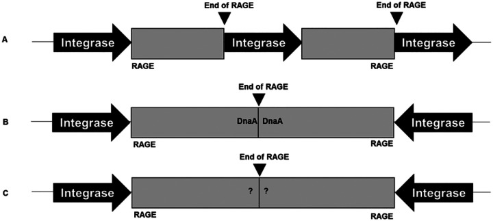
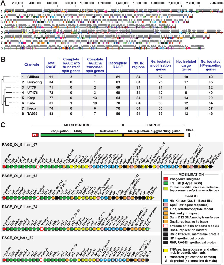
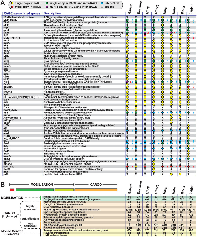
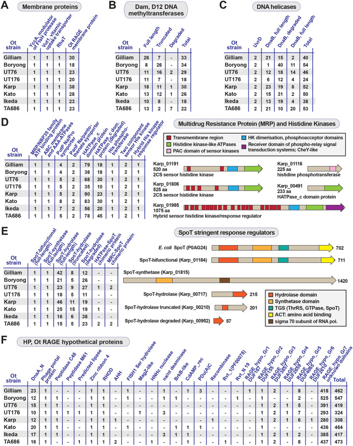
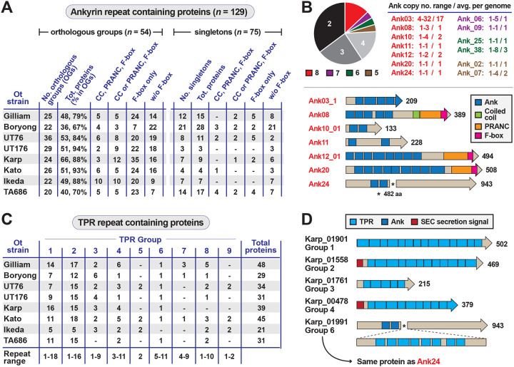
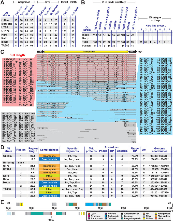
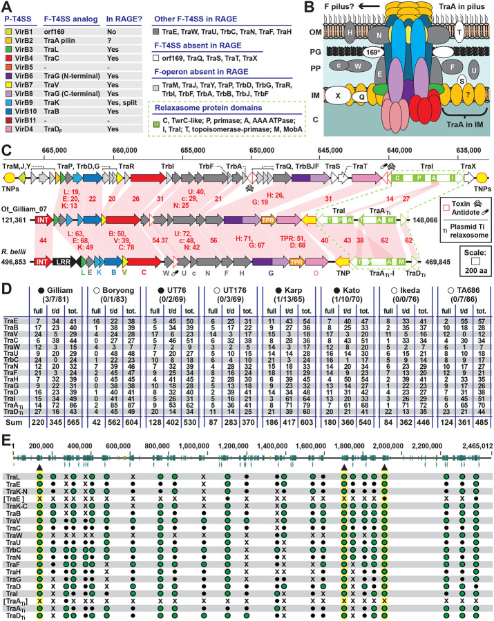
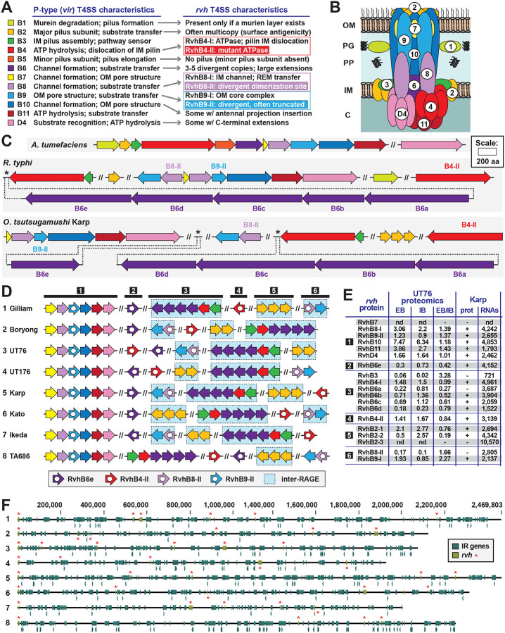
The rickettsial human pathogen Orientia tsutsugamushi (Ot) is an obligate intracellular Gram-negative bacterium with one of the most highly fragmented and repetitive genomes of any organism. Around 50% of its ~2.3 Mb genome is comprised of repetitive DNA that is derived from the highly proliferated Rickettsiales amplified genetic element (RAGE). RAGE is an integrative and conjugative element (ICE) that is present in a single Ot genome in up to 92 copies, most of which are partially or heavily degraded. In this report, we analysed RAGEs in eight fully sequenced Ot genomes and manually curated and reannotated all RAGE-associated genes, including those encoding DNA mobilisation proteins, P-type (vir) and F-type (tra) type IV secretion system (T4SS) components, Ankyrin repeat- and tetratricopeptide repeat-containing effectors, and other piggybacking cargo. Originally, the heavily degraded Ot RAGEs led to speculation that they are remnants of historical ICEs that are no longer active. Our analysis, however, identified two Ot genomes harbouring one or more intact RAGEs with complete F-T4SS genes essential for mediating ICE DNA transfer. As similar ICEs have been identified in unrelated rickettsial species, we assert that RAGEs play an ongoing role in lateral gene transfer within the Rickettsiales. Remarkably, we also identified in several Ot genomes remnants of prophages with no similarity to other rickettsial prophages. Together these findings indicate that, despite their obligate intracellular lifestyle and host range restricted to mites, rodents and humans, Ot genomes are highly dynamic and shaped through ongoing invasions by mobile genetic elements and viruses.

Keywords: Orientia tsutsugamushi; Rickettsiales; bacteriophage; comparative genomics; integrative and conjugative elements; intracellular pathogens; lateral gene transfer; mobile genetic elements; obligate intracellular bacteria.


Conflict of interest statement

Conflict of Interest The authors declare no conflict of interest.

Figures

Figure Methods 1.
Figure Methods 1.
Identification of RAGE termination positions.
Fig. Methods 2.
Fig. Methods 2.
The overview of domain of histidine protein kinases. Figure is modified from ref.
Fig.Methods 3
Fig.Methods 3
Consensus term for Ank domain by SMART.
Figure 1.
Figure 1.. Ot RAGE and IR elements.
A. An overview of the genomes of eight Ot strains with genes classified into RAGE and IR regions. Numbers at left refer to Ot strains listed in panel B. Grey arrows = RAGE regions; colored arrows = IR regions. The colors correspond to conserved IR regions between strains and demonstrate the lack of synteny between Ot genomes. B. Table summarizing RAGEs, IR regions and isolated mobile genes, cargo genes and hypothetical genes that could not be classified into RAGE or IR elements. Ot strains are listed accordingly to a previously estimated phylogeny, with numbers corresponding to full genome maps in panel A. C. Organization of genes in the four complete RAGEs found in our analysis. t = truncated (at least one identifiable domain present); d = degraded (no identifiable domains present). Detailed analysis of the reannotation and classification of all genes in the eight genomes are given in Supplementary Dataset 1.
Figure 2.
Figure 2.. Single and multi-copy cargo genes encoded on Ot RAGEs.
A. Single or low-copy cargo genes encoded on Ot RAGE. Summary statistics show whether genes are present in single or multiple copies on RAGEs in different strains, and also in single or multiple copies in IRs. The exact number of copies is given for each gene. Blue text = number of copies in IR; red text = number of copies in RAGE. B. Frequency and distribution of high copy cargo genes (both full length and truncated/degraded) within RAGEs in eight strains of Ot. Numbers in brackets denote additional copies in IRs.
Fig. 3.
Fig. 3.. Analysis of high copy cargo genes on RAGE elements in Ot.
A-C. Frequency and distribution of RAGE cargo genes annotated as (A) membrane proteins, (B) Dam DNA methyltransferases, and (C) DNA helicases. DnaB is a replicative DNA helicase and UvrB is a repair DNA helicase. D. Frequency and distribution of RAGE cargo genes encoding MRP/histidine kinases, with examples of His kinase divergent architectures. E. Frequency and distribution of RAGE cargo genes encoding SpoT stringent response regulators, with examples of divergent architectures. The bifunctional SpoT protein is compared to the canonical SpoT protein of E. coli. E. Frequency and distribution of RAGE cargo genes encoding HPs. DnaA_N, N-terminal domain of DnaA; RHOD, rhodanese homology domain; AHH, adenosyl homocysteine hydrolase; MagZ, nucleoside triphosphate pyrophosphohydrolase; na/nt, nucleic acid/nucleotide deaminase; BrkB-like, YihY/virulence factor BrkB family protein; PDu(A)C, copper chaperone; CdAMP_rec, cyclic diAMP receptor proteins; Rvt_1 (PF00078), reverse transcriptase Pfam PF00078; Rvt_N 19, domain of reverse transcriptase Rvt_N; DUF, domain of unknown function.
Figure 4.
Figure 4.. Putative effectors piggybacking on Ot RAGE.
A. Frequency and distribution of Anks in Ot genomes. Anks are broken down into orthologous groups (OGs, present in two or more genomes) or singletons (unique to a genome). CC, coiled coil; PRANC (Pox proteins Repeats of ANkyrin, C-terminal), domain found at the C terminus of certain Pox virus proteins; F-box, motif of approximately 50 amino acids that functions in protein-protein interactions. B. (top) graphical view of Ank OG strain representation (2–8 genomes). Roughly 25% of Ank OGs are found in five or more strains, with variable levels of conservation in copy number per genome. (bottom) Architectures for Anks present in all Ot genomes, with proteins from Ot strain Karp. C. Frequency and distribution of TPRs in Ot genomes. Nine ortholog groups contain all the TPR s across eight Ot genomes. D. Examples of diverse TPR architectures for six proteins from Ot strain Karp.
Figure 5.
Figure 5.. A diversity of mobile genetic elements associates with Ot RAGEs.
A. Frequency and distribution of Ot RAGE-associated genes encoding integrases, Group II intron-associated reverse transcriptases, and IS elements ISOt3 and ISOt5. B. Frequency and distribution of IS elements in Ikeda and Karp strains. C. Alignment showing classification of ISOt1 elements as full length or degraded. Full length copies of ISOt1 in Karp are shown by red dotted box. D. Overview of prophage elements in Ot genomes as identified by PHASTER search tool. Int = integrase, Tnp = transposase, Env = envelope, Cap = capsid, Pro = protease. E. Overview of predicted phage region in TA686.
Figure 6.
Figure 6.. Characteristics of the F-type T4SS and relaxosome proteins encoded on Ot RAGEs.
A. Composition of the Ot RAGE F-T4SS in relation to the Agrobacterium tumefaciens vir P-T4SS and the Escherichia coli tra/trb F-T4SS from the F operon. Analogues across divergent T4SSs are coloured similarly, with other colours as follows: dark gray, RAGE T4SS proteins found in F-T4SSs but not P-T4SSs; white, E. coli F-T4SS scaffold genes not present in RAGE T4SSs; light gray, other E. coli F operon genes not present in RAGE. For relaxosome proteins (olive green), domains were predicted with SMART. B. Theoretical assembly of the RAGE T4SS in relation to data from other F- and P-type T4SSs. The uncertain synthesis of a pilus is depicted (see text for details). C. Comparison of the E. coli F operon to mobilisation genes of complete RAGEs from Ot str. Gilliam and Rickettsia bellii str. RML369-C. This E. coli strain, K-12 ER3466 (CP010442), has the F operon on a chromosomal segment flanked by transposases (yellow circles). Red shading and numbers indicate % aa identity across pairwise alignments. Dashed lines enclose the relaxosome genes, whose protein domains are described in panel A. INT, integrase; LRR, leucine rich repeat protein. D. Frequency and distribution of of full length and truncated tra/trb genes in Ot strains. Complete circles, genomes containing full sets of tra/trb genes within one or more RAGE; open circles, no complete tra/trb gene sets. Numbers in parentheses: number of complete RAGEs/number of complete RAGE genes containing truncated genes/incomplete RAGEs. Details of truncated genes and gene fusions are given in Supplementary Datasets 1 and 8. E. Genomic location of tra/trb gene clusters in Ot str. Gilliam. Triangles and highlighting depict complete RAGEs. Bracketed TraE and TraATI are commonly occurring pseudogenized duplications. Green circles, complete gene; small black circles, predicted pseudogene; Xs, gene absent with tra/trb gene cluster.
Figure 7.
Figure 7.. Synopsis of Ot P-type (vir-like) T4SS genes.
A. Description of the general rvh T4SS characteristics, summarized from prior studies,,,. B. Theoretical assembly of the RAGE T4SS in relation to data from other P-type T4SSs. There is no synthesis of a T-pilus (see text for details) C. Comparison of genes encoding vir T4SS in Agrobacterium tumefaciens, the archetypal P-T4SS, and those encoding the rvh T4SS in Rickettsia typhi and Ot. D. Arrangement of rvh genes in Ot genomes. Red genes are located in RAGE regions whilst blue are located in IR regions. E. Previously published RNAseq and proteomics data showing relative expression levels of rvh genes in strains UT76 and Karp. These are taken from Atwal et. al 2022 (UT76) and Mika-Gospodorz et. al 2020 (Karp). UT76 data shows relative peptide counts in intracellular bacteria (IB) and extracellular bacteria (EB). Karp data shows presence or absence of detectable peptides from proteomics analysis (+/−) and relative RNA transcripts from RNAseq data (TPM/transcripts per million). F. Distribution of vir genes across Ot genomes showing lack of conservation of absolute position, despite similarities in gene groupings as shown in Fig. 7D.

Similar articles

References

    1. Giannotti D., Boscaro V., Husnik F., Vannini C. & Keeling P. J. The “Other” Rickettsiales: an Overview of the Family “Candidatus Midichloriaceae”. Appl Environ Microbiol 88, e0243221, doi:10.1128/aem.02432-21 (2022). - DOI - PMC - PubMed
    1. Salje J. Cells within cells: Rickettsiales and the obligate intracellular bacterial lifestyle. Nat Rev Microbiol, doi:10.1038/s41579-020-00507-2 (2021). - DOI - PubMed
    1. Schon M. E., Martijn J., Vosseberg J., Kostlbacher S. & Ettema T. J. G. The evolutionary origin of host association in the Rickettsiales. Nat Microbiol 7, 11891199, doi:10.1038/s41564-022-01169-x (2022). - DOI - PMC - PubMed
    1. Castelli M. et al. Deianiraea, an extracellular bacterium associated with the ciliate Paramecium, suggests an alternative scenario for the evolution of Rickettsiales. ISME J 13, 2280–2294, doi:10.1038/s41396-019-0433-9 (2019). - DOI - PMC - PubMed
    1. Ettema T. The evolutionary origin of host association in an ancient bacterial clade. doi:10.7554/eLife.19469.001 (2022). - DOI - PMC - PubMed

Publication types