. 2023 May 4:11:1180774.
doi: 10.3389/fcell.2023.1180774. eCollection 2023.

Unorthodox localization of P2X7 receptor in subcellular compartments of skeletal system cells

Affiliations

Unorthodox localization of P2X7 receptor in subcellular compartments of skeletal system cells

Letizia Penolazzi et al. Front Cell Dev Biol. .

Abstract

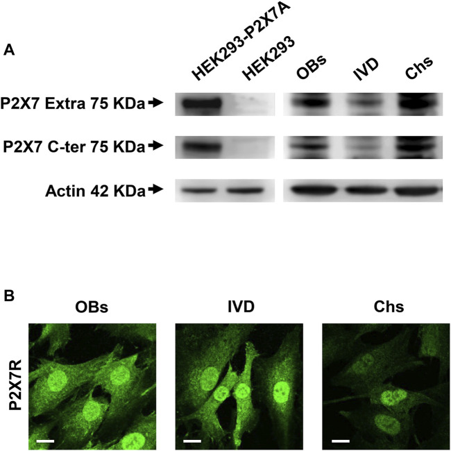
Identifying the subcellular localization of a protein within a cell is often an essential step in understanding its function. The main objective of this report was to determine the presence of the P2X7 receptor (P2X7R) in healthy human cells of skeletal system, specifically osteoblasts (OBs), chondrocytes (Chs) and intervertebral disc (IVD) cells. This receptor is a member of the ATP-gated ion channel family, known to be a main sensor of extracellular ATP, the prototype of the danger signal released at sites of tissue damage, and a ubiquitous player in inflammation and cancer, including bone and cartilaginous tissues. Despite overwhelming data supporting a role in immune cell responses and tumor growth and progression, a complete picture of the pathophysiological functions of P2X7R, especially when expressed by non-immune cells, is lacking. Here we show that human wild-type P2X7R (P2X7A) was expressed in different samples of human osteoblasts, chondrocytes and intervertebral disc cells. By fluorescence microscopy (LM) and immunogold transmission electron microscopy we localized P2X7R not only in the canonical sites (plasma membrane and cytoplasm), but also in the nucleus of all the 3 cell types, especially IVD cells and OBs. P2X7R mitochondrial immunoreactivity was predominantly detected in OBs and IVD cells, but not in Chs. Evidence of subcellular localization of P2X7R may help to i. understand the participation of P2X7R in as yet unidentified signaling pathways in the joint and bone microenvironment, ii. identify pathologies associated with P2X7R mislocalization and iii. design specific targeted therapies.

Keywords: P2X7 receptor; chondrocytes; immunogold and electron microscopy; intervertebral disc cells; osteoblasts; purinergic signaling; subcellular localization.


Conflict of interest statement

The authors declare that the research was conducted in the absence of any commercial or financial relationships that could be construed as a potential conflict of interest.

Figures

FIGURE 1
FIGURE 1
Expression of P2X7R in human osteoblasts (OBs), IVD cells (IVD), and chondrocytes (Chs). (A) Representative Western blot analysis with anti-P2X7 extracellular loop and anti-P2X7 C-terminal antibodies; P2X7R stably transfected HEK293 cells (HEK293-P2X7A) and HEK293 wild-type (HEK293) were used as positive and negative control respectively. β-actin was used as loading control. Number of cell samples: OBs = 6, IVD = 4, Chs = 3 (B) Representative immunofluorescence and confocal microscopy analysis with anti-P2X7R C-terminal antibody. Scale bars = 10 μm.
FIGURE 2
FIGURE 2
Subcellular localization of P2X7R by immunofluorescence and confocal microscopy in human osteoblasts (OBs), IVD cells (IVD), and chondrocytes (Chs). (A) Mitochondrial P2X7R localization analysis. The cells were co-labeled with an anti-P2X7R C-terminal antibody (Alexafluor 488, green) and an anti-TOM20 antibody (Alexafluor 546, red). Merge images represent an overlay of the two channels where co-localization is indicated by a color change (yellow). Average Pearson’s and Mander’s co-localization coefficients (±SEM) were evaluated and reported in the graphs. Scale bars = 10 μm. (B) Nuclear P2X7R localization analysis. The cells were co-labeled with an anti-P2X7R C-tail antibody (Alexafluor 488, green) and DAPI (nuclear staining, blue). Co-localization of P2X7R with DAPI was assessed by Pearson’s and Mander’s coefficients (average ± SD). Scale bars = 10 μm.
FIGURE 3
FIGURE 3
Subcellular localization of P2X7R by immunogold labeling. Human osteoblasts (A), IVD cells (B), chondrocytes (C) and HEK293 wild-type cells (D) were subjected to pre-embedding immunogold staining. For each cell type the mean percentage of gold particles distribution was reported. Fifty areas from the extracellular membrane (EM), cytoplasm (C), nucleus (N) and mitochondria (M) were randomly chosen, and gold particles were manually counted using ImageJ software, and expressed as percent of total (±SD). High magnification boxes for each subcellular compartment were reported below. Arrowheads = gold particles. Scale bars = 1 µm.

Similar articles

Cited by

References

    1. Adinolfi E., Callegari M. G., Ferrari D., Bolognesi C., Minelli M., Wieckowski M. R., et al. (2005). Basal activation of the P2X7 ATP receptor elevates mitochondrial calcium and potential, increases cellular ATP levels, and promotes serum-independent growth. Mol. Biol. Cell 16 (7), 3260–3272. 10.1091/mbc.e04-11-1025 - DOI - PMC - PubMed
    1. Adinolfi E., Cirillo M., Woltersdorf R., Falzoni S., Chiozzi P., Pellegatti P., et al. (2010). Trophic activity of a naturally occurring truncated isoform of the P2X7 receptor. FASEB J. 24 (9), 3393–3404. 10.1096/fj.09-153601 - DOI - PubMed
    1. Agrawal A., Gartland A. (2015). P2X7 receptors: Role in bone cell formation and function. J. Mol. Endocrinol. 54 (2), R75–R88. 10.1530/JME-14-0226 - DOI - PubMed
    1. Andrejew R., Oliveira-Giacomelli Á., Ribeiro D. E., Glaser T., Arnaud-Sampaio V. F., Lameu C., et al. (2020). The P2X7 receptor: Central hub of brain diseases. Front. Mol. Neurosci. 13, 124. 10.3389/fnmol.2020.00124 - DOI - PMC - PubMed
    1. Angelozzi M., Penolazzi L., Mazzitelli S., Lambertini E., Lolli A., Piva R., et al. (2017). Dedifferentiated chondrocytes in composite microfibers as tool for cartilage repair. Front. Bioeng. Biotechnol. 5, 35. 10.3389/fbioe.2017.00035 - DOI - PMC - PubMed

LinkOut - more resources