. 2023 May 4:11:1154205.
doi: 10.3389/fcell.2023.1154205. eCollection 2023.

Distinct patterning responses of wing and leg neuromuscular systems to different preaxial polydactylies

Affiliations

Distinct patterning responses of wing and leg neuromuscular systems to different preaxial polydactylies

Maëva Luxey et al. Front Cell Dev Biol. .

Abstract

The tetrapod limb has long served as a paradigm to study vertebrate pattern formation and evolutionary diversification. The distal part of the limb, the so-called autopod, is of particular interest in this regard, given the numerous modifications in both its morphology and behavioral motor output. While the underlying alterations in skeletal form have received considerable attention, much less is known about the accompanying changes in the neuromuscular system. However, modifications in the skeleton need to be properly integrated with both muscle and nerve patterns, to result in a fully functional limb. This task is further complicated by the distinct embryonic origins of the three main tissue types involved-skeleton, muscles and nerves-and, accordingly, how they are patterned and connected with one another during development. To evaluate the degree of regulative crosstalk in this complex limb patterning process, here we analyze the developing limb neuromuscular system of Silkie breed chicken. These animals display a preaxial polydactyly, due to a polymorphism in the limb regulatory region of the Sonic Hedgehog gene. Using lightsheet microscopy and 3D-reconstructions, we investigate the neuromuscular patterns of extra digits in Silkie wings and legs, and compare our results to Retinoic Acid-induced polydactylies. Contrary to previous findings, Silkie autopod muscle patterns do not adjust to alterations in the underlying skeletal topology, while nerves show partial responsiveness. We discuss the implications of tissue-specific sensitivities to global limb patterning cues for our understanding of the evolution of novel forms and functions in the distal tetrapod limb.

Keywords: Silkie chicken; developmental plasticity; limb development; neuromuscular system; polydactyly.


Conflict of interest statement

The authors declare that the research was conducted in the absence of any commercial or financial relationships that could be construed as a potential conflict of interest.

Figures
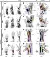
FIGURE 1
FIGURE 1
A comparative 3D-analysis of the developing muscles in control and Silkie wings and legs.Fore- (A–F) and hindlimb (G–L) muscle development in control and Silkie embryos between day 7 and day 9 of development. Red dotted lines indicate the territories of the forming preaxial extra digits in Silkie wings (D–F) and legs (J–L). Nerves and muscles were visualized with antibodies against neurofilament (NF200) and myosin heavy chains (MF20). After 3D-reconstruction, muscle surfaces were rendered using segmentation-based tracing to highlight individual bundles. Individual autopod muscles are pseudo-colored and labeled based on Sullivan, 1962; Kardon, 1998. No individual extra muscles are visible in the preaxial polydactyl territories at day 9 of development, in either dorsal (E,K) or ventral (F,L) of Silkie fore- or hindlimbs (compare to B,H and C,I). Autopod muscle nomenclature: (B) Wing dorsal muscles - EIB: Extensor indicis brevis; EMB: Extensor medius brevis; IOD: Interosseus dorsalis; FDM: Flexor digiti minori, UMD: Ulnimetacarpalis dorsalis. (C) Wing ventral muscles—FDS: Flexor digitorum superficialis; AdI: Adductor indicis; FI: Flexor indicis; AbI: Abductor indicis; AM: Abductor medius; IOP: Interosseus palmaris; FDQ: Flexor digiti quarti. (H) Foot dorsal muscles - EHL: Extensor hallucis longus; AB2: Abductor digit 2; EP3: Extensor propius 3; EB4: Extensor brevis digit 4. (I) Foot ventral muscles - FHB: Flexor halluces brevis; AD2: Adductor digit 2; AB4: Abductor digit 4. A/P, anterior/posterior, Prox/Dist., proximal/distal. Scale bars represent approx. 500 μm.
FIGURE 2
FIGURE 2
A comparative 3D-analysis of the developing nerves in control and Silkie wings and legs.Fore- (A–F) and hindlimb (G–L) nerve development in control and Silkie embryos between day 7 and day 9 of development. Red dotted lines indicate the territories of the forming preaxial extra digits in Silkie wings (D–F) and legs (J–L). Asterisks highlight the lack of invading nerve branches in the wings preaxial extra digit domain (E,F). White arrowheads highlight the stunted defasciculations at the base of the polydactyl territories (D,J) and partial sensory nerve projections occasionally observed in wings (E, F, insets; n = 3/9). From day 8, the preaxial extra digit of the leg shows an innervation pattern similar to the others, with anterior and posterior sensory branches projecting towards the digit tip (K, L; arrowheads). A/P, anterior/posterior; Prox/Dist., proximal/distal. Scale bars represent approx. 500 μm.
FIGURE 3
FIGURE 3
A comparative 3D-analysis of innervation patterns in control, Silkie and RA-induced polydactyl fore- and hindlimbs.Dorsal views of 3D-reconstructed developing nervous systems at day 8 of development in control (A,B), Silkie (C,D) and RA (Retinoic Acid)-treated (E,F) limbs. Red dotted lines indicate the territories of the preaxial extra digits (C–F). The three main motor nerves of the wings (A,C,E) and legs (B,D,F) were surface-rendered using segmentation-based tracing and pseudo-coloring. In the wing, the dorsal radial nerve (cyan) and the two ventral nerves, median and ulnar (yellow and magenta), show a similar branching organization in control and Silkie wings (A,C). In the RA-induced mirror-image polydactyl wing, the radial and the median nerves bifurcate (arrowheads) and innervate the extra-digit territory (red dotted line), whereas the ulnar nerve remains unchanged (E). In the leg, the plantar nerve (purple) innervates the ventral part of the leg in an identical manner in control and Silkie limbs (B,D), while the fusion with an ectopic projection (light green) is present only in RA-treated limbs (F, arrow). At the dorsal level, the lateral fibular nerve (orange) innervates the last two posterior digits in control, Silkie and RA-induced polydactyl limbs (B,D,F). The median fibular nerve (green) shows bifurcation and extra branches innervating the preaxial digit in Silkie and RA-induced polydactyl (D, F; black arrow heads), albeit at different proximal-distal levels (D,F, arrowheads). A/P, anterior/posterior, Prox/Dist., proximal/distal. Images are oriented anterior to the right, distal on top. Scale bars represent approx. 500 μm.

Similar articles

References

    1. Anderson C., Williams V. C., Moyon B., Daubas P., Tajbakhsh S., Buckingham M. E., et al. (2012). Sonic hedgehog acts cell-autonomously on muscle precursor cells to generate limb muscle diversity. Genes. Dev. 26, 2103–2117. 10.1101/gad.187807.112 - DOI - PMC - PubMed
    1. Andreone B. J., Lacoste B., Gu C. (2015). Neuronal and vascular interactions. Annu. Rev. Neurosci. 38, 25–46. 10.1146/annurev-neuro-071714-033835 - DOI - PMC - PubMed
    1. Arisawa K., Yazawa S., Atsumi Y., Kagami H., Ono T. (2006). Skeletal analysis and characterization of gene expression related to pattern formation in developing limbs of Japanese silkie fowl. J. Poult. Sci. 43, 126–134. 10.2141/jpsa.43.126 - DOI
    1. Berki B., Sacher F., Fages A., Tschopp P., Luxey M. (2023). A method to investigate muscle target-specific transcriptional signatures of single motor neurons. Dev. Dyn. 252, 208–219. 10.1002/dvdy.507 - DOI - PMC - PubMed
    1. Bininda-Emonds O. R. P., Jeffery J. E., Sánchez-Villagra M. R., Hanken J., Colbert M., Pieau C., et al. (2007). Forelimb-hindlimb developmental timing changes across tetrapod phylogeny. BMC Evol. Biol. 7, 182. 10.1186/1471-2148-7-182 - DOI - PMC - PubMed

LinkOut - more resources