. 2023 Apr 19:32:522-535.
doi: 10.1016/j.omtn.2023.04.009. eCollection 2023 Jun 13.

Single-swap editing for the correction of common Duchenne muscular dystrophy mutations

Affiliations

Single-swap editing for the correction of common Duchenne muscular dystrophy mutations

Andreas C Chai et al. Mol Ther Nucleic Acids. .

Abstract

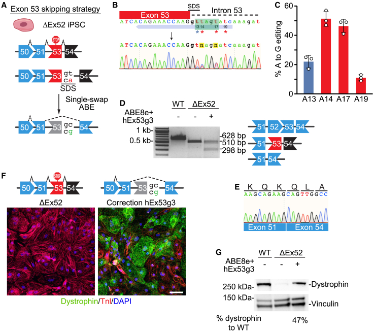
Duchenne muscular dystrophy (DMD) is a fatal X-linked recessive disease of progressive muscle weakness and wasting caused by the absence of dystrophin protein. Current gene therapy approaches using antisense oligonucleotides require lifelong dosing and have limited efficacy in restoring dystrophin production. A gene editing approach could permanently correct the genome and restore dystrophin protein expression. Here, we describe single-swap editing, in which an adenine base editor edits a single base pair at a splice donor site or splice acceptor site to enable exon skipping or reframing. In human induced pluripotent stem cell-derived cardiomyocytes, we demonstrate that single-swap editing can enable beneficial exon skipping or reframing for the three most therapeutically relevant exons-DMD exons 45, 51, and 53-which could be beneficial for 30% of all DMD patients. Furthermore, an adeno-associated virus delivery method for base editing components can efficiently restore dystrophin production locally and systemically in skeletal and cardiac muscles of a DMD mouse model containing a deletion of Dmd exon 44. Our studies demonstrate single-swap editing as a potential gene editing therapy for common DMD mutations.

Keywords: AAV; CRISPR-Cas9; DMD; Duchenne muscular dystrophy; MT: RNA/DNA editing; base editing; exon skipping; gene editing; iPSC; iPSC-CM.


Conflict of interest statement

F.C., R.B.-D., and E.N.O. have filed patent applications related to this work. E.N.O. is a consultant for Vertex Pharmaceuticals and Tenaya Therapeutics.

Figures

None
Graphical abstract
Figure 1
Figure 1
Single-swap editing at the SAS of DMD exon 51 induces beneficial exon reframing in ΔEx48–50 iPSC-CMs (A) Single-swap editing at the canonical 5′AG3′ SAS around human DMD exon 51 activates a cryptic splice acceptor site within exon 51 that causes an 11 nt deletion of the mature transcript. In ΔEx48–50 iPSC-CMs, this 11 nt deletion restores the open reading frame. (B) Schematic of hEx51g2 targeting the SAS of DMD exon 51 and representative chromatogram following editing using ABE8e-nSpCas9-NG. Editable adenines are indicated by asterisks: target adenine is at position 15; bystander adenine, position 20. Editing window is in green. (C) Editing efficiency by Sanger sequencing of hEx51g2 with ABE8e-nSpCas9-NG in ΔEx48–50 iPSCs at editable adenines for DMD exon 51 SAS. Target adenine is colored in blue; editing efficiency is 71.6% ± 0.6%. Bystander adenine is colored in red. n = 3 independent replicates. (D) RT-PCR analysis of mRNA from WT, ΔEx48–50, and ΔEx48–50 edited with ABE8e-nSpCas9-NG and hEx51g2 iPSC-CMs. The cDNA of the WT is 717 bp, of the ΔEx48–50 is 320 bp, and of the ΔEx48–50 with ABE8e-nSpCas9-NG and hEx51g2 is 309 bp. (E) Sanger sequencing of the cDNA from the ΔEx48–50 iPSC-CMs edited with ABE8e-nSpCas9-NG and hEx51g2 reveals splicing of DMD exon 47 to exon 51 with an 11 bp deletion. (F) Immunocytochemistry of ΔEx48–50 iPSC-CMs edited with ABE8e-nSpCas9-NG and hEx51g2 shows restoration of dystrophin protein. Dystrophin is in green; cardiac troponin I (TnI) highlights CMs in red; DAPI stains for nuclei in blue. Scale bar, 50 μm. (G) Western blot of ΔEx48–50 iPSC-CMs edited with ABE8e-nSpCas9 and hEx51g2 shows restoration of dystrophin protein. Vinculin is the loading control. Relative intensity is measured as dystrophin expression normalized to vinculin compared with the WT. Data are mean ± SD.
Figure 2
Figure 2
Single-swap editing at the SAS of DMD exon 45 induces beneficial exon skipping in ΔEx44 iPSC-CMs (A) Single-swap editing at the canonical 5’AG3’ SAS around human DMD exon 45 causes exon skipping. In ΔEx44 iPSC-CMs, skipping of exon 45 restores the open reading frame. (B) Schematic of hEx45g3 targeting the SAS of DMD exon 45 and representative chromatogram following editing using ABE8e-nSpCas9. Editable adenines are indicated by asterisks: target adenine is at position 13; bystander adenine, positions 10, 15, and 20. Editing window is in green. (C) Editing efficiency by Sanger sequencing of hEx45g3 with ABE8e-nSpCas9 in ΔEx44 iPSCs at editable adenines for DMD exon 45 SAS. Target adenine is colored in blue; editing efficiency is 83.3% ± 5.0%. Bystander adenines are colored in red. n = 3 independent replicates. (D) Schematic of hEx45g5 targeting the SAS of DMD exon 45 and representative chromatogram following editing using ABE8e-nSpCas9. Editable adenines are indicated by asterisks: target adenine is at position 19; bystander adenine, positions 10, 15, and 16. Editing window is in green. (E) Editing efficiency by Sanger sequencing of hEx45g5 with ABE8e-nSpCas9 in ΔEx44 iPSCs at editable adenines for DMD exon 45 SAS. Target adenine is colored in blue; editing efficiency is 79.3% ± 4.7%. Bystander adenines are colored in red. n = 3 independent replicates. (F) RT-PCR analysis of mRNA from WT, ΔEx44, and ΔEx44 edited with ABE8e-nSpCas9 and either hEx45g3 or hEx45g5 iPSC-CMS. The cDNA of the WT is 692 bp, of the ΔEx44 is 544 bp, and of the ΔEx44 with exon skipping of exon 45 is 368 bp. (G) Sanger sequencing of the 368 bp cDNA band shows splicing of DMD exon 43 to exon 46. (H) Immunocytochemistry of ΔEx44 iPSC-CMs edited with ABE8e-nSpCas9 and either hEx45g3 or hEx45g5 shows restoration of dystrophin protein. Dystrophin is in green; cardiac troponin I (TnI) highlights CMs in red; DAPI stains for nuclei in blue. Scale bar, 50 μm. (I) Western blot of ΔEx44 iPSC-CMs edited with ABE8e-nSpCas9 and either hEx45g3 or hEx45g5 shows restoration of dystrophin protein. Vinculin is the loading control. Relative intensity is measured as dystrophin expression normalized to vinculin compared with the WT. Data are mean ± SD.
Figure 3
Figure 3
Single-swap editing at the SDS of DMD exon 53 induces beneficial exon skipping in ΔEx52 iPSC-CMs (A) Single-swap editing on the antisense strand of the canonical 5′GT3′ SDS around human DMD exon 53 causes exon skipping. In ΔEx52 iPSC-CMs, skipping of exon 53 restores the open reading frame. (B) Schematic of hEx53g3 targeting the SDS of DMD exon 53 and representative chromatogram following editing using ABE8e-nSpCas9-NG. Editable adenines are indicated by asterisks: the target adenine is at position 13; bystander adenine, positions 14, 17, and 19. Editing window is in green. (C) Editing efficiency by Sanger sequencing of hEx53g3 with ABE8e-nSpCas9-NG in ΔEx52 iPSCs at editable adenines for DMD exon 53 SDS. Target adenine is colored in blue; editing efficiency is 22.0% ± 4.4%. Bystander adenines are colored in red. n = 3 independent replicates. (D) RT-PCR analysis of mRNA from WT, ΔEx52, and ΔEx52 edited with ABE8e-nSpCas9-NG and hEx52g3 iPSC-CMs. The cDNA of the WT is 628 bp, of the ΔEx52 is 510 bp, and of the ΔEx52 with exon skipping of exon 53 is 298 bp. (E) Sanger sequencing of the 298 bp cDNA band shows splicing of DMD exon 51 to exon 54. (F) Immunocytochemistry of ΔEx52 iPSC-CMs edited with ABE8e-nSpCas9-NG and hEx53g3 shows restoration of dystrophin protein. Dystrophin is in green; cardiac troponin I (TnI) highlights CMs in red; DAPI stains for nuclei in blue. Scale bar, 50 μm. (G) Western blot of ΔEx52 iPSC-CMs edited with ABE8e-nSpCas9 and hEx53g3 shows restoration of dystrophin protein. Vinculin is the loading control. Relative intensity is measured as dystrophin expression normalized to vinculin compared with the WT. Data are mean ± SD.
Figure 4
Figure 4
Intramuscular injection of a single-swap editing dual AAV system restores dystrophin protein production in a ΔEx44 mouse model of DMD (A) Schematic for the dual AAV ABE8e system. The CK8e muscle-specific promoter drives expression of ABE8eV106W-nSpCas9 base editor halves and their intein tags for protein trans-splicing. Each viral construct also contains the woodchuck hepatitis post-transcriptional regulatory element (WPRE3), a synthetic mini polyadenylation signal (PolyA), and an hU6 promoter-sgRNA cassette (U6-sgRNA). (B) At P12, ΔEx44 mice received saline in the right leg and the dual AAV ABE8e system in the left leg by intramuscular injection into the TA muscle. Three weeks post-injection, TA muscles were collected. (C) Representative Sanger sequencing chromatograms of genomic DNA from the right leg 3 weeks post-saline injection (top) and from the left leg 3 weeks post-dual AAV ABE8e treatment (bottom). Blue arrow indicates the hmEx45g3-18nt sgRNA. Target adenine is at position 13. Bystander adenines are at positions 10 and 15. Editing window is in green. (D) Editing efficiency of target adenine and indel frequency by amplicon deep sequencing in genomic DNA from the left TA 3 weeks post-dual AAV ABE8e treatment. Target adenine is A13 (blue); editing efficiency is 29.5% ± 2.7% in the left leg. Indel frequency is 0.2% ± 0.1% in the left leg. n = 4 mice. (E) Immunohistochemistry for dystrophin expression from the saline-injected right leg and the dual AAV ABE8e-injected left leg from a ΔEx44 mouse and the TA of a WT mouse. Scale bar, 100 μm. Dystrophin is stained in green. (F) Western blot and (G) quantification of dystrophin protein expression from the left leg of a WT mouse, a ΔEx44 mouse, and four dual AAV ABE8e-injected ΔEx44 mice (47.1% ± 3.5%). Vinculin is the loading control. Relative intensity is measured as dystrophin expression normalized to vinculin compared with the WT. n = 1–4 mice. Data are mean ± SD.
Figure 5
Figure 5
Systemic injection of a single-swap editing dual AAV system in a ΔEx44 mouse model of DMD restores dystrophin protein production in skeletal and cardiac muscles (A) At P2, ΔEx44 mice received systemically either a low dose or a high dose of the dual AAV ABE8e system via injection into the temporal facial vein. Tissues were collected 8 weeks later. (B) Editing efficiency for target adenine and indel frequency by amplicon sequencing in genomic DNA from the TA (left, ΔEx44, 0.2% ± 0.0%; low dose, 5.5% ± 1.2%; high dose, 8.1% ± 3.0%) and heart (right, ΔEx44, 0.2% ± 0.0%; low dose, 22.0% ± 2.2%; high dose, 26.2% ± 4.4%) of ΔEx44 mice following saline or dual AAV ABE8e treatment at the two doses. n = 2–3 mice. (C) Sanger sequencing of cDNA of heart tissue from a dual AAV ABE8e-treated ΔEx44 mouse showing splicing of Dmd exon 43 to exon 46. (D) Efficiency of exon skipping in mature mRNA from the TA (left, ΔEx44, 1.4%; low dose, 20.7% ± 1.7%; high dose, 36.7% ± 2.0%) and heart (right, ΔEx44, 0.2%; low dose, 52.4% ± 7.2%; high dose, 55.5% ± 3.6%) following dual AAV ABE8e treatment at the two doses. n = 1–3 mice. (E) Western blot and (F) quantification of dystrophin protein expression from the TA (left in F, ΔEx44, 3.0%; low dose, 19.3% ± 2.5%; high dose, 31.0% ± 5.6%) and heart (right in F, ΔEx44, 2.0%; low dose, 36.0% ± 1.0%; high dose, 65.0% ± 14.4%) of a WT mouse, a ΔEx44 mouse, and three dual AAV ABE8e-injected ΔEx44 mice each at the low and high dose. Vinculin is the loading control. Relative intensity is measured as dystrophin expression normalized to vinculin compared with the WT. Data are mean ± SD. n = 1–3 mice.
Figure 6
Figure 6
Single-swap editing restores functional dystrophin protein that rescues muscular dystrophy and weakness in a ΔEx44 mouse model of DMD (A) Immunohistochemistry for dystrophin from the TA and heart and (B) H&E staining from the TA of a WT mouse, a ΔEx44 mouse, and a dual AAV ABE8e-injected ΔEx44 mouse at the low and high dose. Scale bar, 100 μm. Dystrophin is stained in green. From WT mice, ΔEx44 mice, and dual AAV ABE8e-injected ΔEx44 mice at the low and high dose, quantification of (C) dystrophin-positive fibers in TA muscles (WT, 100.0% ± 0.0%; ΔEx44, 1.3% ± 0.6%; low dose, 62.3% ± 6.0%; high dose, 75.7% ± 6.5%) and heart (WT, 100.0% ± 0.0%; ΔEx44, 0.1% ± 0.0%; low dose, 95.2% ± 0.2%; high dose, 96.1% ± 0.4%), (D) centrally nucleated fibers in TA muscles (WT, 1.3% ± 1.2%; ΔEx44, 64.0% ± 1.0%; low dose, 25.3% ± 7.8%; high dose, 10.0% ± 3.0%), and (E) grip strength (WT, 7.7 ± 1.2 grams-force/grams body weight [gf/g]; ΔEx44, 1.7 ± 0.9 gf/g; low dose, 4.2 ± 0.3 gf/g; high dose, 4.9 ± 0.3 gf/g). Data are mean ± SD. n = 3 mice. ∗p < 0.05, ∗∗p < 0.01, ∗∗∗p < 0.0001 by ordinary one-way ANOVA.

References

    1. Birnkrant D.J., Bushby K., Bann C.M., Apkon S.D., Blackwell A., Brumbaugh D., Case L.E., Clemens P.R., Hadjiyannakis S., Pandya S., et al. DMD Care Considerations Working Group Diagnosis and management of Duchenne muscular dystrophy, part 1: diagnosis, and neuromuscular, rehabilitation, endocrine, and gastrointestinal and nutritional management. Lancet Neurol. 2018;17:251–267. doi: 10.1016/S1474-4422(18)30024-3. - DOI - PMC - PubMed
    1. Birnkrant D.J., Bushby K., Bann C.M., Alman B.A., Apkon S.D., Blackwell A., Case L.E., Cripe L., Hadjiyannakis S., Olson A.K., et al. DMD Care Considerations Working Group Diagnosis and management of Duchenne muscular dystrophy, part 2: respiratory, cardiac, bone health, and orthopaedic management. Lancet Neurol. 2018;17:347–361. doi: 10.1016/S1474-4422(18)30025-5. - DOI - PMC - PubMed
    1. Hoffman E.P., Brown R.H., Jr., Kunkel L.M. Dystrophin: the protein product of the Duchenne muscular dystrophy locus. Cell. 1987;51:919–928. doi: 10.1016/0092-8674(87)90579-4. - DOI - PubMed
    1. Bladen C.L., Salgado D., Monges S., Foncuberta M.E., Kekou K., Kosma K., Dawkins H., Lamont L., Roy A.J., Chamova T., et al. The TREAT-NMD DMD Global Database: analysis of more than 7,000 Duchenne muscular dystrophy mutations. Hum. Mutat. 2015;36:395–402. doi: 10.1002/humu.22758. - DOI - PMC - PubMed
    1. Min Y.L., Li H., Rodriguez-Caycedo C., Mireault A.A., Huang J., Shelton J.M., McAnally J.R., Amoasii L., Mammen P.P.A., Bassel-Duby R., Olson E.N. CRISPR-Cas9 corrects Duchenne muscular dystrophy exon 44 deletion mutations in mice and human cells. Sci. Adv. 2019;5:eaav4324. doi: 10.1126/sciadv.aav4324. - DOI - PMC - PubMed