. 2022 Sep 22;3(4):515-537.
doi: 10.1162/nol_a_00075. eCollection 2022.

Left Frontal White Matter Links to Rhythm Processing Relevant to Speech Production in Apraxia of Speech

Affiliations

Left Frontal White Matter Links to Rhythm Processing Relevant to Speech Production in Apraxia of Speech

Rose Bruffaerts et al. Neurobiol Lang (Camb). .

Abstract

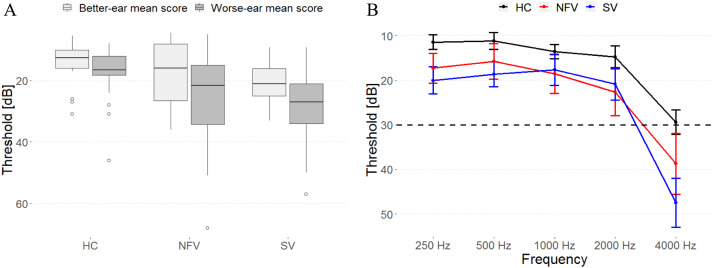
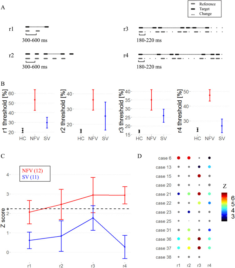
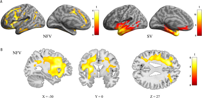
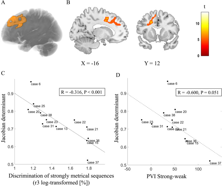
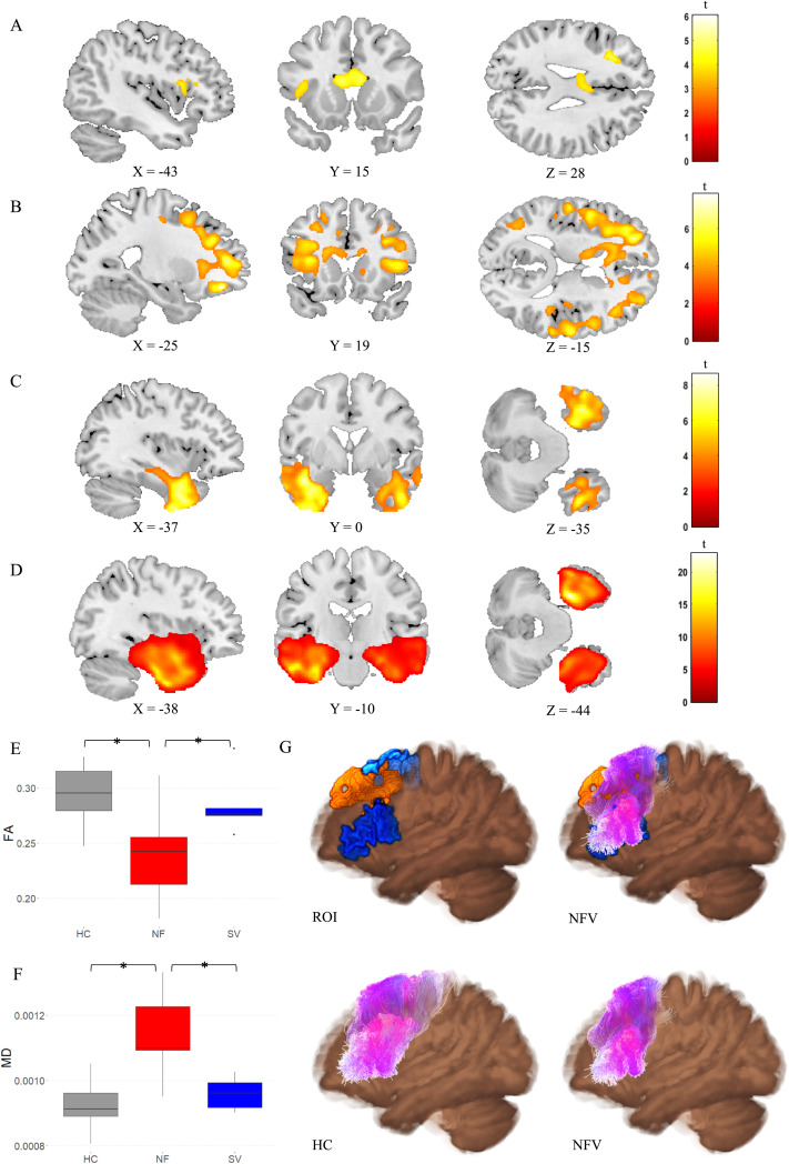
Recent mechanistic models argue for a key role of rhythm processing in both speech production and speech perception. Patients with the non-fluent variant (NFV) of primary progressive aphasia (PPA) with apraxia of speech (AOS) represent a specific study population in which this link can be examined. Previously, we observed impaired rhythm processing in NFV with AOS. We hypothesized that a shared neurocomputational mechanism structures auditory input (sound and speech) and output (speech production) in time, a "temporal scaffolding" mechanism. Since considerable white matter damage is observed in NFV, we test here whether white matter changes are related to impaired rhythm processing. Forty-seven participants performed a psychoacoustic test battery: 12 patients with NFV and AOS, 11 patients with the semantic variant of PPA, and 24 cognitively intact age- and education-matched controls. Deformation-based morphometry was used to test whether white matter volume correlated to rhythmic abilities. In 34 participants, we also obtained tract-based metrics of the left Aslant tract, which is typically damaged in patients with NFV. Nine out of 12 patients with NFV displayed impaired rhythmic processing. Left frontal white matter atrophy adjacent to the supplementary motor area (SMA) correlated with poorer rhythmic abilities. The structural integrity of the left Aslant tract also correlated with rhythmic abilities. A colocalized and perhaps shared white matter substrate adjacent to the SMA is associated with impaired rhythmic processing and motor speech impairment. Our results support the existence of a temporal scaffolding mechanism structuring perceptual input and speech output.

Keywords: apraxia of speech; psychoacoustics; rhythm; speech production; structural MRI.


Conflict of interest statement

Competing Interests: The authors have declared that no competing interests exist.

Figures

<b>Figure 1.</b>
Figure 1.
Pure-tone audiograms of all participants. (A) Mean composite ear and frequency score (250–4000 Hz) data for each participant group. (B) Mean thresholds (and standard error of the mean) for detection of tones at frequencies of 250, 500, 1000, 2000, and 4000 Hz for each participant group. HC: healthy controls. NFV: participants with non-fluent variant of primary progressive aphasia. SV: participants with semantic variant of primary progressive aphasia.
<b>Figure 2.</b>
Figure 2.
Psychoacoustic tasks. (A) Experimental design: audio samples in supplementary information. (B) Mean raw thresholds (and standard error of the mean) across controls, NFV, and SV patients. (C) Mean z scores (and standard error of the mean) across primary progressive aphasia (PPA) subtypes. Dotted line represents the z cut-off for Bonferroni-corrected p < 0.05. (D) z scores for psychoacoustic tests in NFV (case numbers refer to Table 1). The size and color of dots reflect z score values; nonsignificant values are gray. Missing data indicates that the patient was unable to perform the task. HC: healthy controls. NFV: participants with non-fluent variant of primary progressive aphasia. SV: participants with semantic variant of PPA.
<b>Figure 3.</b>
Figure 3.
Suprasegmental timing of speech. (A) Frequency of words with a strong-weak pattern and a weak-strong pattern in the three participant groups. (B–C) Vowel durations for words with (B) a strong-weak pattern and (C) a weak-strong pattern. (D) PVI of words with a strong-weak and a weak-strong pattern. (E–F) Correlation of PVI for (E) strong-weak and (F) weak-strong words and strongly metrical sequence thresholds (r3, log-transformed, %); regression line indicates a significant correlation at the subgroup level. PVI: pairwise variability index. HC = healthy controls. NFV: participants with non-fluent variant of primary progressive aphasia. SV: participants with semantic variant of primary progressive aphasia.
<b>Figure 4.</b>
Figure 4.
Deformation-based morphometry (DBM) analysis: comparison of controls, the NFV, and SV patients. (A) Renderings shows atrophy of 12 NFV and 11 SV compared to 24 controls (cluster-level FWE-corrected p < 0.05). (B) Slices in NFV. NFV: participants with non-fluent variant of primary progressive aphasia. SV: participants with semantic variant of primary progressive aphasia.
<b>Figure 5.</b>
Figure 5.
Deformation-based morphometry (DBM) of rhythmic abilities in NFV cases. (A) Rendering of the DBM result (orange) projected onto structural template. (B) Slices. (C) Correlation between volume loss and strongly metrical sequence thresholds (r3, log-transformed, %) in NFV patients in the region of interest (A–B), exploratory plot for illustrative purposes. (D) Correlation between volume loss and PVI for words with a strong-weak pattern in NFV patients in the region of interest (A–B) (dotted regression line since there was a trend for significance). Case numbers refer to Table 1. PVI: pairwise variability index.
<b>Figure 6.</b>
Figure 6.
Diffusion tensor imaging (DTI) metrics. (A) Fractional anisotropy (FA) and (B) Mean diffusivity (MD) in NFV patients versus controls (cluster-level FWE-corrected p < 0.05). (C) FA and (D) MD in SV patients versus controls (cluster-level FWE-corrected p < 0.05). Comparison of (E) FA and (F) MD in the left frontal Aslant tract between the NFV and SV subtypes and healthy controls (HC). (G) 3D visualization of the left Aslant tract projected on the average FA map—Top left: seed regions (blue) and result of deformation-based morphometry (DBM) (orange); Top right: seed regions (blue) and result of DBM (orange) combined with Aslant tract streamlines of NFV patients (purple); Bottom left: streamlines of controls (purple); Bottom right: streamlines of NFV patients (purple). ROI: region of interest. HC: healthy controls. NFV: participants with non-fluent variant of primary progressive aphasia. SV: participants with semantic variant of primary progressive aphasia.

References

    1. Aichert, I., Späth, M., & Ziegler, W. (2016). The role of metrical information in apraxia of speech. Perceptual and acoustic analyses of word stress. Neuropsychologia, 82, 171–178. 10.1016/j.neuropsychologia.2016.01.009, - DOI - PubMed
    1. Andersson, J. L. R., Skare, S., & Ashburner, J. (2003). How to correct susceptibility distortions in spin-echo echo-planar images: Application to diffusion tensor imaging. NeuroImage, 20(2), 870–888. 10.1016/S1053-8119(03)00336-7, - DOI - PubMed
    1. Andersson, J. L. R., & Sotiropoulos, S. N. (2016). An integrated approach to correction for off-resonance effects and subject movement in diffusion MR imaging. NeuroImage, 125, 1063–1078. 10.1016/j.neuroimage.2015.10.019, - DOI - PMC - PubMed
    1. Assaneo, M. F., & Poeppel, D. (2018). The coupling between auditory and motor cortices is rate-restricted: Evidence for an intrinsic speech-motor rhythm. Science Advances, 4(2), Article eaao3842. 10.1126/sciadv.aao3842, - DOI - PMC - PubMed
    1. Assaneo, M. F., Ripollés, P., Orpella, J., Lin, W. M., de Diego-Balaguer, R., & Poeppel, D. (2019). Spontaneous synchronization to speech reveals neural mechanisms facilitating language learning. Nature Neuroscience, 22(4), 627–632. 10.1038/s41593-019-0353-z, - DOI - PMC - PubMed

LinkOut - more resources