

Misdiagnosis of scalp angiosarcoma: A case report
- PMID: 37215409
- PMCID: PMC10198081
- DOI: 10.12998/wjcc.v11.i13.3099
Misdiagnosis of scalp angiosarcoma: A case report
Abstract
Background: Angiosarcoma is a rare malignant tumor. Owing to the lack of specific clinical manifestations of this disease, it is difficult to achieve early diagnosis and start early treatment.
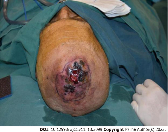
Case summary: A 78-year-old male patient was admitted to the hospital because of a bump on his head that did not heal for 4 mo. The patient was diagnosed with a refractory head wound. The patient underwent neoplasm resection and skin grafting surgery in the Plastic Surgery. The neoplasm was sent for pathological examination during the operation. The final pathological results were confirmed scalp angiosarcoma.
Conclusion: Our research suggests that pathological examination should be performed for refractory ulcers of the scalp, and physical factor therapy should be used with caution before the diagnosis is clear.
Keywords: Case report; Pathological examination; Refractory head wound; Scalp angiosarcoma.
©The Author(s) 2023. Published by Baishideng Publishing Group Inc. All rights reserved.
Conflict of interest statement
Conflict-of-interest statement: All the authors report no relevant conflicts of interest for this article.
Figures


References
-
- Qitao Huang, Haiming Wei, Lili Li. Angiosarcoma: A case report. Journal of Clinical Dermatology . 2015;44:810–811.
-
- Cuda J, Mirzamani N, Kantipudi R, Robbins J, Welsch MJ, Sundram UN. Diagnostic utility of Fli-1 and D2-40 in distinguishing atypical fibroxanthoma from angiosarcoma. Am J Dermatopathol. 2013;35:316–318. - PubMed
-
- Yu Liang, Jianzhong Cao, Hong Li. Rh-endostatin combined with radiotherapy in treatment of scalp angiosarcoma: Report of one case and review of literatur. Cancer Res . 2017;29:273–275.
Publication types
LinkOut - more resources
Full Text Sources
