An unusual ectopic thymoma clonal evolution analysis: A case report
- PMID: 37215501
- PMCID: PMC10199323
- DOI: 10.1515/biol-2022-0600
An unusual ectopic thymoma clonal evolution analysis: A case report
Abstract
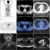
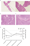
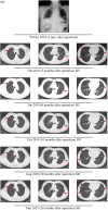
Thymomas and thymic carcinomas are rare and primary tumors of the mediastinum which is derived from the thymic epithelium. Thymomas are the most common primary anterior mediastinal tumor, while ectopic thymomas are rarer. Mutational profiles of ectopic thymomas may help expand our understanding of the occurrence and treatment options of these tumors. In this report, we sought to elucidate the mutational profiles of two ectopic thymoma nodules to gain deeper understanding of the molecular genetic information of this rare tumor and to provide guidance treatment options. We presented a case of 62-year-old male patient with a postoperative pathological diagnosis of type A mediastinal thymoma and ectopic pulmonary thymoma. After mediastinal lesion resection and thoracoscopic lung wedge resection, the mediastinal thymoma was completely removed, and the patient recovered from the surgery and no recurrence was found by examination until now. Whole exome sequencing was performed on both mediastinal thymoma and ectopic pulmonary thymoma tissue samples of the patient and clonal evolution analysis were further conducted to analyze the genetic characteristics. We identified eight gene mutations that were co-mutated in both lesions. Consistent with a previous exome sequencing analysis of thymic epithelial tumor, HRAS was also observed in both mediastinal lesion and lung lesion tissues. We also evaluated the intratumor heterogeneity of non-silent mutations. The results showed that the mediastinal lesion tissue has higher degree of heterogeneity and the lung lesion tissue has relatively low amount of variant heterogeneity in the detected variants. Through pathology and genomics sequencing detection, we initially revealed the genetic differences between mediastinal thymoma and ectopic thymoma, and clonal evolution analysis showed that these two lesions originated from multi-ancestral regions.
Keywords: case report; clonal evolution; ectopic pulmonary thymoma; heterogeneity; immunohistochemical.
© 2023 the author(s), published by De Gruyter.
Conflict of interest statement
Conflict of interest: Authors state no conflict of interest.
Figures


References
-
- Álvarez-Velasco R, Gutiérrez-Gutiérrez G, Trujillo JC, Martínez E, Segovia S, Arribas-Velasco M, et al. Clinical characteristics and outcomes of thymoma-associated myasthenia gravis. Eur J Neurol. 2021;28(6):2083–91. - PubMed
-
- Moran CA. Thymoma Staging: An Analysis of the Different Schemas. Adv Anat Pathol. 2021;28(5):298–306. - PubMed
-
- Ao Y-Q, Jiang J-H, Gao J, Wang H-K, Ding J-Y. Recent thymic emigrants as the bridge between thymoma and autoimmune diseases. Biochim Biophys Acta Rev Cancer. 2022;1877(3):188730. - PubMed
Publication types
LinkOut - more resources
Full Text Sources
Research Materials
Miscellaneous
