. 2023 Apr 29;13(8):2721-2733.
doi: 10.7150/thno.83543. eCollection 2023.

MOF-derived bimetallic nanozyme to catalyze ROS scavenging for protection of myocardial injury

Affiliations

MOF-derived bimetallic nanozyme to catalyze ROS scavenging for protection of myocardial injury

Kaiyan Xiang et al. Theranostics. .

Abstract

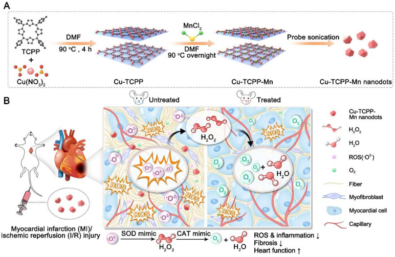
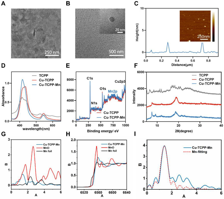
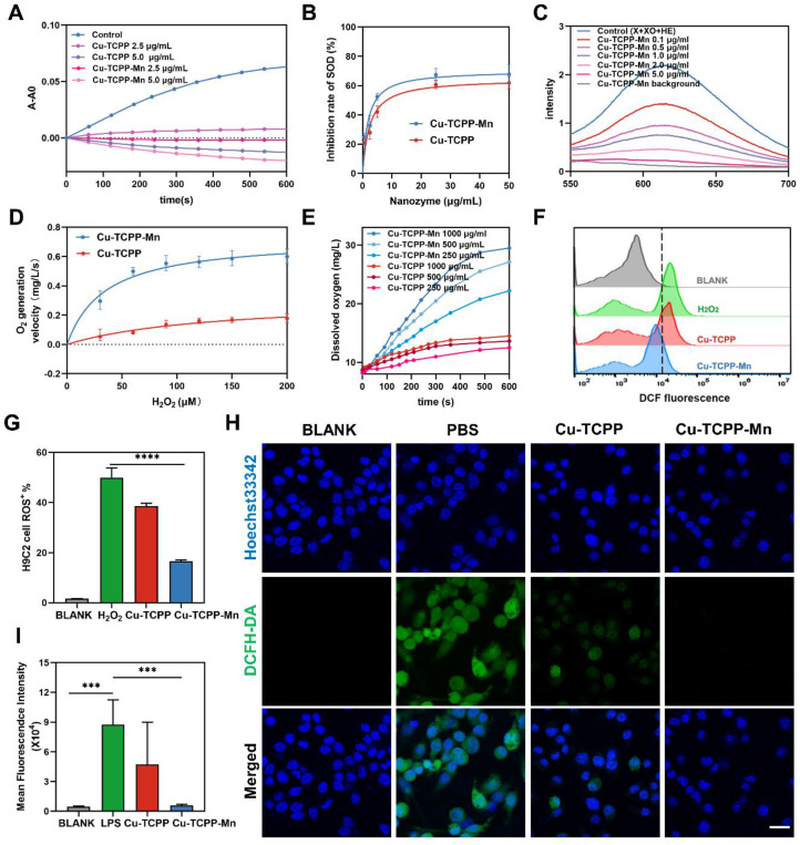
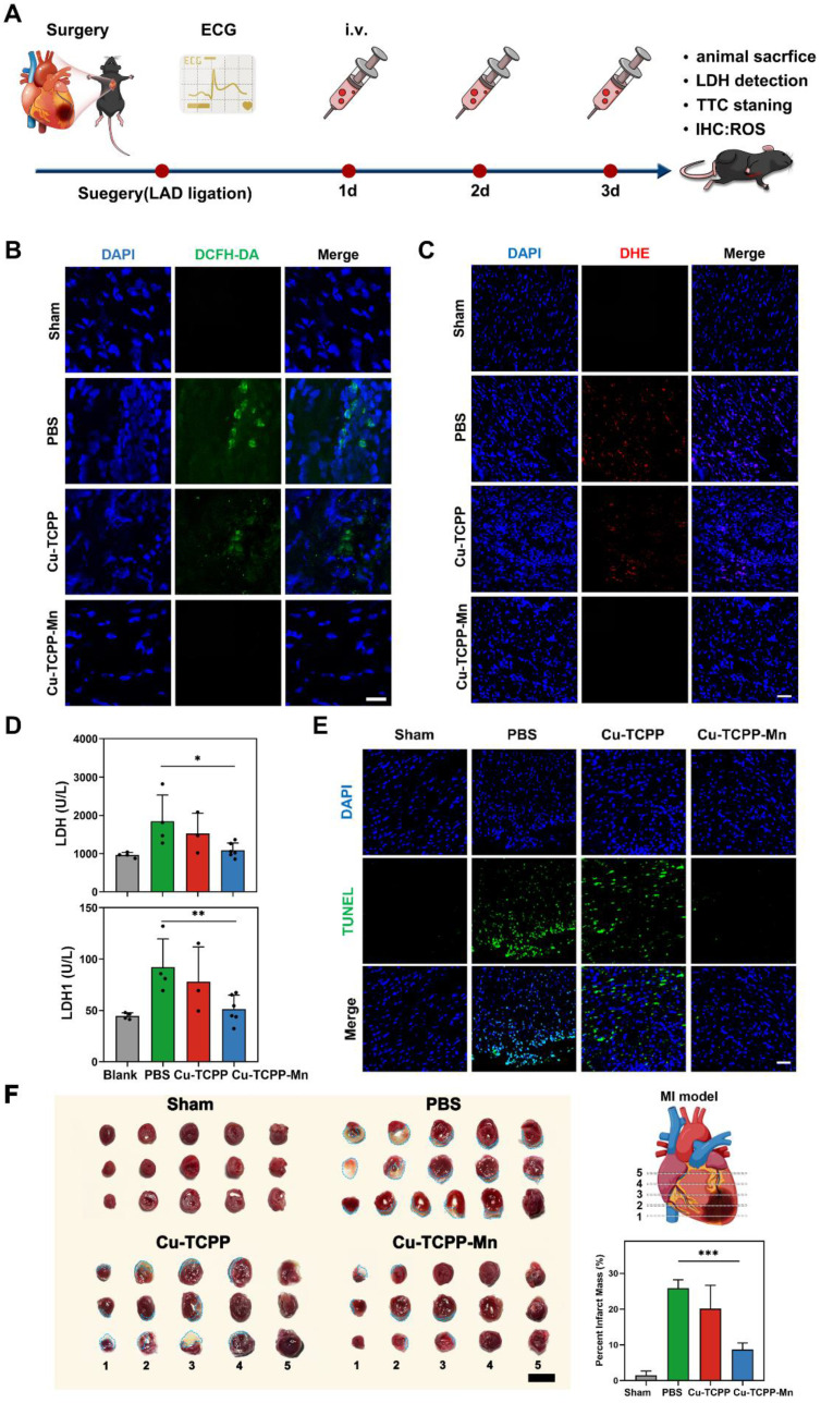
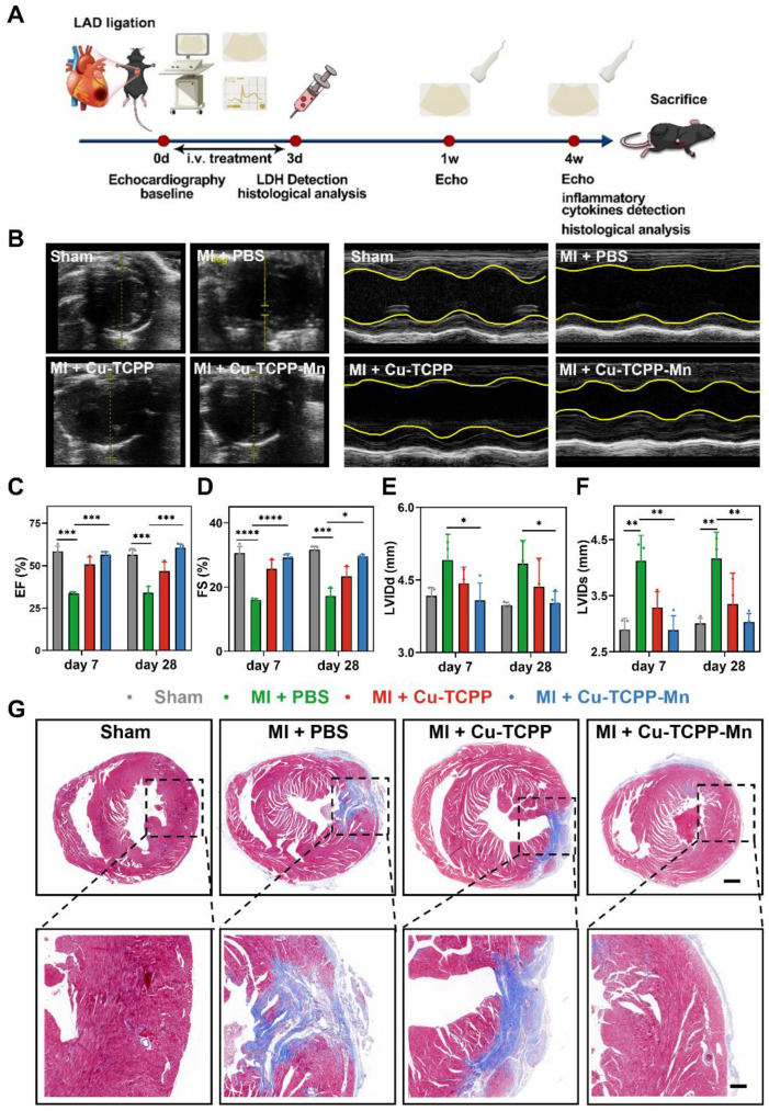
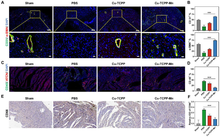
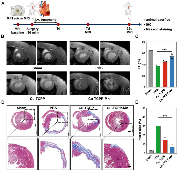
Rationale: Myocardial injury triggers intense oxidative stress, inflammatory response, and cytokine release, which are essential for myocardial repair and remodeling. Excess reactive oxygen species (ROS) scavenging and inflammation elimination have long been considered to reverse myocardial injuries. However, the efficacy of traditional treatments (antioxidant, anti-inflammatory drugs and natural enzymes) is still poor due to their intrinsic defects such as unfavorable pharmacokinetics and bioavailability, low biological stability, and potential side effects. Nanozyme represents a candidate to effectively modulate redox homeostasis for the treatment of ROS related inflammation diseases. Methods: We develop an integrated bimetallic nanozyme derived from metal-organic framework (MOF) to eliminate ROS and alleviate inflammation. The bimetallic nanozyme (Cu-TCPP-Mn) is synthesized by embedding manganese and copper into the porphyrin followed by sonication, which could mimic the cascade activities of superoxide dismutase (SOD) and catalase (CAT) to transform oxygen radicals to hydrogen peroxide, followed by the catalysis of hydrogen peroxide into oxygen and water. Enzyme kinetic analysis and oxygen-production velocities analysis were performed to evaluate the enzymatic activities of Cu-TCPP-Mn. We also established myocardial infarction (MI) and myocardial ischemia-reperfusion (I/R) injury animal models to verify the ROS scavenging and anti-inflammation effect of Cu-TCPP-Mn. Results: As demonstrated by kinetic analysis and oxygen-production velocities analysis, Cu-TCPP-Mn nanozyme possesses good performance in both SOD- and CAT-like activities to achieve synergistic ROS scavenging effect and provide protection for myocardial injury. In both MI and I/R injury animal models, this bimetallic nanozyme represents a promising and reliable technology to protect the heart tissue from oxidative stress and inflammation-induced injury, and enables the myocardial function to recover from otherwise severe damage. Conclusions: This research provides a facile and applicable method to develop a bimetallic MOF nanozyme, which represents a promising alternative to the treatment of myocardial injuries.

Keywords: metal-organic framework; myocardial injury; nanomedicine; nanozyme; reactive oxygen species.


Conflict of interest statement

Competing Interests: The authors have declared that no competing interest exists.

Figures

Figure 1
Figure 1
Schematic illustration of the design and synthesis of Cu-TCPP-Mn nanozyme for myocardial injury treatment. (A) The bimetallic Cu-TCPP-Mn nanozyme was fabricated by embedding manganese and copper into the porphyrin via solvothermal method, followed by sonication into small MOF nanodots. (B) Cu-TCPP-Mn nanozyme retained cascade activity that has been shown to scavenge ROS, inhibit inflammation, reduce myocardium fibrosis and promote constructive remodeling and vascularization in MI and I/R injury animal models.
Figure 2
Figure 2
Characterization of Cu-TCPP-Mn nanozyme. (A) Representative TEM image of Cu-TCPP-Mn nanozyme with sheet-like morphology. (B) Representative TEM images of Cu-TCPP-Mn nanodots. (C) Quantitative analysis of the thickness of Cu-TCPP-Mn nanodots, and representative AFM image of Cu-TCPP-Mn nanodots (inserted). (D) UV-vis absorption spectra of TCPP, Cu-TCPP and Cu-TCPP-Mn. (E) XPS of Cu-TCPP and Cu-TCPP-Mn. (F) PXRD pattern of the TCPP, Cu-TCPP and Cu-TCPP-Mn. (G) Normalized Mn K-edge XANES spectra of different samples; (H) Fourier transform EXAFS spectra (k3-weighted) of Cu-TCPP-Mn; (I) Mn K-edge EXAFS of Cu-TCPP-Mn in the R space and the fitting curves without correcting for the scattering phase shift.
Figure 3
Figure 3
In vitro ROS-scavenging activities of Cu-TCPP-Mn nanozyme. (A) Kinetic curves of A-A0 (550 nm) with X and XO treated with different concentrations of Cu-TCPP-Mn by monitoring the reduction of NBT. (B) Inhibition rate of SOD of Cu-TCPP and Cu-TCPP-Mn calculated by NBT kinetic assay. (C) Fluorescent spectra of the mixture of DHE, X, and XO treated with different concentrations of Cu-TCPP-Mn. (D) Kinetics of O2 generation velocity for CAT-like activity of Cu-TCPP and Cu-TCPP-Mn nanozyme. (E) Typical kinetic curves of dissolved oxygen generated from the decomposition of H2O2 after treatment with different concentrations of Cu-TCPP-Mn (250, 500, 1000 μg). (F) ROS evaluation in H2O2 (100 μM) pre-treated H9C2 cells by flow cytometry with the treatment of Cu-TCPP and Cu- TCPP-Mn (5 μg/mL). (G) Quantitative analysis of ROS production in H9C2 cells treated with Cu-TCPP and Cu-TCPP-Mn (5 μg/mL). (H) Representative fluorescence images of ROS staining (DCFH-DA, green fluorescence) of RAW 264.7 cells treated with Cu-TCPP and Cu-TCPP-Mn and (I) quantitative analysis of mean fluorescence intensity. Scale bar, 20 μm. Data are presented as mean ± standard deviation (S.D.) (n = 5). The comparisons between samples were operated by one-way ANOVA. *** indicates P < 0.001; **** indicates P < 0.0001.
Figure 4
Figure 4
In vivo anti-inflammation effect of Cu-TCPP-Mn nanozyme on MI mice. (A) The schematic of the study design. Representative ROS staining images of heart tissues harvested from MI mice models treated with PBS, Cu-TCPP and Cu-TCPP-Mn using DCFH-DA probe (B, Scale bar, 25 μm) and DHE probe (C, Scale bar, 50 μm). (D) the LDH and LDH1 concentrations of mouse serum with different treatment. (E) Immunofluorescent staining of TUNEL in the ischemic heart after different treatments. Scale bar, 50 μm. (F) Representative TTC staining images and quantitative data of Sham or MI heart slices treated with PBS, Cu-TCPP and Cu-TCPP-Mn, with the infarct area shown in blue dashed line. Scale bar, 100 mm. Data are presented as mean ± S.D. (n = 5). The comparisons between samples were operated by one-way ANOVA. * indicates P < 0.05; ** indicates P < 0.01; *** indicates P < 0.001; ns indicates P > 0.05 with no significance.
Figure 5
Figure 5
Therapeutic efficacy of Cu-TCPP-Mn on MI mice. (A) The schematic of the study design. (B) Representative echocardiographic images and corresponding M-mode images of MI mouse models at day 7 post treatments. The yellow wavy line (right panel) in the top and bottom area indicates left ventricular wall thickness and left ventricular posterior wall thickness. Distance between two wavy lines indicates left ventricular dimension. (C) Ejection fraction (EF) and (D) fractional shortening (FS) were evaluated by echocardiography after different treatments at one week and four weeks, respectively (n = 3-4 per group). (E) Left ventricular end diastolic dimension (LVIDd) and (F) left ventricular end systolic dimension (LVIDs) were evaluated by echocardiography after different treatments at one week and four weeks, respectively (n = 3-4 per group). (G) Representative Masson's trichrome staining images of infarcted hearts 4 weeks after injection (blue represents scar tissue; red represents viable myocardium). Scale bar, 500 μm (top), 150 μm (bottom). Data are presented as mean ± S.D. The comparisons between samples were operated by one-way ANOVA. * indicates P < 0.05; ** indicates P < 0.01; *** indicates P < 0.001; ns indicates P > 0.05 with no significance.
Figure 6
Figure 6
Anti-inflammatory and angiogenesis activity of Cu-TCPP-Mn nanozyme in MI mice. (A) Representative immunofluorescence images and (B) quantitative analysis of heart tissues co-stained with CD31 (vessels, green), α-SMA (α-smooth muscle actin, red), and DAPI (cell nucleus, blue) in Sham and MI mice treated with PBS, Cu-TCPP, and Cu-TCPP-Mn. Scale bar, 200 μm (top), 20 μm (bottom). (C) Representative immunofluorescence images and (D) quantification analysis of heart tissues co-stained with CD45 (neutrophil cell, green), ACTN (Actinin, red), DAPI (cell nucleus, bule) in Sham and MI mice treated with PBS, Cu-TCPP, and Cu-TCPP-Mn. Scale bar, 100 μm. (E) Representative heart tissue sections stained with CD68 (brown) and DAPI (blue) (F) and quantification analysis in MI mice treated with PBS, Cu-TCPP, Cu-TCPP-Mn. Scale bar, 100 μm.
Figure 7
Figure 7
In vivo anti-inflammation of Cu-TCPP-Mn nanozyme on cardiac I/R injury rat models. (A) The schematic of the study design. (B) Representative cine images of infarcted hearts in systolic and diastolic period collected from I/R injury rats scanned by a 9.4 T MRI. (C) EF calculated from 9.4 T MRI images after one week of treatments (n = 3-6 per group). (D) Representative Masson's trichrome-stained and (E) quantitative analysis of infarcted hearts in SD rats 4 weeks after treatment with PBS, Cu-TCPP, and Cu-TCPP-Mn. Data are presented as mean ± S.D. The comparisons between samples were operated by one-way ANOVA. * indicates P < 0.05; ** indicates P < 0.01; *** indicates P < 0.001; ns indicates P > 0.05 with no significance.

References

    1. Zhang Z, Dalan R, Hu Z, Wang JW, Chew N, Poh KK. et al. Reactive oxygen species scavenging nanomedicine for the treatment of ischemic heart disease. Adv Mater. 2022;34(35):e2022169. - PubMed
    1. Yao J, Huang K, Zhu D, Chen T, Jiang Y, Zhang J. et al. A minimally invasive exosome spray repairs heart after myocardial infarction. ACS Nano. 2021;15(7):11099–111. - PubMed
    1. Lee JR, Park BW, Kim J, Choo YW, Kim HY, Yoon JK. et al. Nanovesicles derived from iron oxide nanoparticles-incorporated mesenchymal stem cells for cardiac repair. Sci Adv. 2020;6(18):eaazo0952. - PMC - PubMed
    1. Li HL, Zhuo ML, Wang D, Wang AB, Cai H, Sun LH. et al. Targeted cardiac overexpression of A20 improves left ventricular performance and reduces compensatory hypertrophy after myocardial infarction. Circulation. 2007;115(14):1885–94. - PubMed
    1. Frangogiannis NG. The inflammatory response in myocardial injury, repair, and remodelling. Nat Rev Cardiol. 2014;11(5):255–65. - PMC - PubMed

Publication types