. 2023 Nov;42(11):3219-3228.
doi: 10.1109/TMI.2023.3278269. Epub 2023 Oct 27.

Panretinal Optical Coherence Tomography

Panretinal Optical Coherence Tomography

Shuibin Ni et al. IEEE Trans Med Imaging. 2023 Nov.

Abstract

We introduce a new concept of panoramic retinal (panretinal) optical coherence tomography (OCT) imaging system with a 140° field of view (FOV). To achieve this unprecedented FOV, a contact imaging approach was used which enabled faster, more efficient, and quantitative retinal imaging with measurement of axial eye length. The utilization of the handheld panretinal OCT imaging system could allow earlier recognition of peripheral retinal disease and prevent permanent vision loss. In addition, adequate visualization of the peripheral retina has a great potential for better understanding disease mechanisms regarding the periphery. To the best of our knowledge, the panretinal OCT imaging system presented in this manuscript has the widest FOV among all the retina OCT imaging systems and offers significant values in both clinical ophthalmology and basic vision science.


Figures

Fig. 1
Fig. 1
Schematic of contact handheld panretinal SS-OCT system. Swept-source laser (SVM10F-0210, Thorlabs, Inc., USA); Split ratio of fiber coupler: FC1 (10/90), FC2 (50/50), FC3 (50/50), FC4 (20/80); Galvanometer scanner (Pangolin Laser System, Inc. USA); Balanced detector (PDB482C-AC, Thorlabs, Inc., USA); Linear stage (X-LHM100A-E03, Zaber Technologies, Inc., Canada).
Fig. 2
Fig. 2
(a) 3D layout of telescope eyepiece design after the slow axis of the galvanometer scanner (GSY). (b) Spot diagrams of FOV from 0° to 140°. Black circle is the Airy disk. The complex double aspherical ocular lens induced some irregular beam profile on the retina. (c) Photograph of the fully assembled portable probe. (d) Photograph of contact handheld SS-OCT being used to image premature infants in the OHSU neonatal intensive care unit. (e) SolidWorks rendering of phantom eye model for FOV calibration (radius = 12 mm). (f) Cross-sectional view of the phantom eye. Seven circular rings extruded from the surface were marked. The interval between the rings was 40° measured from the center of the phantom eye (point “C”). (g) En face OCT image from the phantom eye. (h) Selected cross-sectional scan corresponding to the location of red line in (g). Scale bars in (h) are 1 mm.
Fig. 3
Fig. 3
(a) Cross-sectional scan of the meniscus lens and corneal surface. (b). Schematic diagram of the human eye. P: scanning pivot point. θ: scanning angle. β: FOV measured from the center of the eyeball. C: center of the eye. r: radius of the eye. (c) Cross-sectional scan of the retina. (d) Schematic of the reference arm indicating with the linear stage, stopped at the corneal position (P1) and retinal position (P2). Scale bars in (a) and (c) are 200 μm.
Fig. 4
Fig. 4
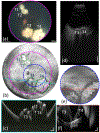
(a) RetCam color fundus image from a patient with multifocal retinoblastoma (tumors labeled as T1-T4). (b) En face OCT image obtained by our contact handheld SS-OCT system. (c) Selected B-scan image corresponding to the location of dashed cyan line. Scale bars are 1 mm (horizontally) and 500 μm (vertically). (d) Selected ultrasound B-scan image. Scale bar is 5 mm. (e) En face OCT image taken by Leica handheld OCT (Envisu C2300, Leica Microsystems, Germany). The imaging area was marked by dashed blue circle in (b). (f) Selected B-scan image corresponding to the location of dashed red line. Scale bars are 500 μm (horizontally) and 200 μm (vertically).
Fig. 5
Fig. 5
(a)-(b) RetCam color fundus images from an infant with ROP stage 3 at different locations. (c) En face OCT image from the same infant acquired by contact handheld panretinal SS-OCT imaging system. Yellow arrows in (a) and (c) point to the area of extraretinal neovascular proliferation.
Fig. 6
Fig. 6
(a) En face OCT image from an infant with ROP stage 1. Blue arrows point to the demarcation line with the retinal vascular-avascular junction. (b) En face OCT image from an infant with ROP stage 2. The gray streak visible in the image is a motion artifact that occurred during image acquisition. (c)-(d) En face OCT images from the infant with ROP stage 3 at different locations after laser treatment. Orange arrows in (b) and (d) point to the area of ora serrata.
Fig. 7
Fig. 7
(a) En face OCT image from a patient with Coats disease (yellow arrows). (b) Selected cross-sectional scan with the regions of extramacular exudation and retinal thickening corresponding to the location of dashed blue line in (a). Scale bars in (b) are 1 mm (horizontally) and 500 μm (vertically).
Fig. 8
Fig. 8
Morphological changes in the retina of the patient with retinal detachment with different therapeutic interventions. The images on the first row are en face OCT images. The images on the second row are selected cross-sectional scans. The dashed cyan lines indicate location of the corresponding cross-sectional scans. (a)-(b) One week after panretinal photocoagulation laser treatment. (c)-(d) Three weeks after panretinal photocoagulation laser treatment. (e)-(f) Two weeks after PPV surgery. (g)-(h) Six weeks after PPV surgery. (i)-(j) Seven weeks after PPV surgery. The PPV surgery was performed six weeks after the panretinal photocoagulation laser treatment. Scale bars are 1 mm (horizontally) and 500 μm (vertically).

References

    1. Nguyen T-TP et al., “Advantages of Widefield Optical Coherence Tomography in the Diagnosis of Retinopathy of Prematurity,” Front. Pediatr, vol. 9, p. 797684, Jan. 2022. - PMC - PubMed
    1. Koulisis N et al., “Correlating Changes in the Macular Microvasculature and Capillary Network to Peripheral Vascular Pathologic Features in Familial Exudative Vitreoretinopathy,” Ophthalmol. Retina, vol. 3, no. 7, pp. 597–606, Jul. 2019. - PubMed
    1. Scruggs BA et al., “Peripheral OCT Assisted by Scleral Depression in Retinopathy of Prematurity,” Ophthalmol. Sci, vol. 2, no. 1, p. 100094. Mar. 2022. - PMC - PubMed
    1. Blair JP, Ulrich JN, Hartnett ME, and Shapiro MJ, “Peripheral retinal nonperfusion in fellow eyes in coats disease,” Retina, vol. 33, no. 8, pp. 1694–1699, Sep. 2013. - PubMed
    1. Wang SK, Callaway NF, Wallenstein MB, Henderson MT, Leng T, and Moshfeghi DM, “SUNDROP: six years of screening for retinopathy of prematurity with telemedicine,” Can. J. Ophthalmol, vol. 50. no. 2. pp. 101–106. Apr. 2015. - PubMed

Publication types

MeSH terms