. 2023 May 30;120(22):e2220979120.
doi: 10.1073/pnas.2220979120. Epub 2023 May 22.

Therapeutic antagonism of the neurokinin 1 receptor in endosomes provides sustained pain relief

Affiliations

Therapeutic antagonism of the neurokinin 1 receptor in endosomes provides sustained pain relief

Alan Hegron et al. Proc Natl Acad Sci U S A. .

Abstract

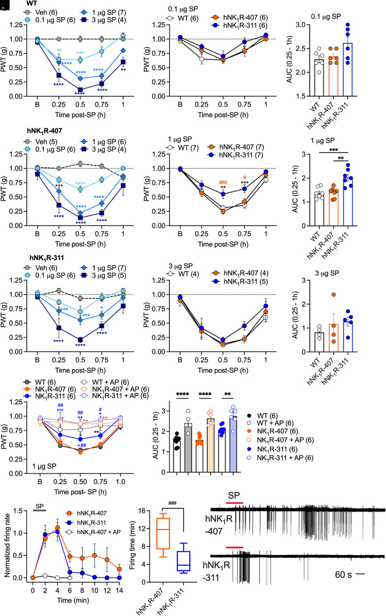
The hypothesis that sustained G protein-coupled receptor (GPCR) signaling from endosomes mediates pain is based on studies with endocytosis inhibitors and lipid-conjugated or nanoparticle-encapsulated antagonists targeted to endosomes. GPCR antagonists that reverse sustained endosomal signaling and nociception are needed. However, the criteria for rational design of such compounds are ill-defined. Moreover, the role of natural GPCR variants, which exhibit aberrant signaling and endosomal trafficking, in maintaining pain is unknown. Herein, substance P (SP) was found to evoke clathrin-mediated assembly of endosomal signaling complexes comprising neurokinin 1 receptor (NK1R), Gαq/i, and βarrestin-2. Whereas the FDA-approved NK1R antagonist aprepitant induced a transient disruption of endosomal signals, analogs of netupitant designed to penetrate membranes and persist in acidic endosomes through altered lipophilicity and pKa caused sustained inhibition of endosomal signals. When injected intrathecally to target spinal NK1R+ve neurons in knockin mice expressing human NK1R, aprepitant transiently inhibited nociceptive responses to intraplantar injection of capsaicin. Conversely, netupitant analogs had more potent, efficacious, and sustained antinociceptive effects. Mice expressing C-terminally truncated human NK1R, corresponding to a natural variant with aberrant signaling and trafficking, displayed attenuated SP-evoked excitation of spinal neurons and blunted nociceptive responses to SP. Thus, sustained antagonism of the NK1R in endosomes correlates with long-lasting antinociception, and domains within the C-terminus of the NK1R are necessary for the full pronociceptive actions of SP. The results support the hypothesis that endosomal signaling of GPCRs mediates nociception and provides insight into strategies for antagonizing GPCRs in intracellular locations for the treatment of diverse diseases.

Keywords: endocytosis; pain; receptors; signaling.


Conflict of interest statement

N.W.B. is a founding scientist of Endosome Therapeutics Inc. Research in N.W.B.’s laboratory is funded, in part, by Takeda Pharmaceuticals International.

Figures

Fig. 1.
Fig. 1.
Assembly of NK1R signalosomes in HEK293T cells. (A) NanoBiT-BRET (nbBRET) uses a nanoluciferase split into two fragments (natural peptide, NP, and LgBiT) to detect BRET between receptor (NK1R), mini Gα proteins (mGα) or βarrestin (βARR), and proteins resident to the plasma membrane (PM, CAAX) or early endosome (EE, FYVE). (BK) Substance P (SP, 100 nM) induced nbBRET between NK1R-NP, LgBiT-CAAX, or LgBiT-FYVE as well as Venus-mGαsq, Venus-mGαsi, Venus-mGαs, or βARR2-YFP. (EH) Effect of dominant-negative dynamin (DnmK44A; E and F) or hypertonic sucrose (0.45 M, 30 min, G and H). (IK) Effect of aprepitant (AP, 100 nM, 30 min). AUC, area under curve. Mean ± SEM. N = 5 to 7 independent experiments. *P < 0.05, **P < 0.01, ***P < 0.001, ****P < 0.0001. One-way ANOVA with Dunnett’s test (D, F, H, and K).
Fig. 2.
Fig. 2.
Development of netupitant analogs. (AC) Effect of graded concentrations of netupitant (NT, 30 min) on substance P (SP)–induced recruitment of mini Gαsq (mGαq) to the plasma membrane (CAAX, A) or early endosome (FYVE, B) on NanoBiT-BRET (nbBRET) with natural peptide (NP)–tagged NK1R in HEK293T cells. (DG) Design and synthesis of NT analogs. (D) Chemical formula of NT. (E) NK1R crystal structure (PDB ID: 6HLP) in complex with NT (magenta) overlaid with the docked pose of PS29 (blue). Residues are displayed in a range of 8 Å relative to NT and PS29. F. NT (PDB ID: 6HLP) in complex with the NK1R crystal structure (PDB ID: 6HLP) presented together with the docked pose of PS29. Receptor surface is clipped between the transmembrane helices 4, 5, and 6 to give better insight into the binding pocket and to show the outward reaching piperazine substituent of NT and PS29 (green circle), showing surface atoms and hetero atoms [oxygen (red), nitrogen (blue), sulfur (yellow)]. (G) Synthetic approach toward carba-NT derivatives. Structural changes compared to NT are highlighted in blue. Mean ± SEM. N = 4 to 9 independent experiments.
Fig. 3.
Fig. 3.
Antagonism of NK1R endosomal signaling in HEK293T cells. (AF) FRET assays of substance P (SP)–induced activation of nuclear ERK activity measured using a nuclear EKAR biosensor. (AC) Cells were preincubated (30 or 90 min) with graded concentrations of aprepitant (AP), netupitant (NT), PS15, PS29, PS34, or PS49 prior to SP (100 nM) challenge. (DF) Duration of inhibition following antagonist removal and recovery. Cells were preincubated with antagonist for 30 min (AP, 100 nM; NT, 300 nM) or 90 min (NT analogs, 1 µM), washed, and recovered in agonist-free medium for 2 h (AP, NT) or 4 h (NT analogs; SI Appendix, Fig. S3), prior to stimulation with SP (100 nM). (G and H) Enhanced bystander BRET (ebBRET) measuring proximity between mini Gαsq (Venus-mGαsq) and a Renilla luciferase–tagged endosomal marker (tdRGFP-Rab5a). Cells were stimulated with SP (100 nM, 5 min), then challenged with vehicle or saturating concentrations of NT, PS15, or PS34 (10 µM). EbBRET was measured for 40 min. Kinetics were fitted as a nonlinear exponential decay from antagonist addition to quantify the rate of antagonism. Mean±SEM. N = 4 to 9 independent experiments. 1-way ANOVA with Holm-Šídák's (E), Dunnett’s (F) test against SP, or Dunnett’s against NT (H). (I) Localization of fluorescent TAMRA-NT (PS68, 100 nM, 10 min, magenta) in HEK293T cells expressing NK1R-eGFP (green), with or without preincubation with unlabeled SP (100 nM, 30 min). Arrow heads denote TAMRA-NT and NK1R-eGFP colocalization at the plasma membrane. Asterisks denote uptake of TAMRA-NT into cells lacking NK1R-eGFP. (Scale bar, 20 µm). Representative images, N = 3 experiments.
Fig. 4.
Fig. 4.
Generation of knockin mice expressing hNK1R-407. (A) NK1R-407 Southern strategy and targeting construct comprising tdTomato, a T2A peptide cleavage site, Flag-hNK1R-407, a downstream phosphoglycerine kinase neomycin cassette flanked by FRT sites, and downstream AflII and BglII sites. (B and C) Southern blots of selected clones with 5′, 3′, and internal probes confirming correct targeting. (D) Southern blot of F1 mice confirming NK1R-407 expression. WT, wild-type.
Fig. 5.
Fig. 5.
Characterization of mice expressing hNK1R-407 or hNK1R-311. (A) PCR gels of the spinal cord from NK1R-407, NK1R-311, and WT mice probed with a primer (#148) specific for human NK1R-407 or a primer (#82) that would detect both human NK1R-407 and NK1R-311. (B and C) qRT-PCR analysis of extracts of the spinal cord (B) and colon (C) probed with primers to mouse NK1R, both human NK1R-407 and NK1R-311, or hNK1R-407 alone. N = 4 mice. (D) RNAScope® in situ hybridization of sections of the spinal cord from NK1R-407, NK1R-311, or WT mice using probes to both human NK1R-407 and NK1R-311 or to mouse NK1R, highlighting neurons with a Nissl stain (magenta). Representative images from n = 3 mice. Scale bar, 50 µm, and in detail box, 20 µm. L, lamina.
Fig. 6.
Fig. 6.
SP-evoked nociception in hNK1R-407, hNK1R-311, and wild-type mice. (AC) Substance P (SP; 0.1, 1, 3 µg/5 µL, intrathecal)–evoked mechanical allodynia in WT (A), NK1R-407 (B), and NK1R-311 (C) mice. Mechanical allodynia was measured 0.25 to 1 h after SP injection. (DF) Mechanical allodynia to the same dose of SP in the three strains of mice. Paw withdrawal threshold (PWT) to stimulation with calibrated von Frey filaments was measured to assess mechanical allodynia. (GI) AUC in the three strains of mice. (J and K) Effects of intrathecal injection of aprepitant (AP, 100 nM/5 µL) or vehicle on SP (1 µg/5 µL)-evoked mechanical allodynia in NK1R-407, NK1R-311, and WT mice. (LN) Activation of spinal neurons in slice preparations of spinal cord from NK1R-407 and NK1R-311 mice after transient stimulation with SP (10 nM, 2 min). (L) Normalized firing rate. (M) Firing time. (N) Representative traces. Mean ± SEM; (N) denotes number of mice studied. *< 0.05, **< 0.01, ***< 0.001, ****< 0.0001 vs. Veh, WT; #< 0.05, ##< 0.01, ###< 0.001 vs. NK1R-407. Two-way ANOVA, Sídák’s multiple comparison test (A–F and J), one-way ANOVA, Tukey’s multiple comparison test (GI and K), or parametric unpaired two-tailed t test (M).
Fig. 7.
Fig. 7.
Antagonism of capsaicin-evoked nociception in hNK1R-407 mice. (A) Effects of intrathecal (i.t.) injection of aprepitant (AP), netupitant (NT), or NT analogs (PS15, PS49, PS34, PS29; 100 nM/5 µL) on capsaicin-induced mechanical allodynia in hNK1R-407 mice. (B) AUC in the same groups of mice. (CE) Dose–response effects of AP (30, 100, 300 nM/5 µL, i.t.), NT (30, 100, 300 nM/5 µL, i.t.), or PS34 (10, 30, 100 nM/5 µL, i.t.) on capsaicin-induced mechanical allodynia. (F) AUC in the same groups of mice. (N) denotes number of mice studied. *< 0.05, **< 0.01, ***< 0.001, ****< 0.0001 vs. Veh; &< 0.05, &&< 0.01, &&&&< 0.0001 vs. AP, one-way ANOVA, Tukey’s multiple comparison test (B and F) or two-way ANOVA, Sídák’s multiple comparison test (C, D, and E).

References

    1. Yang D., et al. , G protein-coupled receptors: Structure- and function-based drug discovery. Signal Transduct. Target. Ther. 6, 7 (2021). - PMC - PubMed
    1. Gurevich V. V., Gurevich E. V., GPCR signaling regulation: The role of GRKs and arrestins. Front. Pharmacol. 10, 125 (2019). - PMC - PubMed
    1. Dicker F., Quitterer U., Winstel R., Honold K., Lohse M. J., Phosphorylation-independent inhibition of parathyroid hormone receptor signaling by G protein-coupled receptor kinases. Proc. Natl. Acad. Sci. U.S.A. 96, 5476–5481 (1999). - PMC - PubMed
    1. Calebiro D., et al. , Persistent cAMP-signals triggered by internalized G-protein-coupled receptors. PLoS Biol. 7, e1000172 (2009). - PMC - PubMed
    1. DeFea K. A., et al. , The proliferative and antiapoptotic effects of substance P are facilitated by formation of a beta -arrestin-dependent scaffolding complex. Proc. Natl. Acad. Sci. U.S.A. 97, 11086–11091 (2000). - PMC - PubMed

Publication types

LinkOut - more resources