. 2023 May 30;120(22):e2219686120.
doi: 10.1073/pnas.2219686120. Epub 2023 May 22.

Primary cilia TRP channel regulates hippocampal excitability

Affiliations

Primary cilia TRP channel regulates hippocampal excitability

Thuy N Vien et al. Proc Natl Acad Sci U S A. .

Abstract

Polycystins (PKD2, PKD2L1, and PKD2L2) are members of the transient receptor potential family, which form ciliary ion channels. Most notably, PKD2 dysregulation in the kidney nephron cilia is associated with polycystic kidney disease, but the function of PKD2L1 in neurons is undefined. In this report, we develop animal models to track the expression and subcellular localization of PKD2L1 in the brain. We discover that PKD2L1 localizes and functions as a Ca2+ channel in the primary cilia of hippocampal neurons that apically radiate from the soma. Loss of PKD2L1 expression ablates primary ciliary maturation and attenuates neuronal high-frequency excitability, which precipitates seizure susceptibility and autism spectrum disorder-like behavior in mice. The disproportionate impairment of interneuron excitability suggests that circuit disinhibition underlies the neurophenotypic features of these mice. Our results identify PKD2L1 channels as regulators of hippocampal excitability and the neuronal primary cilia as organelle mediators of brain electrical signaling.

Keywords: TRP channels; channelopathy; ciliopathy; polycystins; primary cilia.


Conflict of interest statement

The authors declare no competing interest.

Figures

Fig. 1.
Fig. 1.
Loss of PKD2L1 causes enhanced seizure susceptibility in mice. (A) Examples of 1 min EEG traces in mice at baseline before (control) and after kainic acid injection (kainic acid). Top row of kainic acid traces indicates 1 min results from WT (Left) and PKD2L1−/− (Right) mice during the first epileptiform activity following kainic acid. Middle and bottom traces are 1 min results during the epileptiform activity observed at the onset of SE in WT (Left) and PKD2L1−/− (Right) mice. (B) The time to first epileptiform activity (first latency, Top), onset of SE (onset of SE, Middle), and total percentage of time spent seizing (% time seizing, Bottom) were measured for WT and PKD2L1−/− mice. Sample sizes are indicated within parenthesis. Error bars are equal to SD. P values from paired Student’s t tests are indicated above the graphs. Asterisk indicates significant P values < 0.05.
Fig. 2.
Fig. 2.
Loss of PKD2L1 expression impairs primary ciliary maturation of hippocampal neurons. (A) Global stitched confocal images of sectioned hippocampal slices visualizing primary cilia (transgene ARL13B-EGFP, green) and nuclei labeled with DAPI (staining, blue). (B) Confocal images of the CA1 and CA3 regions of the hippocampus. Inset, expanded views of mature and immature primary cilia taken from PKD2L1+/+ or PKD2L1−/− mice. (C) Analysis of the primary ciliary length from the CA1 (N = 120 for each genotype), CA2 (N = 120 for each genotype), CA3 (N = 140 for each genotype), and dentate gyrus (N = 120 for each genotype). P values from paired t tests for each region of the hippocampus are indicated above the graphs. Average ciliary length is indicated by red lines. (D) Confocal images of fixed cultured PKD2L1+/+ neonatal hippocampal neurons labeled with antibodies for microtubule-associated protein-2 (MAP2) and pan-subtype voltage-gated sodium channels (Pan-Nav). Location of primary cilia (ARL13B-EGFP) sprouting from the soma (DAPI) is indicated by yellow arrows.
Fig. 3.
Fig. 3.
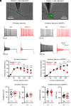
PKD2L1 channel expression contributes to hippocampal neuron excitability. (A) Images of current-clamped hippocampal neurons isolated from PKD2L1+/+: ARL13B-EGFP and PKD2L1−/−: ARL13B-EGFP mice. (B) Example action potential recordings taken from excitatory and inhibitory neurons of both genotypes after 8 to 14 d in culture. (C) Average spike frequency–current relationships recorded from excitatory and inhibitory neurons. P-values (< 0.05) resulting from paired Student’s t tests are indicated. (D) Maximal spike amplitude and resting membrane potential (RMP) of excitatory and inhibitory neurons. Averages are represented by horizontal black line. The number of neurons in each sample size is indicated in the parenthesis. Error bars are equal to SEM.
Fig. 4.
Fig. 4.
PKD2L1 forms an ion channel in hippocampal neuron primary cilia. (A) Hippocampal section histology from the CA1 demonstrating the colocalization of PKD2L1-mCherry (red) into the primary ciliary compartment illuminated by ARL13B-EGFP (green) of neurons. (Scale bar, 20 µm.) (B) Top, images of voltage-clamped mature primary cilia (PKD2L1-mCherry) and immature (PKD2L1−/−) primary cilia from cultured hippocampal neurons. (Scale bar = 5 µm.) Left, example single-channel currents recorded from the cilia of PKD2L1-mCherry:ARL13B-EGFP neurons using 140 mM NaCl within the patch pipette. Open-channel events were detected in 7/9 ciliary patches using Na+ as the external charge carrier and from 4/5 patches using Ca2+ as a charge carrier. Right, current records from immature neuronal cilia isolated from PKD2L1−/−:ARL13B-EGFP mice using the same saline Na+ conditions. No open-channel events were detected from immature ciliary patches (N = 15, total), where N = 12 high-resistance seals were made using Na+ as an external charge carrier and N = 3 using Ca2+. (C) Average single-channel current amplitude–voltage relationships measured from PKD2L1-mCherry:ARL13B-EGFP neuronal primary cilia. Inward single-channel conductances (γ) were estimated from a linear fit of the data generated from experiments using either Na+ or Ca2+ within the recording electrode. (D) Single-channel current open probability (Po)–voltage relationships measured from PKD2L1-mCherry:ARL13B-EGFP (black) and PKD2L1+/+:ARL13B-EGFP (gray) neuronal primary cilia. Half-maximal open channels (V1/2) were estimated by fitting the data to a Boltzmann equation. In each graph, error bars indicate SD and sample sizes are indicated within parenthesis.
Fig. 5.
Fig. 5.
Pharmacological activation of neuronal PKD2L1 increases the frequency of action potential firing. (A) Top, stimulation and inhibition of ciliary single-channel openings by CMZ and gadolinium (Gd3+), respectively, in neurons isolated from PKD2L1-mCherry:ARL13B-EGFP mice. Bottom, lack of single-channel events in control and CMZ conditions in an immature ciliary patch recorded from PKD2L1−/−:ARL13B-EGFP neurons. Ciliary membranes were held at 40 mV in the on-cilia configuration. (B) Left, images of current-clamped neurons isolated from PKD2L1-mCherry:ARL13B-EGFP mice after 4 to 8 d in culture using DIC, GFP, and RFP filters. Right, example hippocampal action potentials activated by 120 pA current injection before (control) and after 1 µM CMZ bath application. (C) Paired scatter plots comparing the maximum number of action potential spikes generated from control and drug treatment conditions. P-value results from a two-tailed Wilcoxon matched-pairs rank test are indicated, and the number of neurons recorded from each condition are indicated within parentheses.

References

    1. Dahl H. A., Fine structure of cilia in rat cerebral cortex. Z Zellforsch Mikrosk Anat. 60, 369–386 (1963), 10.1007/BF00336612. - DOI - PubMed
    1. Duncan D., Electron microscope study of the embryonic neural tube and notochord. Tex. Rep. Biol. Med. 15, 367–377 (1957). https://pubmed.ncbi.nlm.nih.gov/13495942/. - PubMed
    1. Guemez-Gamboa A., Coufal N. G., Gleeson J. G., Primary cilia in the developing and mature brain. Neuron 82, 511–521 (2014), 10.1016/j.neuron.2014.04.024. - DOI - PMC - PubMed
    1. Schou K. B., Pedersen L. B., Christensen S. T., Ins and outs of GPCR signaling in primary cilia. EMBO Rep. 16, 10 (2015), 10.15252/embr.201540530.. - DOI - PMC - PubMed
    1. Hilgendorf K. I., Johnson C. T., Jackson P. K., The primary cilium as a cellular receiver: Organizing ciliary GPCR signaling. Curr. Opin. Cell Biol. 39, 84–92 (2016), 10.1016/j.ceb.2016.02.008. - DOI - PMC - PubMed

Publication types