

Propensity-matched analysis of long-term clinical results after ostial circumflex revascularisation
- PMID: 37217296
- PMCID: PMC10423548
- DOI: 10.1136/heartjnl-2022-322204
Propensity-matched analysis of long-term clinical results after ostial circumflex revascularisation
Abstract
Background: Percutaneous coronary intervention (PCI) of the ostium of the left circumflex artery (LCx) is technically challenging. The aim of this study was to compare long-term clinical outcomes of ostial PCI located in the LCx versus the left anterior descending artery (LAD) in a propensity-matched population.
Methods: Consecutive patients with a symptomatic isolated 'de novo' ostial lesion of the LCx or LAD treated with PCI were included. Patients with a stenosis of >40% in the left main (LM) were excluded. A propensity score matching was performed to compare both groups. The primary endpoint was target lesion revascularisation (TLR); other endpoints included target lesion failure and an analysis of the bifurcation angles.
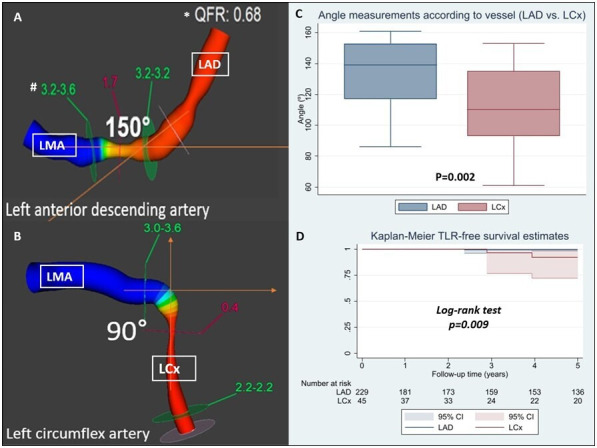
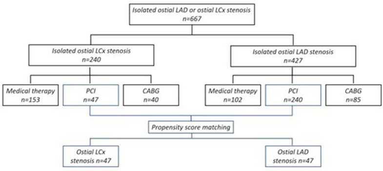
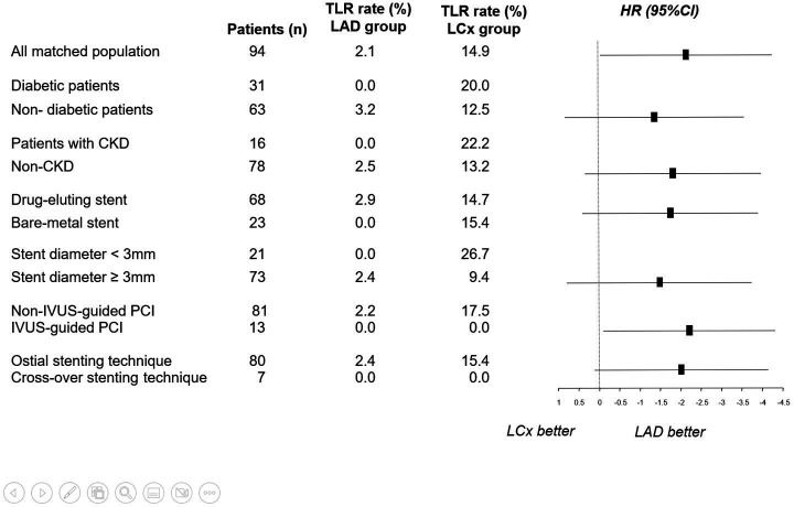
Results: From 2004 to 2018, 287 consecutive patients with LAD (n=240) or LCx (n=47) ostial lesions treated with PCI were analysed. After the adjustment, 47 matched pairs were obtained. The mean age was 72±12 years and 82% were male. The LM-LAD angle was significantly wider than the LM-LCx angle (128°±23° vs 108°±24°, p=0.002). At a median follow-up of 5.5 (IQR 1.5-9.3) years, the rate of TLR was significantly higher in the LCx group (15% vs 2%); with an HR of 7.5, 95% CI 2.1 to 26.4, p<0.001. Interestingly, in the LCx group, TLR-LM occurred in 43% of the TLR cases; meanwhile, no TLR-LM involvement was found in the LAD group.
Conclusions: Isolated ostial LCx PCI was associated with an increase in the rate of TLR compared with ostial LAD PCI at long-term follow-up. Larger studies evaluating the optimal percutaneous approach at this location are needed.
Keywords: acute coronary syndrome; aneurysm, dissecting; atherosclerosis; cardiac catheterization; percutaneous coronary intervention.
© Author(s) (or their employer(s)) 2023. Re-use permitted under CC BY-NC. No commercial re-use. See rights and permissions. Published by BMJ.
Conflict of interest statement
Competing interests: None declared.
Figures
Similar articles
-
The impact of left circumflex coronary artery ostium stenosis on outcomes for patients after percutaneous coronary intervention for unprotected left main disease.Kardiol Pol. 2023;81(9):903-908. doi: 10.33963/KP.a2023.0156. Epub 2023 Jul 25. Kardiol Pol. 2023. PMID: 37489824
-
Dynamic assessment of the left main-left circumflex bending angle: Implications for ostial left circumflex artery in-stent restenosis after successful two-stent PCI.Int J Cardiol. 2023 May 1;378:11-19. doi: 10.1016/j.ijcard.2023.02.030. Epub 2023 Feb 15. Int J Cardiol. 2023. PMID: 36796487
-
Difference in vascular response between sirolimus-eluting- and everolimus-eluting stents in ostial left circumflex artery after unprotected left main as observed by optical coherence tomography.Int J Cardiol. 2017 Mar 1;230:284-292. doi: 10.1016/j.ijcard.2016.12.122. Epub 2016 Dec 23. Int J Cardiol. 2017. PMID: 28065691
-
Optimal Percutaneous Treatment Approach to Unprotected Ostial Left Anterior Descending Artery Disease: A Meta-Analysis and Systematic Review.Heart Lung Circ. 2024 Aug;33(8):1123-1135. doi: 10.1016/j.hlc.2024.02.006. Epub 2024 Apr 12. Heart Lung Circ. 2024. PMID: 38614944
-
Rotational atherectomy to left circumflex ostial lesions: tips and tricks.Cardiovasc Interv Ther. 2023 Oct;38(4):367-374. doi: 10.1007/s12928-023-00941-y. Epub 2023 Jun 10. Cardiovasc Interv Ther. 2023. PMID: 37300802 Review.
Cited by
-
The Role of Intravascular Ultrasound in the Evaluation and Treatment of Free-Floating Stent Struts Following Inadequate Ostial Circumflex Stenting: A Case Report.Medicina (Kaunas). 2024 Sep 24;60(10):1563. doi: 10.3390/medicina60101563. Medicina (Kaunas). 2024. PMID: 39459351 Free PMC article.
References
-
- Chin K. An approach to Ostial lesion management. Curr Interv Cardiol Rep 2001;3:87–9. - PubMed
MeSH terms
LinkOut - more resources
Full Text Sources
Medical
Miscellaneous