. 2023 May 22;14(1):2921.
doi: 10.1038/s41467-023-38571-w.

Functional specialization and interaction in the amygdala-hippocampus circuit during working memory processing

Affiliations

Functional specialization and interaction in the amygdala-hippocampus circuit during working memory processing

Jin Li et al. Nat Commun. .

Abstract

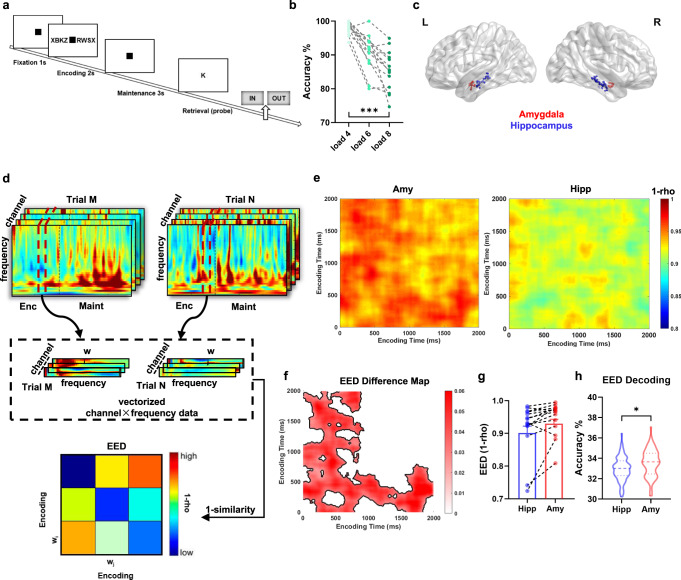
Both the hippocampus and amygdala are involved in working memory (WM) processing. However, their specific role in WM is still an open question. Here, we simultaneously recorded intracranial EEG from the amygdala and hippocampus of epilepsy patients while performing a WM task, and compared their representation patterns during the encoding and maintenance periods. By combining multivariate representational analysis and connectivity analyses with machine learning methods, our results revealed a functional specialization of the amygdala-hippocampal circuit: The mnemonic representations in the amygdala were highly distinct and decreased from encoding to maintenance. The hippocampal representations, however, were more similar across different items but remained stable in the absence of the stimulus. WM encoding and maintenance were associated with bidirectional information flow between the amygdala and the hippocampus in low-frequency bands (1-40 Hz). Furthermore, the decoding accuracy on WM load was higher by using representational features in the amygdala during encoding and in the hippocampus during maintenance, and by using information flow from the amygdala during encoding and that from the hippocampus during maintenance, respectively. Taken together, our study reveals that WM processing is associated with functional specialization and interaction within the amygdala-hippocampus circuit.


Conflict of interest statement

The authors declare no competing interests.

Figures

Fig. 1
Fig. 1. Study framework.
To investigate the function of the amygdala and hippocampus in WM processing, we performed a series of analyses. a First, we calculated the encoding-encoding representational dissimilarity (EED) between trial pairs; b then, the encoding-maintenance similarity (EMS) in the same trial was computed; c next, we measured the directional information flow between the amygdala and the hippocampus; d decoding analysis: we used machine-learning analyses to investigate whether WM load (load 4, 6, and 8) could be predicted by encoding-encoding dissimilarity (EED), encoding-maintenance similarity (EMS), or phase slope index (PSI) features. The brain figure was visualized by BrainNet Viewer toolbox (www.nitrc.org/projects/bnv/) Xia et al..
Fig. 2
Fig. 2. Working memory task, recording sites, representational dissimilarity analysis and decoding analysis during encoding.
a In each trial, a set of consonants was presented (encoding 2 s) followed by a delay (maintenance 3 s). Then a probe letter was shown and the participants indicated whether the probe was in the initial set of consonants (retrieval). b Accuracy decreased from load 4 to load 6 and 8 (repeated-measures ANOVA, p < 0.001, F(2,26) = 42.71). A line connects the data from one participant (n = 14). ***p < 0.001. c Channel location across participants in MNI152 space. Recording regions are indicated by different colors (red, amygdala; blue, hippocampus). The brain figure was visualized by BrainNet Viewer toolbox (www.nitrc.org/projects/bnv/) Xia et al.. d Schematic of encoding-encoding dissimilarity (EED) analysis. Warmer color denotes higher dissimilarity and cooler color means higher similarity. e Averaged EED map during the encoding period across all participants, in the amygdala (left column) and the hippocampus (right column). f EED Difference map obtained by subtracting the hippocampus EED from the amygdala EED reveals a significant cluster (p < 0.05, two-sided cluster-based permutation test, outlined in black), indicating that the amygdala represents the working memory information specifically during the encoding period (dark red area, higher EED in the amygdala than the hippocampus). White areas indicate that there was no significant difference (p > 0.05) between the hippocampus and amygdala. g EED values averaged over the significant cluster in b was extracted within the hippocampus (blue, mean ± s.e.m.) and amygdala (red, mean ± s.e.m.) for each participant, respectively. 12 of 14 participants showed higher EED values within the amygdala than within the hippocampus. h Decoding accuracy using the EED patterns within the amygdala (red) is higher than in the hippocampus (blue) from all cross-validations (n = 100; two-sided permutation test: p = 0.01). Dotted lines indicate the median. Broken lines above and below denote the quartiles. *p < 0.05. Source data are provided as a Source data file.
Fig. 3
Fig. 3. Representational stability within the amygdala and the hippocampus and decoding analysis.
a Schematic of encoding-maintenance similarity (EMS) analysis. Warmer color denotes higher similarity and cooler color means less similarity. b Grand average EMS map across all participants in the amygdala and the hippocampus. c The average EMS was higher in the hippocampus (blue, mean ± s.e.m.) than in the amygdala (red, mean ± s.e.m.; two-sided paired t-test: p = 0.0049, t(13) = 3.381). Each dot represents one participant (n = 14). **p < 0.01. d The decoding accuracy by using the EMS patterns in the hippocampus (blue) is higher than in the amygdala (red) from all cross-validations (n = 100; two-sided permutation test: p < 0.001). Dotted lines indicate the median. Broken lines above and below denote the quartiles. ***p < 0.001. Source data are provided as a Source data file.
Fig. 4
Fig. 4. Directional information flow between the hippocampus and the amygdala during encoding and maintenance.
a The z-scored phase slope index (PSI) across 1–40 Hz during encoding. Asterisks in blue denote significant PSI from the hippocampus to the amygdala and these in red denote the opposite direction (significance was thresholded at |z| > 1.96). b The z-scored PSI across 1–40 Hz during maintenance. c Decoding accuracy of WM load by using PSI features of the amygdala leads connectivity (red) was higher than those of the hippocampus leads connectivity (blue) from all cross-validations (n = 100) during encoding (two-sided permutation test: p < 0.001). Dotted lines indicate the median. Broken lines above and below denote the quartiles. ***p < 0.001. d Decoding accuracy of WM load by using PSI features of the hippocampus leads connectivity (blue) was higher than those of the amygdala leads connectivity (red) from all cross-validations (n = 100) during maintenance (two-sided permutation test: p < 0.001). ***p < 0.001. Source data are provided as a Source data file.

References

    1. Baddeley A. Working memory: theories, models, and controversies. Annu. Rev. Psychol. 2012;63:1–29. doi: 10.1146/annurev-psych-120710-100422. - DOI - PubMed
    1. Christophel TB, Klink PC, Spitzer B, Roelfsema PR, Haynes JD. The distributed nature of working memory. Trends Cogn. Sci. 2017;21:111–124. doi: 10.1016/j.tics.2016.12.007. - DOI - PubMed
    1. LaBar KS, Cabeza R. Cognitive neuroscience of emotional memory. Nat. Rev. Neurosci. 2006;7:54–64. doi: 10.1038/nrn1825. - DOI - PubMed
    1. Gothard KM. Multidimensional processing in the amygdala. Nat. Rev. Neurosci. 2020;21:565–575. doi: 10.1038/s41583-020-0350-y. - DOI - PMC - PubMed
    1. Inman CS, et al. Direct electrical stimulation of the amygdala enhances declarative memory in humans. Proc. Natl Acad. Sci. USA. 2018;115:98–103. doi: 10.1073/pnas.1714058114. - DOI - PMC - PubMed

Publication types