. 2023 Oct;44(10):2091-2102.
doi: 10.1038/s41401-023-01098-3. Epub 2023 May 22.

COX17 restricts renal fibrosis development by maintaining mitochondrial copper homeostasis and restoring complex IV activity

Affiliations

COX17 restricts renal fibrosis development by maintaining mitochondrial copper homeostasis and restoring complex IV activity

Sai-Ya Zhu et al. Acta Pharmacol Sin. 2023 Oct.

Abstract

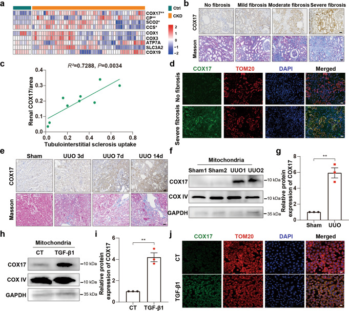
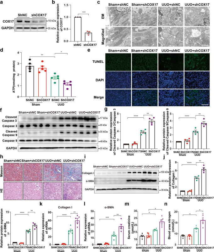
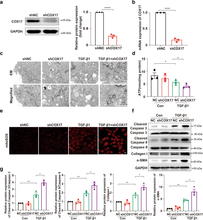
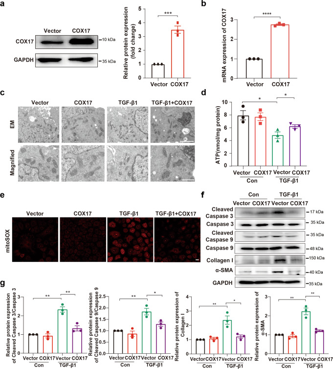
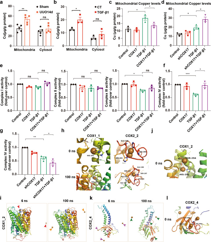
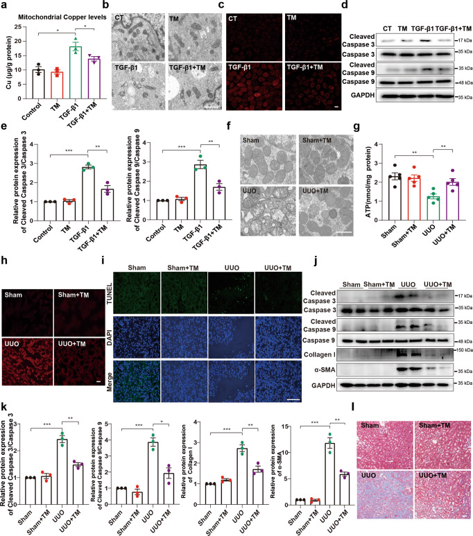
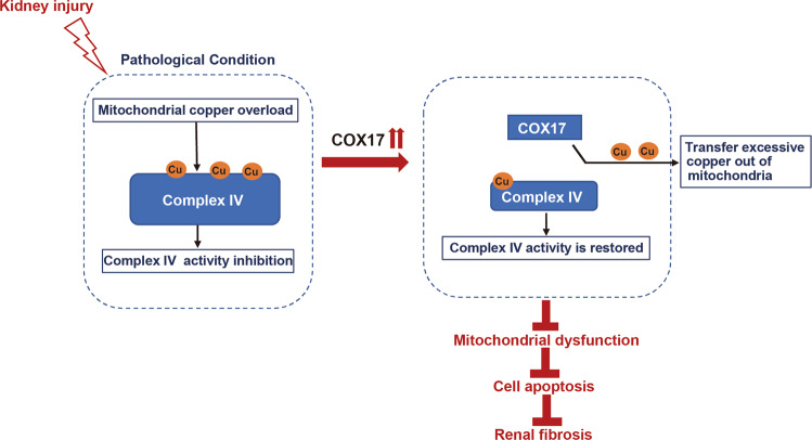
Renal fibrosis relies on multiple proteins and cofactors in its gradual development. Copper is a cofactor of many enzymes involved in renal microenvironment homeostasis. We previously reported that intracellular copper imbalance occurred during renal fibrosis development and was correlated with fibrosis intensity. In this study, we investigated the molecular mechanisms of how copper affected renal fibrosis development. Unilateral ureteral obstruction (UUO) mice were used for in vivo study; rat renal tubular epithelial cells (NRK-52E) treated with TGF-β1 were adapted as an in vitro fibrotic model. We revealed that the accumulation of copper in mitochondria, rather than cytosol, was responsible for mitochondrial dysfunction, cell apoptosis and renal fibrosis in both in vivo and in vitro fibrotic models. Furthermore, we showed that mitochondrial copper overload directly disrupted the activity of respiratory chain complex IV (cytochrome c oxidase), but not complex I, II and III, which hampered respiratory chain and disrupted mitochondrial functions, eventually leading to fibrosis development. Meanwhile, we showed that COX17, the copper chaperone protein, was significantly upregulated in the mitochondria of fibrotic kidneys and NRK-52E cells. Knockdown of COX17 aggravated mitochondrial copper accumulation, inhibited complex IV activity, augmented mitochondrial dysfunction and led to cell apoptosis and renal fibrosis, whereas overexpression of COX17 could discharge copper from mitochondria and protect mitochondrial function, alleviating renal fibrosis. In conclusion, copper accumulation in mitochondria blocks complex IV activity and induces mitochondrial dysfunction. COX17 plays a pivotal role in maintaining mitochondrial copper homeostasis, restoring complex IV activity, and ameliorating renal fibrosis.

Keywords: COX17; NRK-52E cells; copper; cytochrome c oxidase; mitochondria; renal fibrosis.


Conflict of interest statement

The authors declare no competing interests.

Figures

Fig. 1
Fig. 1. COX17 is upregulated in renal interstitial fibrosis.
a Heatmap comparing copper-related gene expression in the kidneys of patients with chronic kidney disease (CKD) with that in control subjects (n = 53). b Immunohistochemical staining of COX17 and Masson staining in kidney sections with different fibrotic degrees. Original magnification, ×200. Bar = 50 μm. c Scatter plots with linear regression show the correlation between COX17 expression levels and the extent of renal fibrosis (R2 = 0.7288, P = 0.0034). d Immunofluorescence co-staining of TOM20 (mitochondrial marker, red) and COX17 (green). Original magnification, ×200. Bar = 20 μm. e Immunohistochemical staining of COX17 and Masson staining in kidney sections. Original magnification, ×200. Bar = 50 μm. f Western immunoblots and g densitometric analysis of COX17 expression in the mitochondria from sham and UUO kidney tissues (n = 5). h Western immunoblots and i densitometric analysis showing COX17 expression in the mitochondria of NRK-52E cells treated with TGF-β1. j Immunofluorescence for COX17 and TOM20, showing COX17 expression in the mitochondria of NRK-52E cells stimulated with TGF-β1. Original magnification, ×200. Bar = 20 μm. Each bar represents the mean ± SEM of at least three independent experiments. *P < 0.05, **P < 0.01.
Fig. 2
Fig. 2. Downregulation of COX17 in vivo aggravates renal fibrosis.
a Western blot and b densitometric analysis showing COX17 expression in kidney tissues transfected with AAV9-shCOX17 or AAV9-Con. c Representative TEM images of mitochondria in tubular cells at low magnification (upper panel: magnified ×7000; bar = 2 μm) and high magnification (bottom panel: magnified ×15,000; bar = 1 μm). d Quantification of ATP levels (n = 5). e Representative images for TUNEL staining of the kidney tissues. Original magnification, ×400. Bar = 100 μm. f Western blot and g densitometric analysis of the expression of caspase 3, caspase 9, cleaved caspase 3 and cleaved caspase 9 in different groups. h Masson and HE staining of renal sections from the indicated groups. Original magnification, ×200. Bar = 50 μm. i Western blot and j densitometric analysis of the expression of collagen I and α-SMA in different groups. k RT–PCR analysis of collagen I and l α-SMA in different groups. m Serum creatinine and n urea nitrogen levels in different groups (n = 5). Data are the mean ± SEM of at least three independent experiments. *P < 0.05, **P < 0.01, ***P < 0.001 and ****P < 0.0001.
Fig. 3
Fig. 3. Downregulation of COX17 aggravates TGF-β1 induced-mitochondrial dysfunction and cell apoptosis.
a Western blot and b RT-PCR showing COX17 expression in COX17 knockdown and control short hairpin RNA (shRNA) cell lines (n = 3). c Representative electron microscopy images of mitochondria in tubular cells among groups, as indicated at low magnification (upper panel: magnified ×7000; bar = 2 μm) and high magnification (bottom panel: magnified ×15,000; bar = 1 μm). d Quantification of intracellular ATP levels (n = 3). e Representative MitoSOX staining micrographs in different groups, as indicated. Original magnification, ×200. Bar = 20 μm. f Immunoblots and g densitometric analysis of caspase 3, caspase 9, cleaved caspase 3, cleaved caspase 9, collagen I and α-SMA expression in tubular cells among different groups. Data are the mean ± SEM of at least three independent experiments. *P < 0.05, **P < 0.01 and ****P < 0.0001.
Fig. 4
Fig. 4. Overexpression of COX17 mitigates mitochondrial dysfunction and cell apoptosis.
a, b The efficiency of COX17 overexpression in NRK-52E cells was detected by Western blot and RT-PCR (n = 3). c EM images showing mitochondria in tubular cells of the indicated groups at low magnification (upper panel: magnified ×7000; bar = 2 μm) and high magnification (bottom panel: magnified ×15,000; bar = 1 μm). d Quantification of intracellular ATP levels (n = 3). e Representative MitoSOX staining micrographs in different groups as indicated. f Immunoblots and g densitometric analysis of caspase 3, caspase 9, cleaved caspase 3, cleaved caspase 9, collagen I and α-SMA expression in tubular cells among different groups. Original magnification, ×200. Bar = 20 μm. Data are the mean ± SEM of at least three independent experiments. *P < 0.05, **P < 0.01, ***P < 0.001 and ****P < 0.0001.
Fig. 5
Fig. 5. COX17 reduces mitochondrial copper content and restores respiratory chain complex IV activity.
a The copper content was detected in the cytosol and mitochondria in the kidneys of the UUO model (n = 5). b Copper content was detected in the mitochondria of NRK-52E cells after 48 h of TGF-β1 treatment (n = 3). c, d Copper content was detected in the mitochondria of control, COX17-overexpressing and knockdown cell lines treated with TGF-β1 (n = 3). e, f The relative activities of respiratory chain complexes I, II, III and IV in control and COX17-overexpressing cell lines treated with TGF-β1 (n = 3). g The relative activity of respiratory chain complex IV in control and COX17-knockdown cell lines treated with TGF-β1 (n = 3). h MD simulations of COX1 and COX2. i MD simulation of COX1_2. j MD simulation of COX1_2 at 0 ns. k MD simulation of COX2_4. l MD simulation of COX2_4 at 0 ns. Data are the mean ± SEM of at least three independent experiments. *P < 0.05, **P < 0.01 and ***P < 0.001. COX1_1, COX1-Cu1+(1); COX1_2, COX1-Cu1+(2); COX2_2, COX2-Cu1+(2); COX2_4, COX2-Cu1+(4). ns indicates no significance.
Fig. 6
Fig. 6. Alleviating mitochondrial copper overload improves mitochondrial dysfunction, cell apoptosis and renal fibrosis.
NRK-52E cells were treated with TGF-β1 (5 ng/mL) or TM (7.5 µmol/L) as indicated. a The copper content was detected in the mitochondria of NRK-52E cells after 48 h of TGF-β1 treatment (n = 3). b Representative electron micrographs of mitochondria in NRK-52E cells (magnified ×15,000; bar = 1 μm). c Representative MitoSOX staining micrographs in different groups as indicated. d Immunoblots and e densitometric analysis of caspase 3, caspase 9, cleaved caspase 3 and cleaved caspase 9 expression in tubular cells among different groups. f Representative TEM images of mitochondria in different groups (magnified ×15,000; bar = 1 μm). g Quantification of ATP levels (n = 5). h Representative MitoSOX staining micrographs in different groups as indicated. i Representative sections of TUNEL-positive cells. Original magnification, ×400. Bar = 100 μm. j Immunoblot and k densitometric analyses of caspase 3, caspase 9, cleaved caspase 3, cleaved caspase 9, collagen I and α-SMA among different groups. l Masson staining of renal sections from the indicated groups. Original magnification, ×200. Bar = 50 μm. Each bar represents the mean ± SEM of at least three independent experiments. *P < 0.05, **P < 0.01 and ***P < 0.001.
Fig. 7
Fig. 7. Diagram depicts the role of COX17 in renal fibrosis.
This study highlights that mitochondrial copper is overloaded in fibrotic kidneys and involved in disrupting respiratory chain complex IV activity, mitochondrial dysfunction, and cell apoptosis. The antifibrotic activity of COX17 in kidneys attenuates mitochondrial copper overload and restores complex IV activity.

References

    1. Panizo S, Martinez-Arias L, Alonso-Montes C, Cannata P, Martin-Carro B, Fernandez-Martin JL, et al. Fibrosis in chronic kidney disease: pathogenesis and consequences. Int J Mol Sci. 2021;22:408–26. - PMC - PubMed
    1. Lutsenko S. Human copper homeostasis: a network of interconnected pathways. Curr Opin Chem Biol. 2010;14:211–7. doi: 10.1016/j.cbpa.2010.01.003. - DOI - PMC - PubMed
    1. Kim BE, Nevitt T, Thiele DJ. Mechanisms for copper acquisition, distribution and regulation. Nat Chem Biol. 2008;4:176–85. doi: 10.1038/nchembio.72. - DOI - PubMed
    1. Janssen R, de Brouwer B, von der Thusen JH, Wouters EFM. Copper as the most likely pathogenic divergence factor between lung fibrosis and emphysema. Med Hypotheses. 2018;120:49–54. doi: 10.1016/j.mehy.2018.08.003. - DOI - PubMed
    1. Yadav A, Kumar L, Misra N, Deepak U, Shiv GC. Kumar, Estimation of serum zinc, copper, and iron in the patients of oral submucous fibrosis. Natl J Maxillofac Surg. 2015;6:190–3. doi: 10.4103/0975-5950.183851. - DOI - PMC - PubMed

LinkOut - more resources