Review
. 2023 Aug;53(9):1799-1828.
doi: 10.1007/s00247-023-05648-z. Epub 2023 May 23.

Tuberculosis revisted: classic imaging findings in childhood

Affiliations
Review

Tuberculosis revisted: classic imaging findings in childhood

Nasreen Mahomed et al. Pediatr Radiol. 2023 Aug.

Abstract

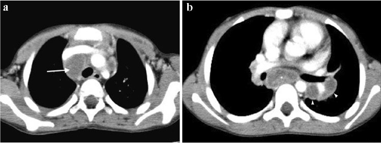
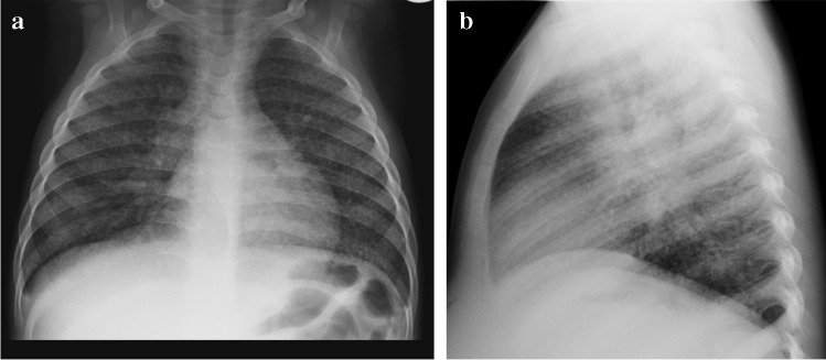
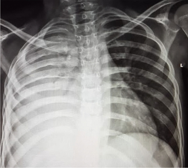
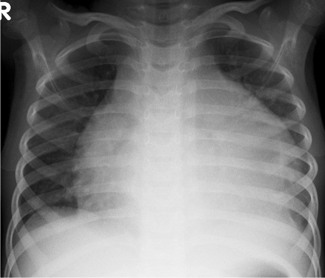
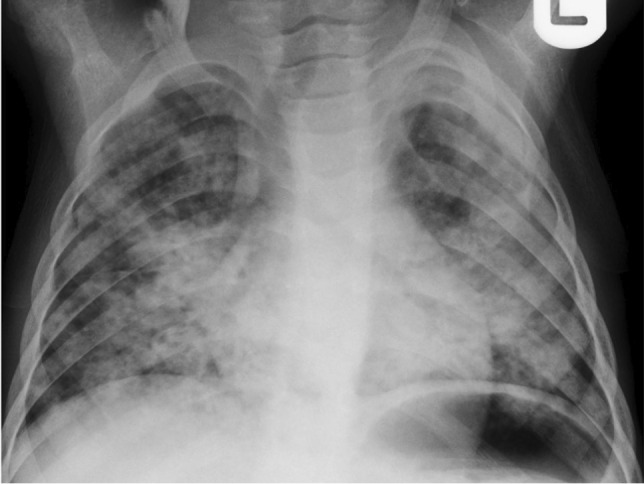
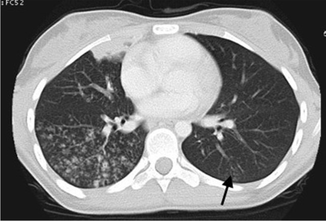
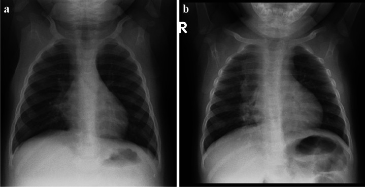
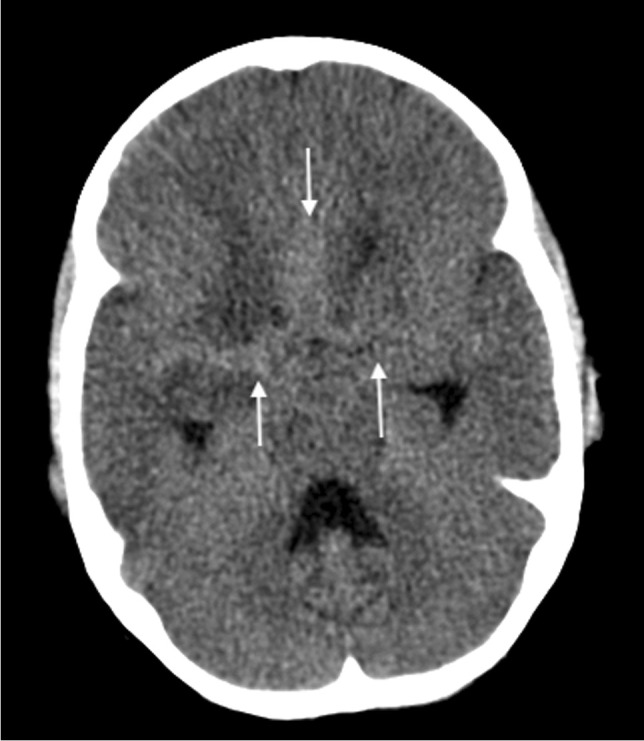
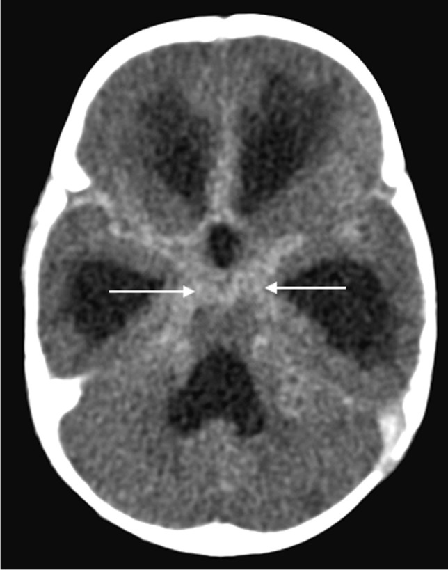
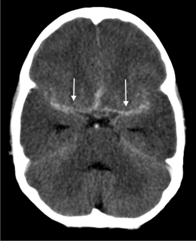
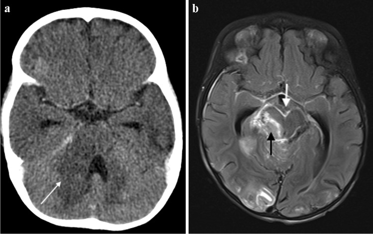
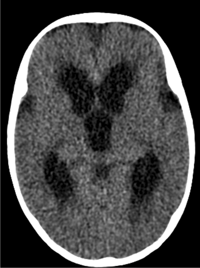
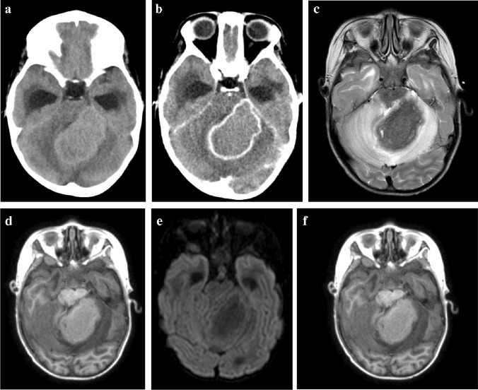
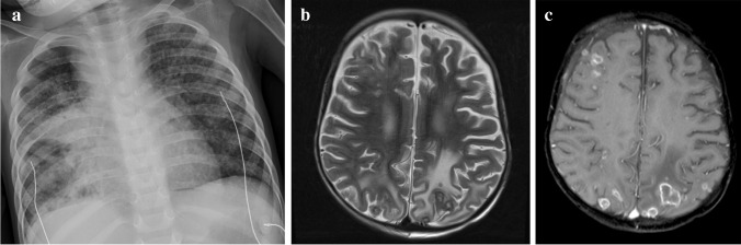
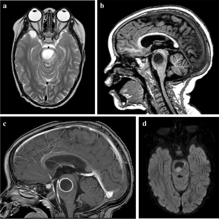
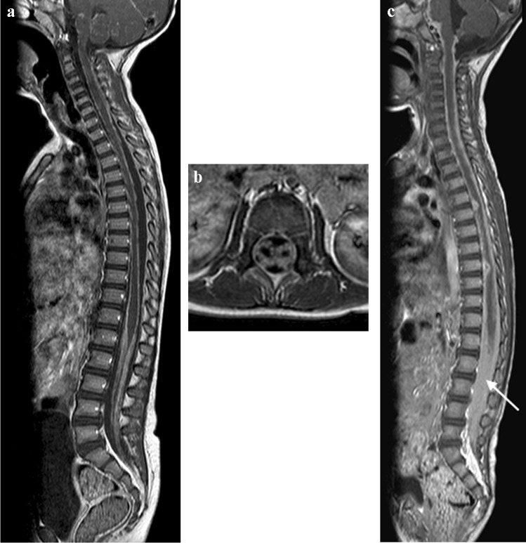
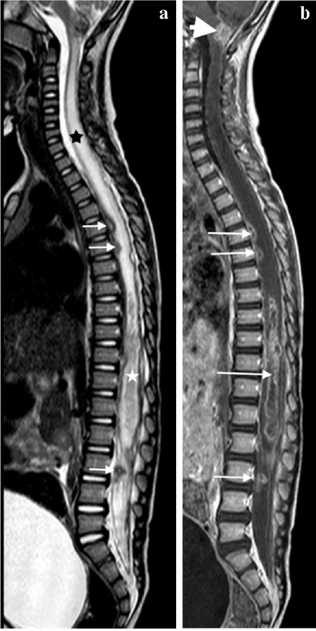
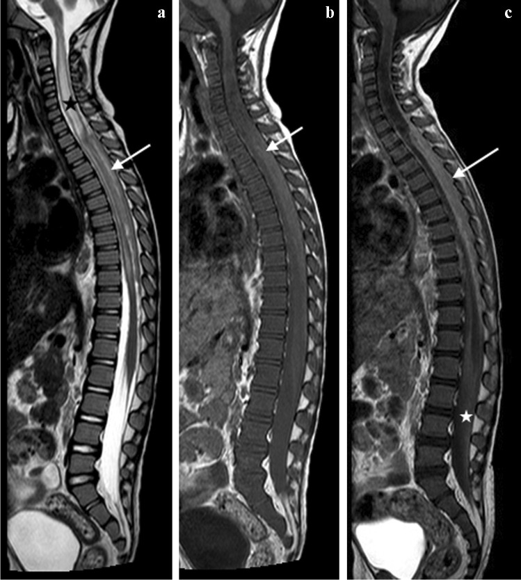
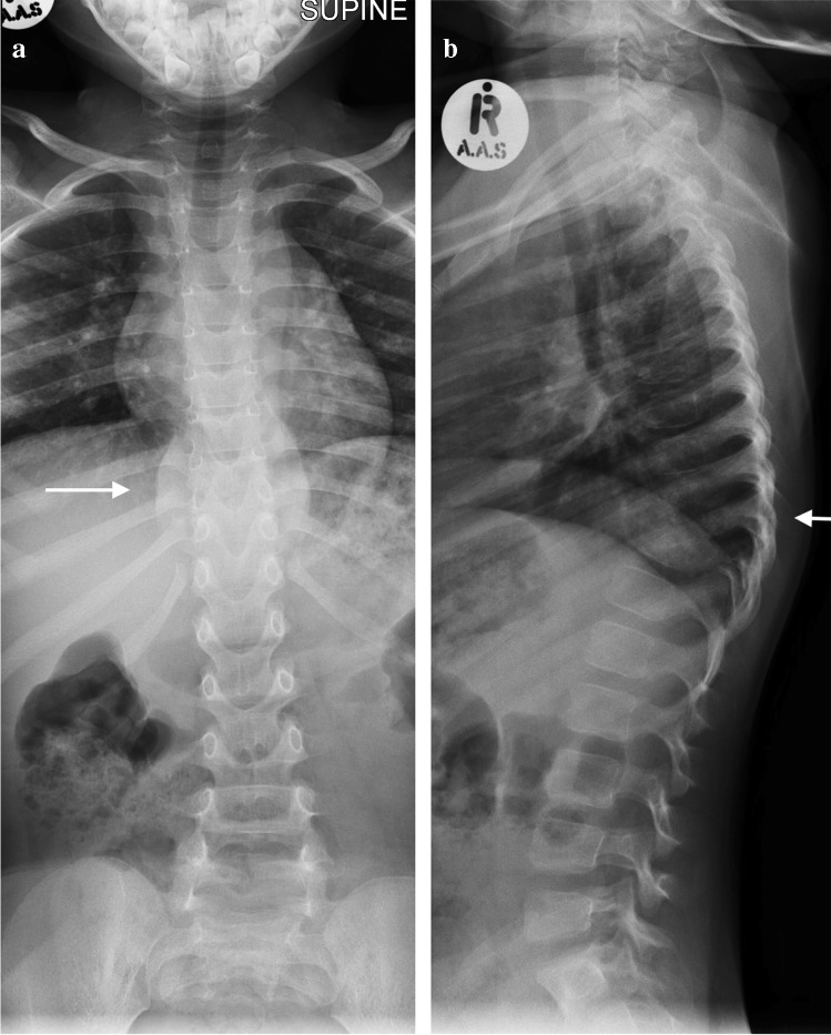
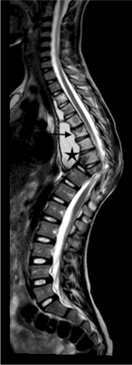
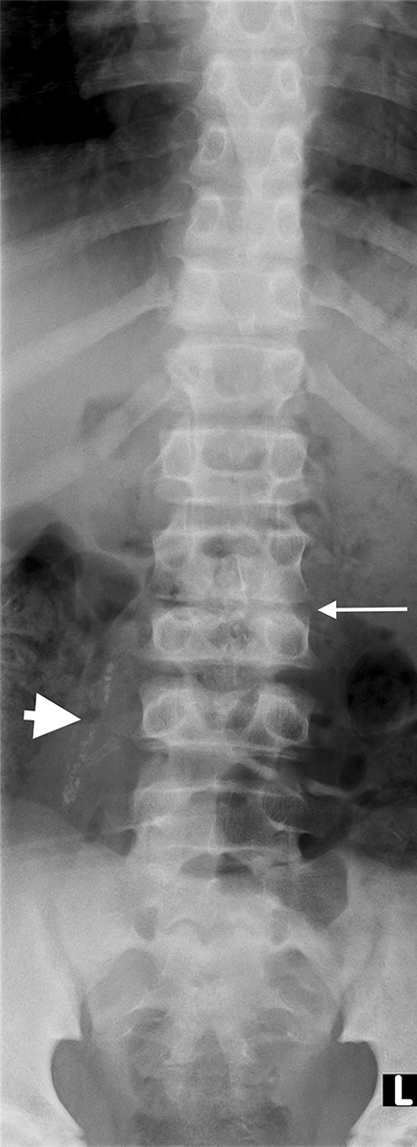
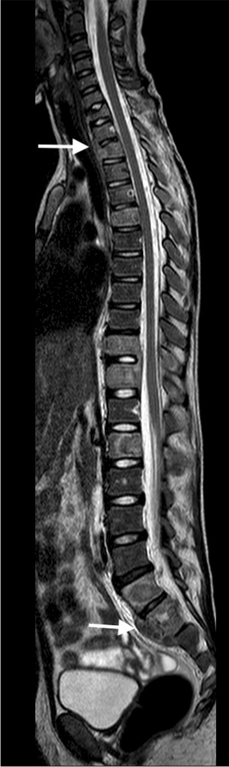
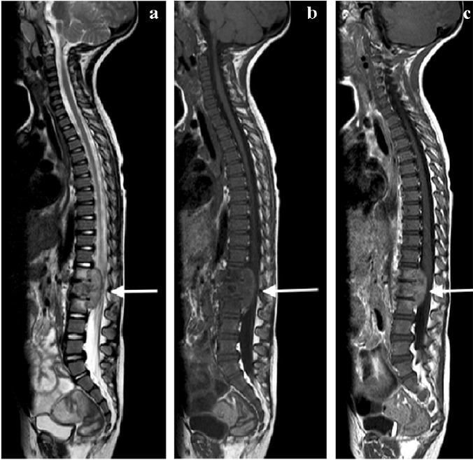
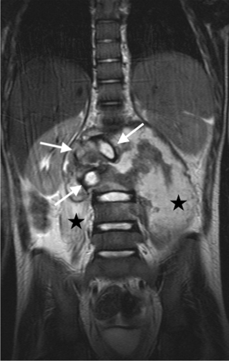
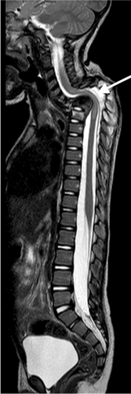
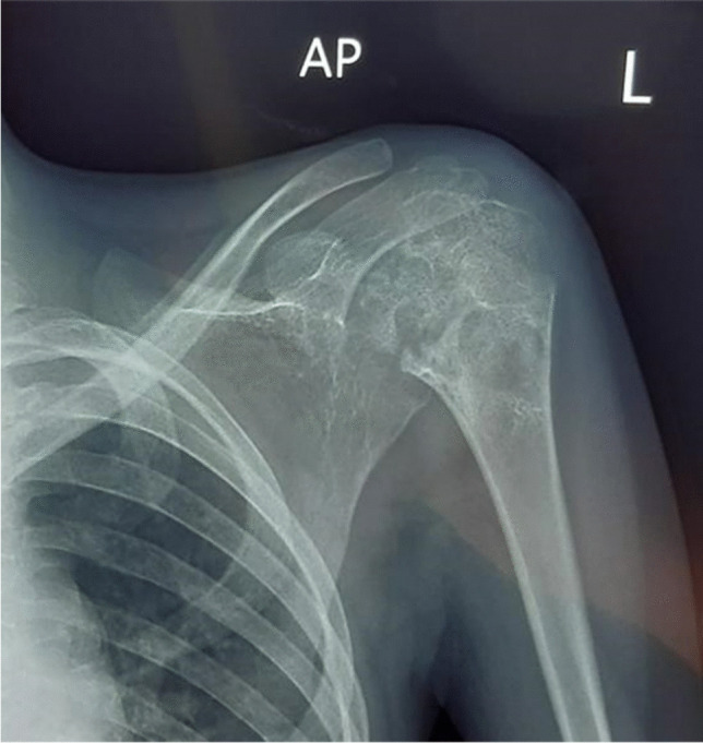
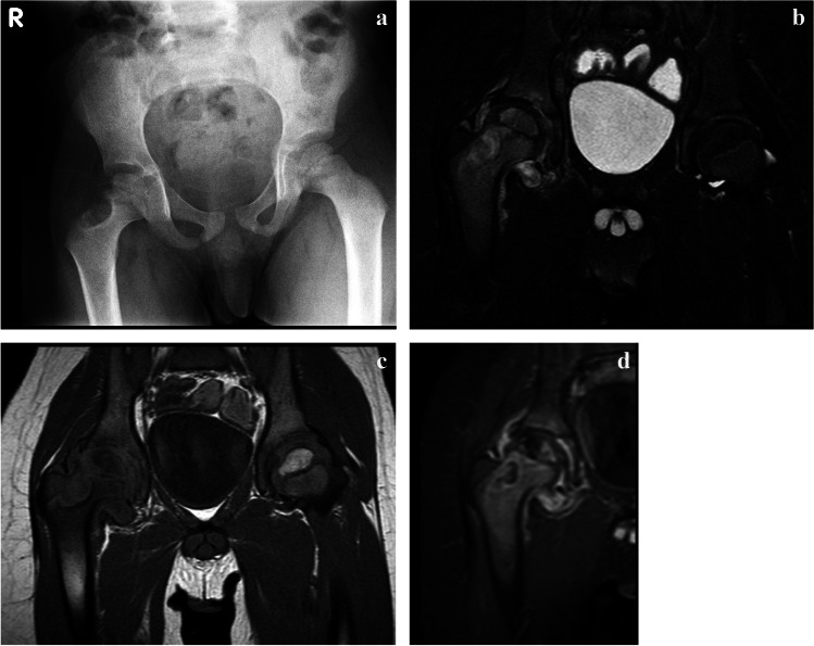
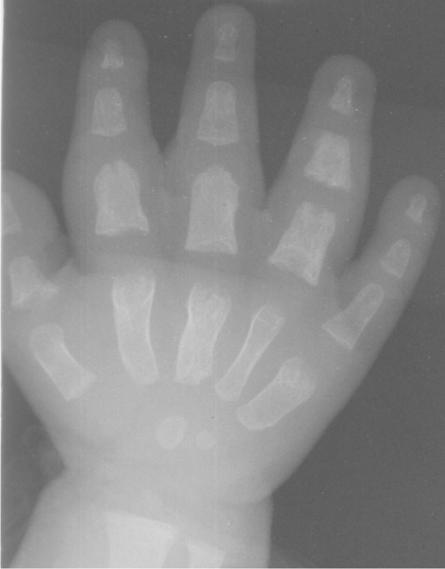
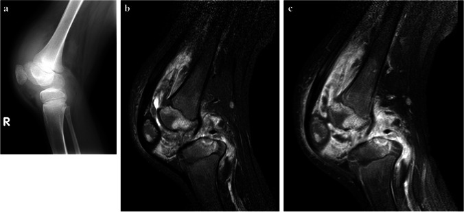
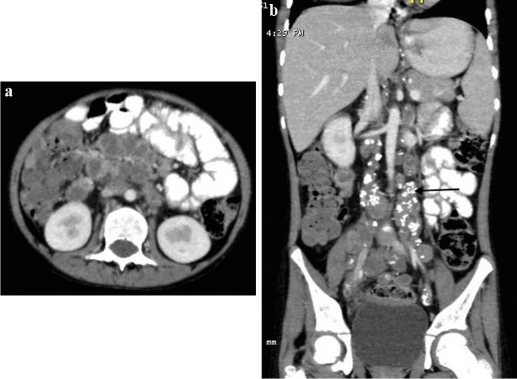
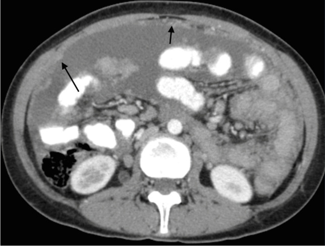
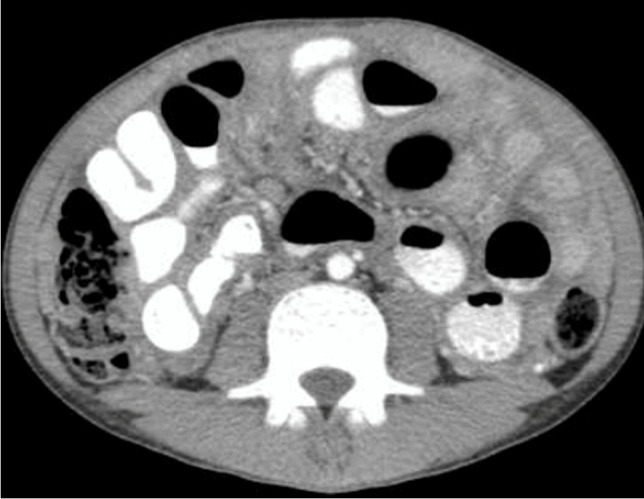
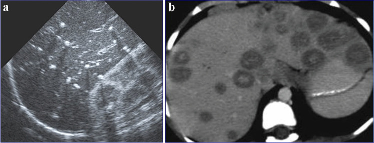
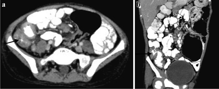
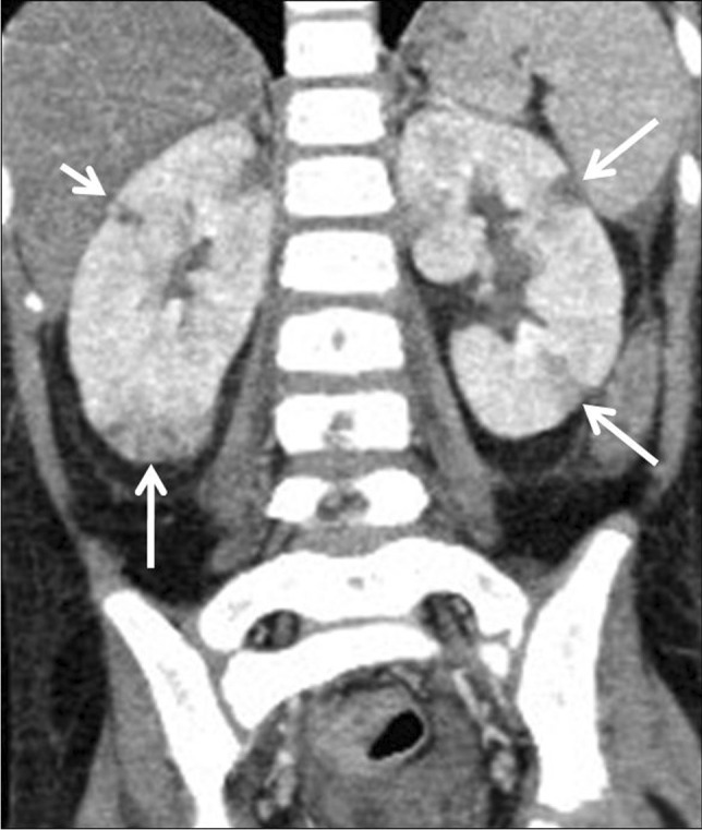
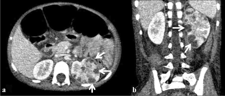
Tuberculosis (TB) remains one of the major public health threats worldwide, despite improved diagnostic and therapeutic methods. Tuberculosis is one of the main causes of infectious disease in the chest and is associated with substantial morbidity and mortality in paediatric populations, particularly in low- and middle-income countries. Due to the difficulty in obtaining microbiological confirmation of pulmonary TB in children, diagnosis often relies on a combination of clinical and radiological findings. The early diagnosis of central nervous system TB is challenging with presumptive diagnosis heavily reliant on imaging. Brain infection can present as a diffuse exudative basal leptomeningitis or as localised disease (tuberculoma, abscess, cerebritis). Spinal TB may present as radiculomyelitis, spinal tuberculoma or abscess or epidural phlegmon. Musculoskeletal manifestation accounts for 10% of extrapulmonary presentations but is easily overlooked with its insidious clinical course and non-specific imaging findings. Common musculoskeletal manifestations of TB include spondylitis, arthritis and osteomyelitis, while tenosynovitis and bursitis are less common. Abdominal TB presents with a triad of pain, fever and weight loss. Abdominal TB may occur in various forms, as tuberculous lymphadenopathy or peritoneal, gastrointestinal or visceral TB. Chest radiographs should be performed, as approximately 15% to 25% of children with abdominal TB have concomitant pulmonary infection. Urogenital TB is rare in children. This article will review the classic radiological findings in childhood TB in each of the major systems in order of clinical prevalence, namely chest, central nervous system, spine, musculoskeletal, abdomen and genitourinary system.

Keywords: Adoloescent; Child; Imaging; Infant; Infection; Radiology; Spina ventosa; Tuberculosis.


Conflict of interest statement

None

Figures

Fig. 1
Fig. 1
An 8-year-old boy on steroids with cough and night sweats. Posteroanterior chest radiograph shows a focal area of opacification in the right upper lobe representing a Ghon focus (arrow) and right pleural effusion. Elevation of the right hemidiaphragm suggests a sub-pulmonic component. Gene expert was positive for pulmonary tuberculosis
Fig. 2
Fig. 2
Chest imaging in a 2-year-old girl with pulmonary tuberculosis. a Posteroanterior chest radiograph (CXR) shows pneumonia of the right upper lobe. Note the displacement of the trachea towards the left and attenuation of the bronchus intermedius suggesting paratracheal lymphadenopathy. b Lateral CXR shows inferior bulging of the horizontal fissure and soft tissue density of lymphadenopathy around the carina. c A post-contrast axial computed tomography image (mediastinal window) demonstrates large volume lymphadenopathy of low density and peripherally enhancing lymph nodes (arrow). The right upper lobe is expanded and of predominantly low density, consistent with caseous necrosis. There is a small associated pleural effusion
Fig. 3
Fig. 3
Anteroposterior chest radiograph in a 1-year-old boy with tuberculosis demonstrating bilateral hilar lymphadenopathy
Fig. 4
Fig. 4
A 1-year-old girl with a cough and known to be a tuberculosis (TB) contact a Posteroanterior chest radiograph shows extensive mediastinal and hilar lymphadenopathy. b Axial post-contrast computed tomography (mediastinal window) performed a few days later confirms peripherally enhancing and low-density anterior mediastinal, pre-vascular, hilar and paratracheal nodes. There is also a pre-tracheal node that shows punctate calcification (arrow). These features are characteristic of TB. Coronal thick slab multiplanar reconstruction (mediastinal window) demonstrates deviation of the trachea to the left but no significant airway compression
Fig. 5
Fig. 5
An 8-year-old boy with pulmonary tuberculosis. Axial post-contrast CT (mediastinal window) demonstrates multiple peripherally enhancing necrotic lymph nodes in the mediastinum (arrow), left hilum (arrowheads) and posterior mediastinum (asterisk)
Fig. 6
Fig. 6
Posteroanterior (a) and lateral (b) chest radiographs in a 5-year-old boy demonstrate a diffuse pattern of miliary nodules in keeping with haematogenous spread of tuberculosis. Note the leftward displacement of the trachea and prominent right hilum, representing paratracheal and hilar lymphadenopathy
Fig. 7
Fig. 7
Posteroanterior chest radiograph in a 9-year-old girl with confirmed pulmonary tuberculosis shows a large right-sided pleural effusion with minimum mediastinal shift due to underlying atelectasis
Fig. 8
Fig. 8
Posteroanterior chest radiograph in a 6-year-old boy demonstrates a tuberculous pericardial effusion  causing globular cardiomegaly
Fig. 9
Fig. 9
Posteroanterior chest radiograph of an 8-month-old girl with proven tuberculosis (TB) shows multiple ill-defined nodules of differing sizes and parenchymal opacification of the right middle and both lower lobes. These features are suggestive of bronchogenic spread of TB
Fig. 10
Fig. 10
A 12-year-old girl with pulmonary tuberculosis. Axial computed tomography image (lung window) demonstrates multiple coalescing centrilobular nodules in the right lung. A small patch of consolidation is also seen. A few ill-defined centrilobular nodules are also seen in the left lung (arrow)
Fig. 11
Fig. 11
A 3-year-old boy newly diagnosed with human immunodeficiency virus infection and started on antiretroviral therapy. a Posteroanterior chest radiograph (CXR) on initiation of therapy shows no radiographic evidence of pulmonary tuberculosis (TB) and the patient was asymptomatic. b Repeat posteroanterior CXR 6 weeks later, on presentation with a cough, shows marked paratracheal, right hilar and subcarinal lymphadenopathy with splaying of the carina and attenuation of the bronchus intermedius and middle/lower lobe bronchi in keeping with TB. This is an example of unmasking TB immune reconstitution inflammatory syndrome
Fig. 12
Fig. 12
Non-contrast axial computed tomography of the brain in a 30-month-old boy with tuberculosis demonstrates hyperdensity (arrows) in the basal cisterns, representing granulation tissue and exudate. Note the bilateral basal ganglia and right temporal lobe low density, indicating established infarcts
Fig. 13
Fig. 13
Axial post-contrast  computed tomography image of the brain in a 30-month-old boy with tuberculosis (same patient as Fig. 12) shows diffuse basal meningeal enhancement filling the cisterns (arrows) and accompanying hydrocephalus
Fig. 14
Fig. 14
Axial post-contrast computed tomography image of the brain in a 30-month-old boy with tuberculosis (same patient as Figs. 12 and 13) shows enhancement at the junction of the suprasellar and middlecerebral artery cisterns (the “Y-sign”) (arrows)
Fig. 15
Fig. 15
a Brain imaging in an 18-month-old girl with newly diagnosed human immunodeficiency virus infection and presumed tuberculous meningitis. a Axial post-contrast computed tomography shows hypodensity with mass effect in the right cerebellar peduncle (arrow), hydrocephalus with temporal horn prominence and absence of basal meningeal enhancement. b Axial T1 post-gadolinium magnetic resonance image clearly demonstrates basal meningeal enhancement, an enhancing mass in the right peduncle (white arrow) and multiple tiny miliary nodules (black arrow)
Fig. 16
Fig. 16
Axial non-contrast computed tomography scan of the brain in a 2-year-old boy with severe hydrocephalus due to tuberculous meningitis
Fig. 17
Fig. 17
Axial non-contrast computed tomography of the  brain in a 4-year-and-6-month-old boy with tuberculous meningitis. There is only mild hydrocephalus despite the child being severely obtunded at presentation
Fig. 18
Fig. 18
Axial non-contrast computed tomography of the brain in a 7-year-old boy with tuberculosis shows infarcts in the “TB zone” of the bilateral medial lenticulostriate and thalamo-perforating vessels (head of the caudate nucleus, anterior limb of the internal capsule and the anteromedial thalamus)
Fig. 19
Fig. 19
Magnetic resonance imaging of the brain in a 12-year-old boy with tuberculous meningitis and persistent decreased level of consciousness. a Axial T2 demonstrates multiple hyperintense foci in the pons (arrow). b Axial diffusion-weighted image demonstrates restricted diffusion (arrow) c Low signal (arrows) on the apparent diffusion coefficient map confirms infarcts. d Coronal magnetic resonance angiogram image shows partial occlusion of the basilar artery (black arrow) and irregularity of both posterior communicating arteries (white arrows)
Fig. 20
Fig. 20
Axial T1 post-gadolinium magnetic resonance image in a 3-year-and-4-month old girl shows enhancement of the right trigeminal nerve (arrow). There is enhancement of the right cerebellar peduncle and there are multiple peripherally enhancing tuberculomas
Fig. 21
Fig. 21
A 5-year-old girl with headaches and vomiting referred with a posterior fossa mass. a, b Pre- (a) and post-contrast (b) axial computed tomography images show an intermediate density mass with peripheral enhancement involving the left cerebellar peduncle and cerebellar hemisphere and complicated by hydrocephalus. c, d Axial magnetic resonance images (MRI). The mass is profoundly hypointense on T2 (c) and shows no diffusion restriction (d). e Magnetic resonance spectroscopy shows  a lactate peak (arrow). f Axial post-gadolinium T1 MRI shows central intermediate intensity and peripheral enhancement. These imaging features are characteristic of a tuberculoma—there was response to anti-tuberculous treatment
Fig. 22
Fig. 22
A 9-month-old boy presenting with a decreased level of consciousness. a Posteroanterior chest radiograph shows paratracheal and hilar lymphadenopathy and a diffuse micronodular pattern in keeping with miliary tuberculosis. b, c Axial T2 (b) and T1 post-gadolinium (c) magnetic resonance imaging  show innumerable lesions at the grey/white matter junction and meninges, some of which are hypointense (caseating tuberculomas) and some of which have T2 hyperintense centres (liquifying caseating tuberculomas) and show surrounding oedema (b). There is peripheral enhancement on the T1 post-gadolinium image (c)
Fig. 23
Fig. 23
Brain magnetic resonance imaging in a 12-year-old boy a Axial T2 demonstrates a hyperintense lesion in the pons with a hypointense rim. b, c Sagittal images show intermediate to low signal on T1 (b) with peripheral enhancement post-gadolinium (c). d Axial diffusion weighted imaging shows some restriction in the wall and content. There was corresponding low signal on apparent diffusion coefficient map (not shown). The differential for these imaging findings includes a tuberculous (TB) abscess or liquifying tuberculoma. The patient’s clinical condition deteriorated requiring drainage; surgery confirmed a TB abscess with the content teeming with TB bacilli
Fig. 24
Fig. 24
Sagittal (a) and axial T1 post-gadolinium (b) magnetic resonance imaging (MRI) in a 6-year-old girl with tuberculous (TB) meningitis show enhancement of the arachnoid with enhancement and clumping of the nerve roots. c Sagittal T1 post-contrast MRI in a different patient with TB meningitis, a 4-year and-10-month old girl who had repeated failed lumbar punctures, shows the spinal canal to be completely ocupied by enhancing tissue (arrow)
Fig. 25
Fig. 25
Sagittal magnetic resonance images in a 3-year-old girl with tuberculous meningitis and paraplegia. a T2 image demonstrates an irregular hypointense intramedullary lesion from T7 (black asterisk) to the conus (white asterisk); additional lesions are seen of the anterior cord at T6/T7 and within the nerve roots (arrows). There is marked cord oedema extending to the cranio-cervical junction. b T1post-gadolinium image demonstrates peripheral enhancement of the lesions (thin arrows) consistent with tuberculomas. Multiple small enhancing tuberculomas are also seen at the cranio-cervical junction (thick arrow)
Fig. 26
Fig. 26
Sagittal magnetic resonance images of the spine in a 5-year-and-3-month-old girl with tuberculous meningitis and progressive lower limb weakness. a T2 image shows a hypointense posterior epidural mass extending from T2 to T9 (arrow) and compressing the cord anteriorly. There is cord oedema proximal to the mass (asterisk). bc  T1 pre- (b) and post- (c) gadolinium show the mass to be homogeneously enhancing (arrows). There is also nerve root enhancement (asterisk in c)
Fig. 27
Fig. 27
Spine radiographs in a 3-year-old boy who presented with a lump on his back and in whom tuberculosis was confirmed. a The anteroposterior radiograph shows a  paravertebral soft tissue mass extending from T8 to T11 (arrow). b The lateral radiograph shows a thoracic kyphosis due to collapse of the T10 vertebral body (arrow)
Fig. 28
Fig. 28
A 6-year-old boy with tuberculous spondylitis. Sagittal T2 magnetic resonance image of the spine shows scalloping of the anterior vertebral bodies (arrow) secondary to an anterior subligamentous collection (asterisk). There is a thoracic kyphosis due to collapse of the T10 vertebral body
Fig. 29
Fig. 29
Anteroposterior radiograph of the lumbar spine in a 10-year-old boy with a history of treated tuberculous spondylitis shows disc space narrowing at L2/L3 and loss of height of L3 with sclerosis of its superior endplate (thin arrow). There is calcification of the right psoas muscle (thick arrow)
Fig. 30
Fig. 30
A sagittal T2 magnetic resonance image of the spine in a 7-year-old girl with tuberculous spondylitis. There is multilevel non-contiguous disease as evidenced by the increased signal in the vertebral bodies; note the anterior subligamentous spread at T1/T2 and S1/S2 (arrows). There is preservation of the disc height at all levels and disc signal at almost all levels
Fig. 31
Fig. 31
A 2-year-old boy with back pain. ac Sagittal T2 (a), T1 (b) and T1 post-gadolinium (c) magnetic resonance images of the spine show the involved T12, L1 and L2 vertebral bodies with a soft tissue mass that enhances homogeneously and protrudes posteriorly into the spinal canal compressing the cauda equina (arrows). Note the enhancing vertebral bodies and the T2 hypointense non-enhancing discs, also the involvement of the dens of C2 that was not suspected clinically. Tuberculosis was confirmed
Fig. 32
Fig. 32
A coronal T2 magnetic resonance image in an 8-year-old girl with tuberculous spondylitis shows expanded discs with normal signal (arrows) subsumed within a large paraspinal collection that extends into both psoas muscles (asterisks)
Fig. 33
Fig. 33
Sagittal T1 pre- (a) and post-gadolinium (b) magnetic resonance images in a 5-year-old girl who presented with kyphosis. There is a hypointense peripherally enhancing anterior subligamentous collection (arrows) causing scalloping of the anterior vertebral bodies. There is kyphosis centred around the destroyed T7 and T8 vertebral bodies (identified by way of their intact posterior elements). Tuberculosis was confirmed
Fig. 34
Fig. 34
Sagittal T2 magnetic resonance image in a 4-year-old boy presenting with kyphosis and tetraparesis. There is cervicothoracic kyphosis and the cord is draped over the apex of the gibbus with localised cord signal abnormality (arrow). The patient made a full recovery post-surgical and anti-tuberculous therapy
Fig. 35
Fig. 35
Anteroposterior shoulder radiograph in a 4-year-old girl presenting with left shoulder pain demonstrates the Phemister triad: juxta-articular osteoporosis, peripheral osseous erosions and narrowing of joint space, typical of tuberculous arthritis. Tuberculosis was confirmed on bone biopsy
Fig. 36
Fig. 36
Cystic tuberculous (TB) osteomyelitis in a 7-year-old boy who had been limping since a minor fall 3 months previously. a An anteroposterior pelvic radiograph shows a well-defined eccentrically located cystic lesion of the lateral right femoral neck with cortical irregularity of the adjacent metaphysis. The femoral head is osteopaenic. b-d Coronal magnetic resonance images. T2 with fat suppression (b) and T1 (c) confirm involvement of the metaphysis and epiphysis and that the process crosses the joint to involve the superolateral acetabulum with a small associated effusion. There is heterogeneous enhancement of the bone and diffuse synovial enhancement post-contrast (d). Bone biopsy–confirmed tuberculosis
Fig. 37
Fig. 37
Dorsopalmar radiograph of the right hand in a 9-month-old girl, presenting with a painless swelling of the fingers for 2 months. There is soft tissue swelling of the second through fourth digits with fusiform expansion of the second, third and fifth metacarpals and fusiform swelling and erosion of the first, second, third and fourth proximal and fourth middle phalanges, typical of tuberculous dactylitis (spina ventosa)
Fig. 38
Fig. 38
Images of the right knee in a 12-year-old boy with tuberculous tenosynovitis and osteomyelitis. There was a history of a swollen knee for 6 months, no history of trauma and although he was not septic, he was noted to be severely malnourished. a A lateral knee radiograph shows a large joint effusion, suspected synovial thickening and osteopaenia of the patella. No periosteal reaction or focal bone lesion is demonstrated. He proceeded to magnetic resonance imaging (MRI) following a failed attempt at joint aspiration. A sagittal short tau inversion recovery MRI shows thickened low signal synovium lining the suprapatellar and popliteal fossa with a small amount of fluid in the suprapatellar space. There is also patchy oedema of the patella and femoral epiphysis with cartilage thinning and a focal erosive lesion of the tibial epiphysis. c A sagittal fat saturated T1 post-contrast MRI shows diffuse synovial and patchy multifocal bone enhancement. Synovial biopsy confirmed tuberculosis
Fig. 39
Fig. 39
A 9-year-old boy with ileocecal tuberculosis. Axial (a) and coronal (b) contrast-enhanced computed tomography images show circumferential mural thickening involving the caecum and terminal ileum. Enlarged calcified lymph nodes are also seen on the coronal image (arrow)
Fig. 40
Fig. 40
An axial computed tomography image of the mid-abdomen following intravenous and oral contrast administration in an 8-year-old boy shows high attenuating free fluid and fine nodularities in the peritoneum (arrows), in keeping with peritoneal tuberculosis of the wet ascitic type
Fig. 41
Fig. 41
An axial computed tomography image of the mid-abdomen following intravenous and oral contrast in a 3-year-and-5-month-old boy shows diffuse thick hazy stranding of the peritoneum separating the intestinal loops, in keeping with peritoneal tuberculosis of the dry plastic type
Fig. 42
Fig. 42
Hepatic tuberculosis (TB) in two children. a A transverse abdominal ultrasound in a 3-year-old boy reveals multiple small (3–5 mm) hyperechoic nodules, some of which exhibit posterior shadowing compatible with micronodular hepatic TB. b An axial contrast-enhanced computed tomography  abdominal image in a 7-year-old girl shows multiple round nodules measuring more than 10 mm. The lesions are low in attenuation with some showing central hyperattenuation. Some of the lesions in the left hepatic lobe coalesce and appear mass-like
Fig. 43
Fig. 43
A 9-year-old boy with ileocecal tuberculosis. Axial (a) and coronal (b) contrast-enhanced computed tomography of the abdomen show circumferential mural thickening involving the caecum (arrow in a) and terminal ileum (arrow in b)
Fig. 44
Fig. 44
Tuberculous pyelonephritis in a 4-year-old boy. A coronal contrast-enhanced computed tomography image of the abdomen shows a striated nephrogram in the bilateral kidneys with wedge-shaped hypodense areas (arrows)
Fig. 45
Fig. 45
Tuberculous renal abscesses in a 5-year-old girl. Contrast-enhanced computed tomography images of the abdomen in axial (a) and coronal (b) planes show multiple peripherally enhancing hypodense lesions in the periphery of the left kidney (arrows)

References

    1. World Health Organisation Global Tuberculosis report 2021. Geneva. https://www.who.int/publications/i/item/9789240037021. Accessed Jan 2023
    1. Nachiappan AC, Rahbar K, Shi X, et al. Pulmonary tuberculosis: role of radiology in diagnosis and management. Radiographics. 2017;37:52–72. - PubMed
    1. George R, Andronikou S, Theron S, et al. Pulmonary infections in HIV-positive children. Pediatr Radiol. 2009;39:545–554. - PubMed
    1. Zar HJ. Chronic lung disease in human immunodeficiency virus (HIV) infected children. Pediatr Pulmonol. 2008;43:1–10. - PubMed
    1. Jeena PM, Coovadia HM, Thula SA, et al. Persistent and chronic lung disease in HIV-1 infected and uninfected African children. AIDS. 1998;12:1185–1193. - PubMed

Publication types

LinkOut - more resources