. 2023 Jul;15(7):998-1005.
doi: 10.1038/s41557-023-01205-1. Epub 2023 May 22.

In vitro selection of macrocyclic peptide inhibitors containing cyclic γ2,4-amino acids targeting the SARS-CoV-2 main protease

Affiliations

In vitro selection of macrocyclic peptide inhibitors containing cyclic γ2,4-amino acids targeting the SARS-CoV-2 main protease

Takashi Miura et al. Nat Chem. 2023 Jul.

Abstract

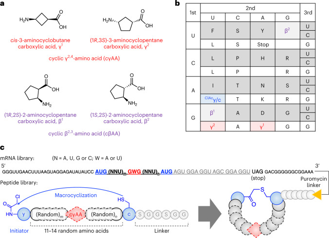
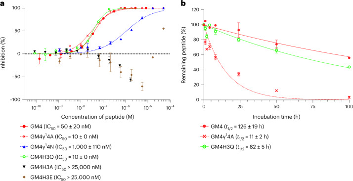
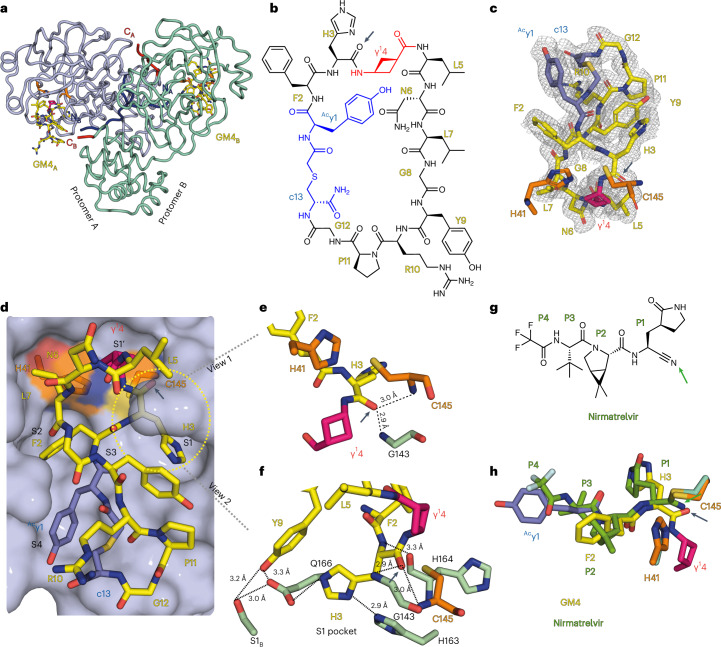
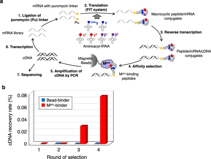
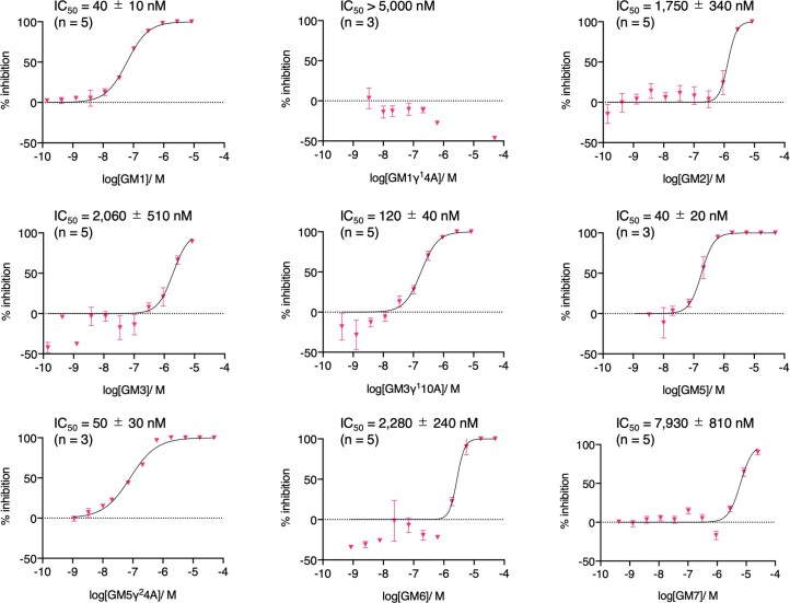
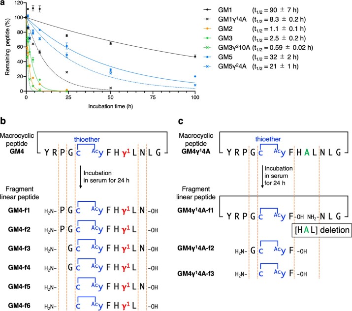
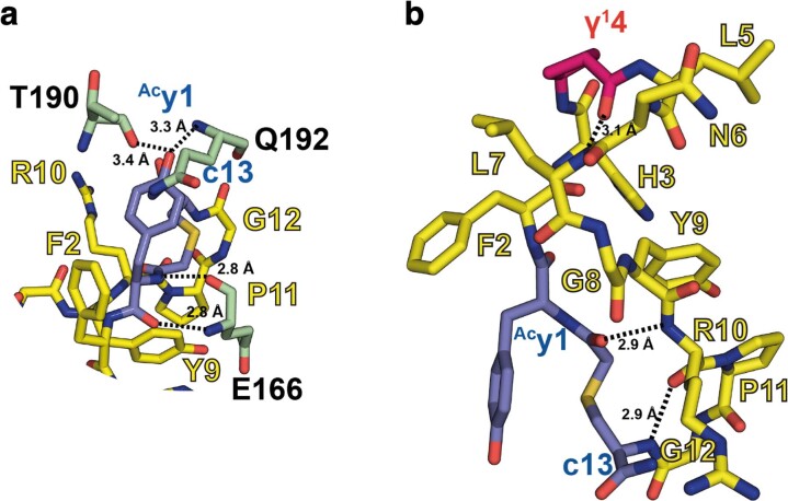
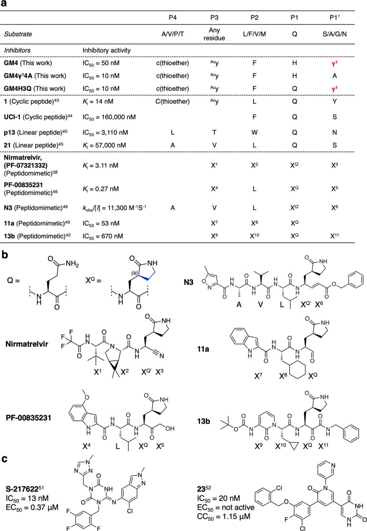
γ-Amino acids can play important roles in the biological activities of natural products; however, the ribosomal incorporation of γ-amino acids into peptides is challenging. Here we report how a selection campaign employing a non-canonical peptide library containing cyclic γ2,4-amino acids resulted in the discovery of very potent inhibitors of the SARS-CoV-2 main protease (Mpro). Two kinds of cyclic γ2,4-amino acids, cis-3-aminocyclobutane carboxylic acid (γ1) and (1R,3S)-3-aminocyclopentane carboxylic acid (γ2), were ribosomally introduced into a library of thioether-macrocyclic peptides. One resultant potent Mpro inhibitor (half-maximal inhibitory concentration = 50 nM), GM4, comprising 13 residues with γ1 at the fourth position, manifests a 5.2 nM dissociation constant. An Mpro:GM4 complex crystal structure reveals the intact inhibitor spans the substrate binding cleft. The γ1 interacts with the S1' catalytic subsite and contributes to a 12-fold increase in proteolytic stability compared to its alanine-substituted variant. Knowledge of interactions between GM4 and Mpro enabled production of a variant with a 5-fold increase in potency.


Conflict of interest statement

The authors declare no competing interests.

Figures

Fig. 1
Fig. 1. Ribosomal incorporation of cγAAs into a macrocyclic peptide library.
a, Structures of the cγAAs and cβAAs used in this study. b, The reprogrammed codon table that contains ClAcy at the initiator AUG codon, and c, γ1, γ2, β1 and β2 at the elongator AUG, GAG, GUG, GUU and UGU codons, respectively. NNA codons are omitted because they were not used in the mRNA library. c, Construction of the random mRNA library and the corresponding peptide library. Peptides spontaneously macrocyclized between ClAcy and c via a thioether bond. The mRNA and peptide were covalently linked via a puromycin linker. Source data
Fig. 2
Fig. 2. Mpro inhibitory activity and serum stability of GM4 and its mutants.
a, Dose response analysis of peptides against Mpro. The Mpro inhibitory activities of peptides were investigated by solid-phase extraction purification coupled to MS analysis using a RapidFire 365 high-throughput sampling robot (Agilent) connected to an iFunnel Agilent 6550 accurate mass quadrupole TOF mass spectrometer. IC50 values were determined by the mean of three or five independent replicates each performed in technical duplicate. Data are presented as mean values ± standard deviation, s.d. (n = 5 for GM4, GM4γ14A and GM4H3Q; n = 3 for GM4γ14N, GM4H3A and GM4H3E). Extended Data Fig. 2 shows other peptides. b, Serum stability assay of macrocyclic peptides. GM4, GM4γ14A and GM4H3Q were co-incubated with an internal standard peptide in human serum (37 °C). At each time point, the relative intensity of each peptide to the standard peptide was estimated by LC/MS. The relative intensity at 0 h was defined as 100%. Half-lives (t1/2) were determined by analysing the mean of three technical replicates of each sample by nonlinear regression using GraphPad Prism 9. Data are presented as mean values ± s.d. (n = 3). Extended Data Fig. 3 shows results for other peptides.
Fig. 3
Fig. 3. Crystallographic studies reveal the binding mode of the GM4 macrocyclic peptide at the Mpro active site.
a, GM4 binds in the substrate binding cleft of both protomers A and B in the Mpro dimer. b, Structure of GM4; the H3GM4 carbonyl O is indicated with an arrow. c, Polder omit map of GM4 contoured at a level of ±1.5 standard deviation (σ). d, View of GM4 at the active site. The H41 and C145 catalytic dyad is in orange. γ14GM4 (magenta) occupies the S1′ pocket; the side chains of H3GM4, F2GM4 and Acy1GM4 occupy the S1, S2 and S4 pockets, respectively. e,f, Close-ups of the S1 pocket, showing the H3GM4 backbone carbonyl in the oxyanion hole (C145, G143 backbone amides). The backbone NH of H3GM4 is positioned to interact with the H164 backbone CO. The H3GM4 imidazole is positioned to hydrogen bond with the H163 side chain and to interact with the E166 side chain, which interacts with S1 of protomer B, as observed in apo-Mpro structures. Y9GM4 is 3.1 Å and 3.3 Å from the E166 and S1B side chains, respectively. g, Nirmatrelvir with the residues occupying the S1−S4 subsites labelled P1−P4 (the reactive nitrile is indicated with an arrow). h, Superimposition of views from crystal structures of Mpro with GM4 and nirmatrelvir (Protein Data Bank 7VH8 (ref. )). GM4 non-covalently interacts at the active site, while the nitrile of nirmatrelvir reacts with C145. Note the similar locations of the nirmatrelvir nitrile-derived N and the H3GM4 carbonyl O.
Extended Data Fig. 1
Extended Data Fig. 1. RaPID selection against Mpro using a macrocyclic peptide library containing cγAA.
a, Schematic depiction of RaPID display. 1) Puromycin linker ligation to the 3′-end of the mRNA library. 2) Translation of peptides using the reprogrammed genetic code, followed by spontaneous macrocyclization of peptide via a thioether bond. 3) Reverse transcription of mRNA into cDNA. 4) Binding selection of peptides against Mpro immobilized on magnetic beads. 5) Recovery of the bound fraction and amplification of cDNA by PCR. 6) Transcription of cDNA library into mRNA library. 7) Deep sequencing analysis of cDNA library. b, Recovery rate of cDNA after the binding selection at each round. Red and blue bars indicate the recovery rate of Mpro binders and magnetic beads binders, respectively. Bead-binder selection was not performed in the first round.
Extended Data Fig. 2
Extended Data Fig. 2. Representative dose response analyses of inhibition of Mpro by macrocycles.
Conditions: 75 nM Mpro, 4 µM substrate peptide, macrocyclic peptides in 20 mM HEPES, pH 7.5, 50 mM NaCl. See Methods for details. Table 1 gives the recorded IC50 values as the mean of 3 or 5 independent replicates each performed in technical duplicate. Data are presented as mean values ± SD. Number of replicates is indicated above the graph. See Fig. 2 for GM4 and its variants.
Extended Data Fig. 3
Extended Data Fig. 3. Serum stability assay of macrocyclic peptides.
a, Each of GM1, GM1γ14A, GM2, GM3, GM3γ210A, GM5, and GM5γ24A were co-incubated with an internal standard peptide in human serum at 37 °C. At each time point (0, 0.5, 1, 2, 4, 8, 24, 50, and 100 h), the relative intensity of each peptide with respect to the standard peptide was estimated by LC/MS. The relative intensity at 0 h was defined as 100%. Half-lives (t1/2) were determined by analyzing the mean of 3 technical replicates of each sample by non-linear regression using GraphPad Prism 9. Data are presented as mean values ± SD (n = 3). b,c, Fragments of GM4 (GM4-f1−6) and GM4γ14A (GM4γ14A-f1−3), respectively, after 24 h incubation in human serum as analyzed by LC/MS.
Extended Data Fig. 4
Extended Data Fig. 4. Views from crystallographic studies.
a, Interaction of the y1GM4 residue at the Mpro active site. The phenolic sidechain of y1GM4 forms polar interactions with T190 and Q192. The backbone NH of y1GM4 is positioned to form H-bonds with the mainchain carbonyl and NH groups of E166, as is typical of P3 residues in peptidomimetic Mpro inhibitors (Ref. ). b, Intramolecular hydrogen bonds in GM4; H-bonds are formed between γ14GM4 and L7GM4, Acy1GM4 and R10 GM4, and P11GM4 and c13GM4. H-bonds are indicated with dashed black lines; associated distances are in Ångström.
Extended Data Fig. 5
Extended Data Fig. 5. Comparison of GM4 and its variants with Mpro substrates and reported Mpro inhibitors.
a, Amino acid sequences around the cleavage site of the Mpro substrate and inhibitors. Superscript numbers indicate references. b, Structures of glutamine analog XQ and peptidomimetics. c, Structures and activities of non-covalent small molecule inhibitors of Mpro.

References

    1. Hintermann T, Gademann K, Jaun B, Seebach D. γ-Peptides forming more stable secondary structures than α-peptides: synthesis and helical NMR-solution structure of the γ-hexapeptide analog of H-(Val-Ala-Leu)2-OH. Helv. Chim. Acta. 1998;81:983–1002. doi: 10.1002/hlca.19980810514. - DOI
    1. Seebach D, Brenner M, Rueping M, Jaun B. γ2-, γ3-, and γ2,3,4-amino acids, coupling to γ-hexapeptides: CD spectra, NMR solution and X-ray crystal structures of γ-peptides. Chem. Eur. J. 2002;8:573–584. doi: 10.1002/1521-3765(20020201)8:3<573::AID-CHEM573>3.0.CO;2-H. - DOI - PubMed
    1. Chatterjee S, et al. Expanding the peptide β-turn in αγ hybrid sequences: 12 atom hydrogen bonded helical and hairpin turns. J. Am. Chem. Soc. 2009;131:5956–5965. doi: 10.1021/ja900618h. - DOI - PubMed
    1. Vasudev PG, Chatterjee S, Shamala N, Balaram P. Gabapentin: a stereochemically constrained γ amino acid residue in hybrid peptide design. Acc. Chem. Res. 2009;42:1628–1639. doi: 10.1021/ar9001153. - DOI - PubMed
    1. Basuroy K, et al. Unconstrained homooligomeric γ-peptides show high propensity for C14 helix formation. Org. Lett. 2013;15:4866–4869. doi: 10.1021/ol402248s. - DOI - PubMed

Publication types