. 2023 May 22;21(1):341.
doi: 10.1186/s12967-023-04135-1.

ISG15 targets glycosylated PD-L1 and promotes its degradation to enhance antitumor immune effects in lung adenocarcinoma

Affiliations

ISG15 targets glycosylated PD-L1 and promotes its degradation to enhance antitumor immune effects in lung adenocarcinoma

Tongyuan Qu et al. J Transl Med. .

Abstract

Background: Immunocheckpoint inhibitors (ICIs) have been widely used in the clinical treatment of lung cancer. Although clinical studies and trials have shown that patients can benefit significantly after PD-1/PD-L1 blocking therapy, less than 20% of patients can benefit from ICIs therapy due to tumor heterogeneity and the complexity of immune microenvironment. Several recent studies have explored the immunosuppression of PD-L1 expression and activity by post-translational regulation. Our published articles demonstrate that ISG15 inhibits lung adenocarcinoma progression. Whether ISG15 can enhance the efficacy of ICIs by modulating PD-L1 remains unknown.

Methods: The relationship between ISG15 and lymphocyte infiltration was identified by IHC. The effects of ISG15 on tumor cells and T lymphocytes were assessed using RT-qPCR and Western Blot and in vivo experiments. The underlying mechanism of PD-L1 post-translational modification by ISG15 was revealed by Western blot, RT-qPCR, flow cytometry, and Co-IP. Finally, we performed validation in C57 mice as well as in lung adenocarcinoma tissues.

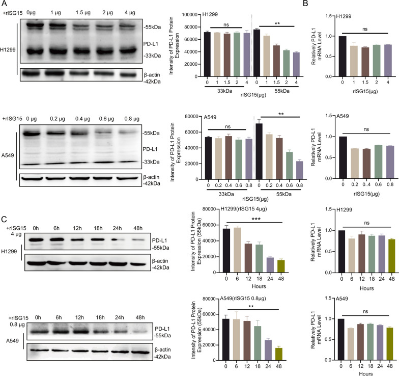
Results: ISG15 promotes the infiltration of CD4+ T lymphocytes. In vivo and in vitro experiments demonstrated that ISG15 induces CD4+ T cell proliferation and invalidity and immune responses against tumors. Mechanistically, we demonstrated that the ubiquitination-like modifying effect of ISG15 on PD-L1 increased the modification of K48-linked ubiquitin chains thus increasing the degradation rate of glycosylated PD-L1 targeting proteasomal pathway. The expression of ISG15 and PD-L1 was negatively correlated in NSCLC tissues. In addition, reduced accumulation of PD-L1 by ISG15 in mice also increased splenic lymphocyte infiltration as well as promoted cytotoxic T cell infiltration in the tumor microenvironment, thereby enhancing anti-tumor immunity.

Conclusions: The ubiquitination modification of PD-L1 by ISG15 increases K48-linked ubiquitin chain modification, thereby increasing the degradation rate of glycosylated PD-L1-targeted proteasome pathway. More importantly, ISG15 enhanced the sensitivity to immunosuppressive therapy. Our study shows that ISG15, as a post-translational modifier of PD-L1, reduces the stability of PD-L1 and may be a potential therapeutic target for cancer immunotherapy.

Keywords: Anticancer immunity; Glycosylated PD-L1; ISG15/ISGylation; Lung adenocarcinoma.


Conflict of interest statement

The authors declare that they have no conflict of interest.

Figures

Fig. 1
Fig. 1
Expression of ISG15 and lymphcytes in lung adenocarcinoma. A Activation of memory CD4+ T cells, infiltration of CD8a+ T cells and expression of CD8+ T lymphocytotoxic factors in high/low ISG15 expressing tumour tissues obtained from the TCGA collection. B Representative IHC images showing the expression of ISG15, CD3, CD4, CD8. Scale bars 100 μm. Magnification, ×100, ×400. C The relationship between CD3+, CD4+, CD8+ T cells and ISG15 was analysed by IHC staining of LUAD
Fig. 2
Fig. 2
ISG15 promotes the infiltration of immune cells in vivo. A Western blot analysis of ISG15 in LLC with over-expressed ISG15 lentiviruses. B The bar graph exhibited the tumor weight of wild-type (WT) and over-expressed ISG15 (Lv-ISG15) LLC cells in immunocompetent C57BL/6 mice. C General views of the lungs and HE staining remove from the mice in WT and LvSG15 groups. Black arrows represent images of metastasis in the lungs. Magnification, ×100, ×400. D Survival curves demonstrating the WT and Lv-ISG15 groups of C57BL/6 mice during one experimental cycle (30 days). E The curve graph exhibited the tumor volume formation of WT and Lv-ISG15 LLC cells in C57BL/6 mice. F Representative images of tumors from C57BL/6 mice with HE and immunohistochemical staining of CD31, KI67, CD3, CD4, and CD8. Scale bars 100 μm. Magnification, ×400. G Populations of tumor-infiltrating CD3+ T cells and relative ratio of CD4+IFN-γ+ and CD8+GzmB+ cells in CD3+ TILs in WT and Lv-ISG15 groups detected by flow cytometry. H Populations of CD4+ and CD8+ T cells in spleen from the mice in WT and Lv-ISG15 groups detected by flow cytometry. The data are shown as the mean ± SD. *P < 0.05, **P < 0.01, ***P < 0.001
Fig. 3
Fig. 3
ISG15 enhances the anti-tumor immunity of CD4T lymphocytes, which is related to PD-L1. A Growth of control or overexpressed with ISG15 (Lv-ISG15) or shRNA-ISG15 (Sh-ISG15) A549/ H1299 cells before and after co-culture with CD4+T cells. The bar chart below indicates the number of cells. B Western blot analysis the expression of apoptosis-related proteins in tumor cells after co-culture. C The IFN-γ secreted by CD4+ T cells co-cultured with A549 and H1299 under the three treatment conditions were evaluated by ELISA. D The expansion of CD4+ T cells co-cultured with irradiated A549/ H1299 cells or A549/ H1299 cells overexpressed/shRNA of ISG15 were assessed by CCK-8. E The secretion of ISG15 in the supernatant of tumor cells under three treatment conditions detected by ELISA. The data are shown as the mean ± SD. F Typical images of IHC staining for ISG15, granzyme B, perforin and PD-L1 in two patients with LUAD. Scale bars 100 μm. Magnification, ×400. G Number of cells positive for Granzyme B or Perforin in each high magnification (×400) field of view. H, correlation analysis of PD-L1 and ISG15 expression in 40 lung adenocarcinoma tissues. *P < 0.05, **P < 0.01, ***P < 0.001
Fig. 4
Fig. 4
ISG15 down-regulates the expression of glycosylated PD-L1. A Western blot analysis of the protein level of PD-L1 in control or Lv-ISG15 or Sh-ISG15 A549, H1299 and LLC cells. B After 5 ng/ml IFN-γ treatment of cells in the control group or Lv-ISG15 group for 12 h, mRNA levels of ISG15 were measured by quantitative qRT-PCR and protein expression levels of PD-L1 were measured by Western blot. C Cellular immunofluorescence staining was used to observe the expression of PD-L1 in Sh-ISG15 and Lv-ISG15 or the control A549/H1299 cells. D The plasma membrane PD-L1 in control or sh-ISG15 A549/H1299 cells was detected by flow cytometry. The data are shown as the mean ± SD. Ns means no significance. *P < 0.05, **P < 0.01, ***P < 0.001
Fig. 5
Fig. 5
Exogenous ISG15 also reduces the expression of glycosylated PD-L1. A Western blot and RT-qPCR were used to detect the expression of different molecular weights of PD-L1 at concentrations of 0 μg, 1 μg, 1.5 μg, 2 μg, and 4 μg of exogenous ISG15 in A549 and H1299. B H1299 or A549 treated with 4 μg or 0.8 μg of exogenous ISG15, the changes of PD-L1 with different molecular weights (left panel) and mRNA level (right panel) in 0–48 h.The data are shown as the mean ± SD. Ns means no significance. *P < 0.05, **P < 0.01, ***P < 0.001
Fig. 6
Fig. 6
ISG15 and PD-L1 form ISGylation and promote its ubiquitination for proteasome-dependent degradation. A Quantitative qRT-PCR analysis of the mRNA level of PD-L1 in Sh-ISG15 and Lv-ISG15 or the control A549/H1299 cells. B Western blotting was performed to detect the expression level of glycosylated PD-L1 at different times in A549 and H1299, after CHX (25 μg/ml) treatment. The data were normalized for comparative purposes (bottom panel). C Ubiquitination level of PD-L1 detected by Immunoprecipitation (IP) assay in Lv-ISG15 or control cells after treated with MG132 (20 μmol) for 8 h. D Immunoprecipitation of ISG15 and PD-L1 at lysine48-linked ubiquitination sites after H1299 and Pc-9 treated with 20 μM MG132 6 h. E Western blot was used to examine the effect of bafilomycin or MG132 on PD-L1 levels in the presence of CHX in Lv-ISG15 cells. F IP experiment detects the interaction between ISG15 and PD-L1. G Co-IP assay detects whether the interaction between ISG15 and PD-L1 is specific to ISGylation and whether it is dissociated by USP18 in H1299 and Pc-9 cells co-transfected ISG15 and USP18 or control plasmids
Fig. 7
Fig. 7
ISG15 elevation-induced destabilization of PD-L1 improve PD-L1-targeted immunotherapy

References

    1. Siegel RL, Miller KD, Fuchs HE, Jemal A. Cancer statistics. CA Cancer J Clin. 2021;71(2021):7–33. doi: 10.3322/caac.21654. - DOI - PubMed
    1. Socinski MA, Obasaju C, Gandara D, Hirsch FR, Bonomi P, Bunn P, Kim ES, Langer CJ, Natale RB, Novello S, Paz-Ares L, Pérol M, Reck M, Ramalingam SS, Reynolds CH, Spigel DR, Stinchcombe TE, Wakelee H, Mayo C, Thatcher N. Clinicopathologic features of advanced squamous NSCLC. J Thorac Oncol. 2016;11:1411–1422. doi: 10.1016/j.jtho.2016.05.024. - DOI - PubMed
    1. Ettinger DS, Akerley W, Borghaei H, Chang AC, Cheney RT, Chirieac LR, D'Amico TA, Demmy TL, Govindan R, Grannis FW, Jr, Grant SC, Horn L, Jahan TM, Komaki R, Kong FM, Kris MG, Krug LM, Lackner RP, Lennes IT, Loo BW, Jr, Martins R, Otterson GA, Patel JD, Pinder-Schenck MC, Pisters KM, Reckamp K, Riely GJ, Rohren E, Shapiro TA, Swanson SJ, Tauer K, Wood DE, Yang SC, Gregory K, Hughes M. Non-small cell lung cancer, version 2. J Natl Compr Canc Netw. 2013;11:645–653. doi: 10.6004/jnccn.2013.0084. - DOI - PubMed
    1. Kovacs SA, Gyorffy B. Transcriptomic datasets of cancer patients treated with immune-checkpoint inhibitors: a systematic review. J Transl Med. 2022;20:249. doi: 10.1186/s12967-022-03409-4. - DOI - PMC - PubMed
    1. Carlisle JW, Steuer CE, Owonikoko TK, Saba NF. An update on the immune landscape in lung and head and neck cancers. CA Cancer J Clin. 2020;70:505–517. doi: 10.3322/caac.21630. - DOI - PubMed

Publication types