. 2023 May 15;136(10):jcs261216.
doi: 10.1242/jcs.261216. Epub 2023 May 23.

Coupled protein quality control during nonsense-mediated mRNA decay

Affiliations

Coupled protein quality control during nonsense-mediated mRNA decay

Alison J Inglis et al. J Cell Sci. .

Abstract

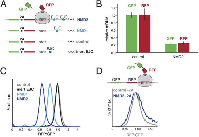
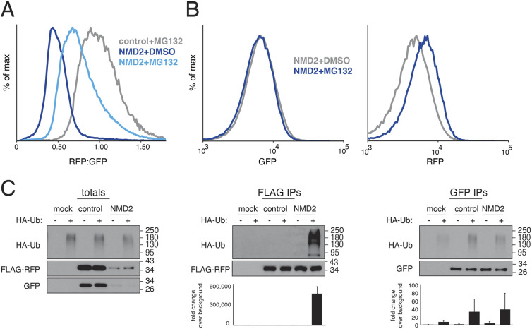
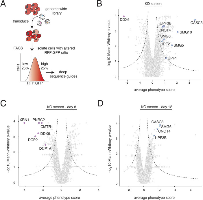
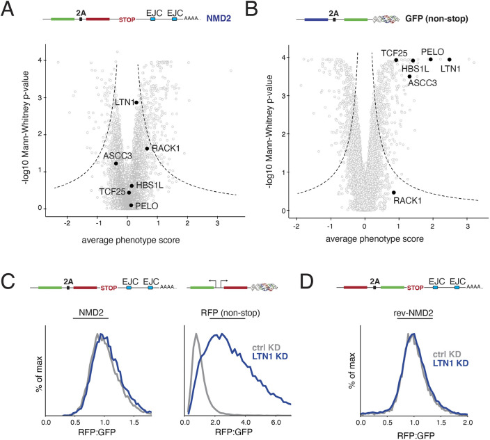
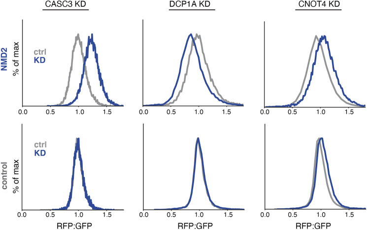
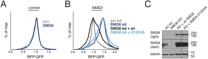
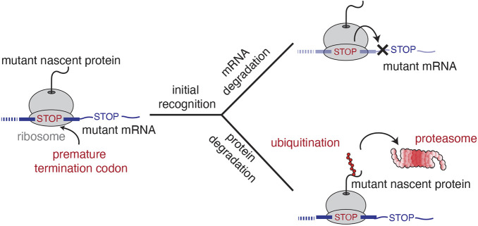
Translation of mRNAs containing premature termination codons (PTCs) results in truncated protein products with deleterious effects. Nonsense-mediated decay (NMD) is a surveillance pathway responsible for detecting PTC containing transcripts. Although the molecular mechanisms governing mRNA degradation have been extensively studied, the fate of the nascent protein product remains largely uncharacterized. Here, we use a fluorescent reporter system in mammalian cells to reveal a selective degradation pathway specifically targeting the protein product of an NMD mRNA. We show that this process is post-translational and dependent on the ubiquitin proteasome system. To systematically uncover factors involved in NMD-linked protein quality control, we conducted genome-wide flow cytometry-based screens. Our screens recovered known NMD factors but suggested that protein degradation did not depend on the canonical ribosome-quality control (RQC) pathway. A subsequent arrayed screen demonstrated that protein and mRNA branches of NMD rely on a shared recognition event. Our results establish the existence of a targeted pathway for nascent protein degradation from PTC containing mRNAs, and provide a reference for the field to identify and characterize required factors.

Keywords: Nonsense-mediated decay; Quality control; Ubiquitin-proteasome pathway; mRNA.


Conflict of interest statement

Competing interests J.S.W. declares outside interest in 5 AM Venture, Amgen, Chroma Medicine, KSQ Therapeutics, Maze Therapeutics, Tenaya Therapeutics, Tessera Therapeutics, and Third Rock Ventures. R.M.V. declares outside interest in Gate Biosciences.

Figures

Fig. 1.
Fig. 1.
Destabilization of nascent proteins from PTC-containing mRNAs. (A) Schematic of the reporter strategy used to monitor protein and mRNA degradation in NMD. GFP and RFP are encoded in a single open reading frame separated by a viral 2A sequence. Positioning an intron within the 3′ UTR results in deposition of an exon junction complex (EJC) upon splicing, triggering NMD when compared to a matched control (control). Either one or two introns derived from the β-globin gene are inserted after the stop codon (NMD1 and NMD2, respectively). To control for the documented stimulation in translation that results from the presence of an EJC (Nott et al., 2004), we created a reporter in which the intron was positioned 12 nucleotides after the stop codon, a distance insufficient for recognition as an NMD substrate (inert EJC) (Nagy and Maquat, 1998). (B) T-Rex HEK293 cell lines stably expressing either the control or the NMD2 reporter were induced with doxycycline for 24 h and the total mRNA was then purified. Relative mRNA levels were determined by RT-qPCR using two sets of primers that anneal to the very 5′ region of the GFP and 3′ region of the RFP open reading frames respectively. The results were normalized to the control and the mean±s.d. from three independent experiments is displayed. (C) T-Rex HEK293 cell lines stably expressing the indicated reporters were analyzed by flow cytometry. The ratio of RFP:GFP fluorescence, normalized to the control reporter, is depicted as a histogram and quantified in Fig.S2E. (D) HEK293T cells were transiently transfected with versions of the control and NMD2 reporters in which the 2A sequence was scrambled, resulting in tethering of both GFP and RFP to the ribosome at the stop codon. Cells were analyzed by flow cytometry after 24 h and quantified in Fig. S2E.
Fig. 2.
Fig. 2.
NMD-dependent protein degradation occurs via the ubiquitin-proteasome pathway. (A) Flow cytometry analysis of HEK293T cells transiently transfected with either the control or NMD2 reporter (Fig. 1A) and treated with the proteasome inhibitor MG132 or DMSO for 6 h. See quantification in Fig. S3A. (B) K562 CRISPRi cells stably expressing an inducible NMD2 reporter were treated with either MG132 or DMSO after induction of the reporter and analyzed by flow cytometry. Shown are the GFP (left) and RFP (right) channels for the indicated conditions displayed as a histogram, with fold change quantified in Fig. S3B. (C) HEK293T cells, stably expressing an HA-tagged ubiquitin (HA-Ub) were transiently transfected with either the control or NMD2 reporter (modified to incorporate a 3×FLAG tag at the N-terminus of RFP). To stabilize ubiquitylated species, cells were treated with MG132 prior to lysis. RFP was immunoprecipitated with anti-FLAG resin and GFP was purified using a GFP nanobody coupled to streptavidin resin (Pleiner et al., 2020). Ubiquitylated species were detected by western blotting for HA-Ub. The quantification of three independent replicates is shown below, with the mean±s.d. plotted.
Fig. 3.
Fig. 3.
Systematic characterization of factors required for NMD-coupled protein quality control. (A) Schematic of the workflow used to carry out the FACS-based reporter screens to identify factors involved in NMD-linked nascent chain degradation. K562 reporter cell lines contained a tet-inducible NMD2 reporter and were infected with either a whole-genome CRISPRi sgRNA library or a CRISPR-KO library. Reporter expression was induced with doxycycline for 24 h prior to cell sorting. Cells were sorted based on ratiometric changes in RFP relative to GFP, and the sgRNAs expressed in those cells were identified using deep sequencing. The CRISPR knockout screen was sorted on days 8, 10 and 12 post-library infection to account for drop out of essential genes. The CRISPRi screen was sorted on day 8. (B) Volcano plot of the RFP:GFP stabilization phenotype (log2 for the three strongest sgRNAs per gene) and Mann–Whitney P-values from the genome-wide CRISPRi screen, with each point representing one gene. Genes falling outside the dashed lines are statistically significant. Each gray point represents a gene. Notable hits causing an increase in the RFP to GFP ratio are shown in light blue and include known NMD factors (UPF1, UPF2, UPF3B, SMG5, SMG6 and SMG10), the splicing factor CASC3 and the E3 ligase CNOT4. DDX6, a known suppressor of NMD, which causes a lower RFP to GFP ratio, is shown in purple. (C) Volcano plot as in B for the genome-wide CRISPR knock-out screen sorted at the day 8 timepoint. In purple are highlighted factors that cause a decrease in RFP relative to GFP. These include genes involved in mRNA de-capping (PNRC1, CMTR1, DCP1A and DCP2), DDX6 and the 5′-3′ exonuclease XRN1. (D) As in C but for day 12. In blue are shown known NMD factors (CASC3, SMG6 and UPF3B) and the E3 ligase CNOT4. Highlighted genes can be tracked across the 3 days of screening in Fig. S4A. The full datasets can be found in Tables S1 and S2.
Fig. 4.
Fig. 4.
NMD-linked protein degradation is not mediated by the canonical RQC pathway. (A) Volcano plot of the NMD2 reporter CRISPRi screen as in Fig. 3A. Highlighted in black are factors involved in the canonical RQC (LTN1, RACK1, ASCC3, HBS1L, TCF25 and PELO). (B) For comparison, RQC factors are highlighted in black on a volcano plot for an earlier CRISPRi screen using a non-stop reporter (consisting of a BFP, a viral 2A skipping sequence and a GFP conjugated a triple helix moiety to stabilize the mRNA transcript, which would usually be degraded due to the lack of a stop codon) conducted using identical conditions as in A (Hickey et al., 2020). (C) K562 CRISPRi cells stably expressing either an inducible NMD2 reporter or a constitutively expressed non-stop reporter with matched GFP and RFP fluorophores (in this case, a functionally equivalent non-stop reporter with two separate promoters, one driving GFP, and the other RFP conjugated to the triple helix moiety; as in Hickey et al., 2020) were infected with a sgRNA targeting the E3 ligase LTN1. The RFP to GFP ratios for NMD2, and the GFP to RFP ratio for the non-stop reporter as determined by flow cytometry are displayed as a histogram and are quantified in Fig. S4B. (D) K562 CRISPRi cells expressing a reversed version of the NMD2 reporter (rev-NMD2) were infected with an sgRNA against LTN1 and analyzed as in C and are quantified in Fig. S4C.
Fig. 5.
Fig. 5.
Validation of factors involved in NMD-coupled protein quality control. Factors of interest were individually depleted by sgRNA in K562 Zim3 CRISPRi cells expressing the indicated reporter. Displayed are the RFP:GFP ratios for the NMD2 (top) and control (bottom) reporters as determined by flow cytometry; see Fig. S5A,C,E for quantification. Similar results were obtained using reverse reporters as in Fig. S5B,D,F.
Fig. 6.
Fig. 6.
NMD-coupled protein quality control is dependent on endonucleolytic cleavage of the mRNA by SMG6. (A) HEK293T cells were treated with siRNA against SMG6 for 48 h, then were transiently transfected with the control reporter. The cells were analyzed by flow cytometry after 24 h (see quantification in Fig. S7A). (B) HEK293T cells were treated with siRNA against SMG6 for 48 h, then were transiently transfected with an siRNA-resistant version of either wild-type SMG6 or a PIN domain mutant (D1353A) along with the NMD2 reporter. The cells were analyzed by flow cytometry after 24 h (see quantification in Fig. S7B). Similar results were obtained with reverse reporters (Fig. S7D). (C) Levels of SMG6 in the samples from B were analyzed by western blotting against SMG6. Image representative of three repeats.
Fig. 7.
Fig. 7.
Model for NMD-coupled protein quality control. When the ribosome reaches the stop codon, NMD substrates are recognized in a context-dependent manner. These early recognition steps initiate two parallel pathways that rely on distinct suites of factors to concomitantly degrade the mRNA and nascent protein. We postulate that NMD-coupled quality control results in ubiquitylation of the nascent protein prior to its release from the ribosome where it subsequently degraded by the proteasome.

Similar articles

Cited by

References

    1. Alexandrov, A., Shu, M.-D. and Steitz, J. A. (2017). Fluorescence amplification method for forward genetic discovery of factors in human mRNA degradation. Mol. Cell 65, 191-201. 10.1016/j.molcel.2016.11.032 - DOI - PMC - PubMed
    1. Amrani, N., Ganesan, R., Kervestin, S., Mangus, D. A., Ghosh, S. and Jacobson, A. (2004). A faux 3′-UTR promotes aberrant termination and triggers nonsense-mediated mRNA decay. Nature 432, 112-118. 10.1038/nature03060 - DOI - PubMed
    1. Anczuków, O., Ware, M. D., Buisson, M., Zetoune, A. B., Stoppa-Lyonnet, D., Sinilnikova, O. M. and Mazoyer, S. (2008). Does the nonsense-mediated mRNA decay mechanism prevent the synthesis of truncated BRCA1, CHK2, and p53 proteins? Hum. Mutat. 29, 65-73. 10.1002/humu.20590 - DOI - PubMed
    1. Andersen, C. B. F., Ballut, L., Johansen, J. S., Chamieh, H., Nielsen, K. H., Olliveira, C. L. P., Pedersen, J. S., Seraphin, B., Le Hir, H. E. and Andersen, G. R. (2006). Structure of the exon junction core complex with a trapped DEAD-box ATPase bound RNA. Science 313, 1968-1972. 10.1126/science.1131981 - DOI - PubMed
    1. Arribere, J. A. and Fire, A. Z. (2018). Nonsense mRNA suppression via nonstop decay. Elife 7, e33292. 10.7554/eLife.33292 - DOI - PMC - PubMed

Publication types