. 2023 Jun 7;14(11):1963-1970.
doi: 10.1021/acschemneuro.3c00096. Epub 2023 May 23.

PROTAC-Induced Glycogen Synthase Kinase 3β Degradation as a Potential Therapeutic Strategy for Alzheimer's Disease

Affiliations

PROTAC-Induced Glycogen Synthase Kinase 3β Degradation as a Potential Therapeutic Strategy for Alzheimer's Disease

Melissa Guardigni et al. ACS Chem Neurosci. .

Abstract

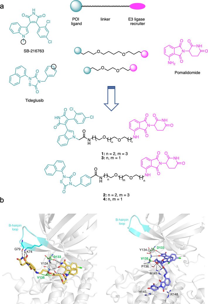
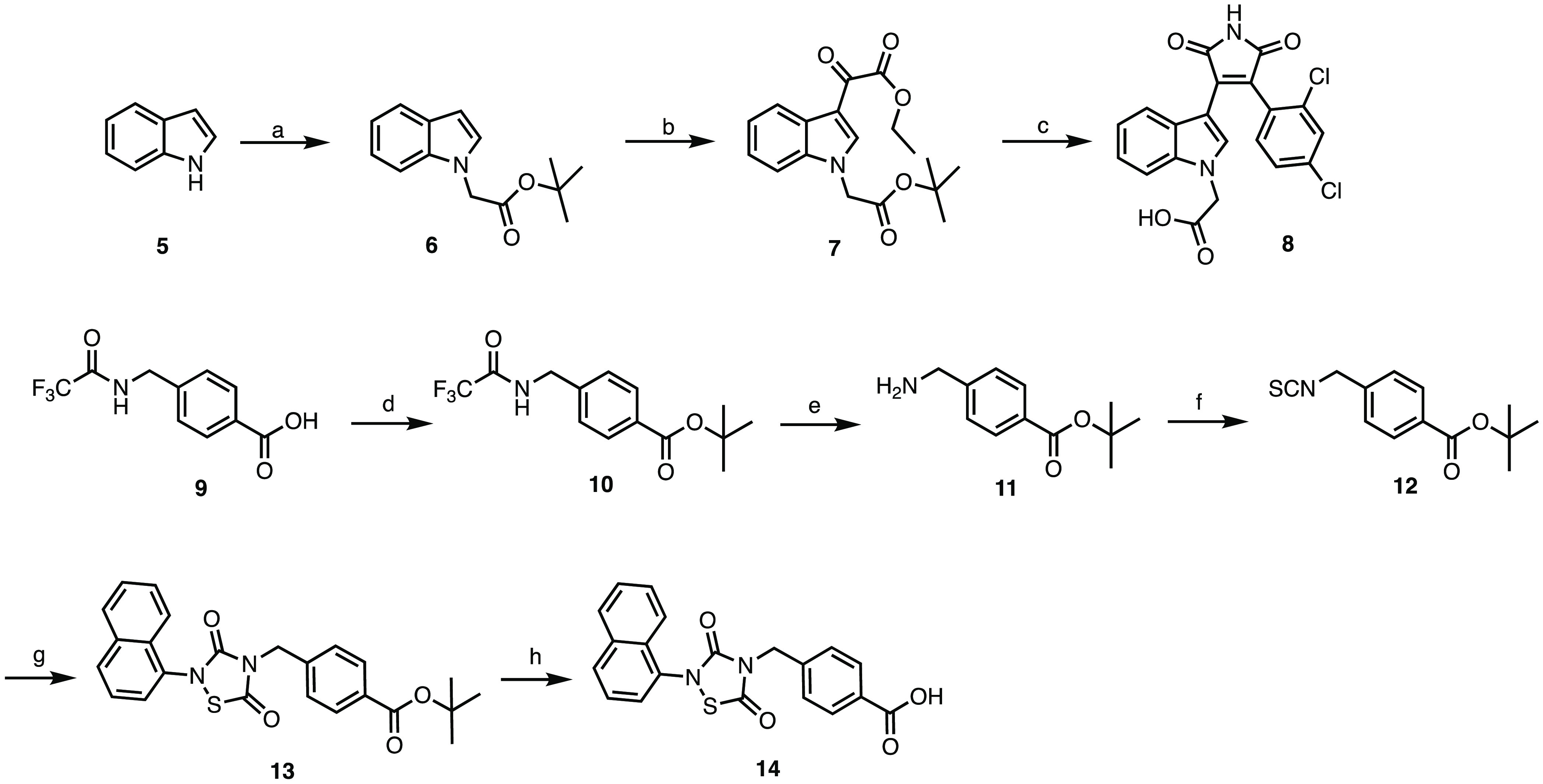
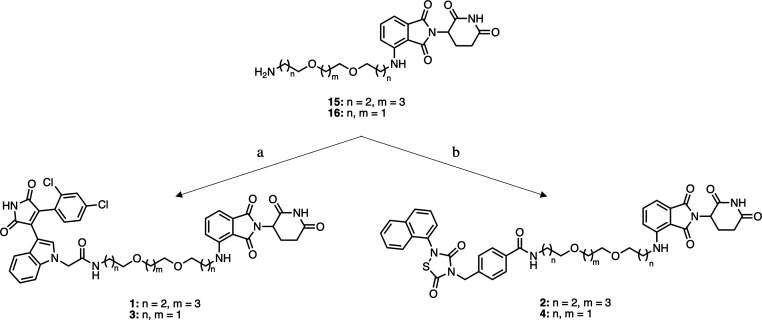
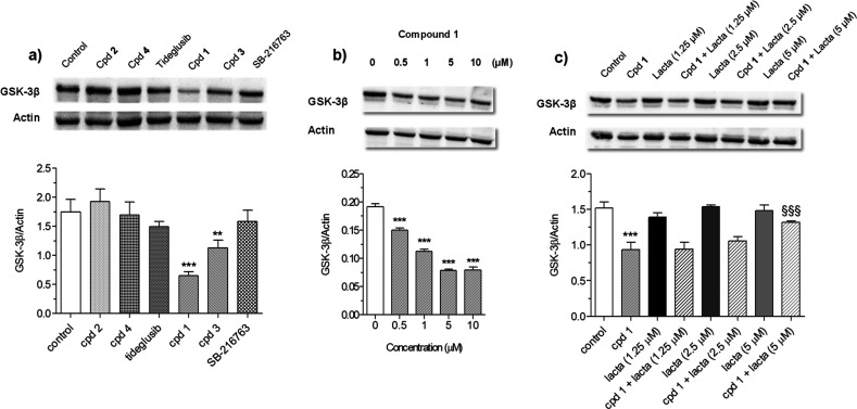
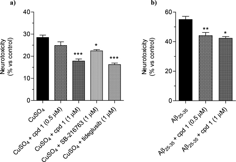
Glycogen synthase kinase 3β (GSK-3β) is a serine/threonine kinase and an attractive therapeutic target for Alzheimer's disease. Based on proteolysis-targeting chimera (PROTAC) technology, a small set of novel GSK-3β degraders was designed and synthesized by linking two different GSK-3β inhibitors, SB-216763 and tideglusib, to pomalidomide, as E3 recruiting element, through linkers of different lengths. Compound 1 emerged as the most effective PROTAC being nontoxic up to 20 μM to neuronal cells and already able to degrade GSK-3β starting from 0.5 μM in a dose-dependent manner. PROTAC 1 significantly reduced the neurotoxicity induced by Aβ25-35 peptide and CuSO4 in SH-SY5Y cells in a dose-dependent manner. Based on its encouraging features, PROTAC 1 may serve as a starting point to develop new GSK-3β degraders as potential therapeutic agents.

Keywords: Alzheimer’s disease; chemical knockdown; glycogen synthase kinase 3β; protein degradation; proteolysis targeting chimeras.


Conflict of interest statement

The authors declare no competing financial interest.

Figures

Figure 1
Figure 1
(a) Design strategy leading to GSK-3β-directed PROTAC 14. Black circles represent tethering points. (b) MD-extracted structure of the 1–GSK-3β (left) and 3–GSK-3β (right) complexes showing H-bond contacts with kinase residues.
Scheme 1
Scheme 1. Synthesis of the Precursors 8 and 14
Reagents and conditions. (a) tert-butyl bromoacetate, K2CO3, acetone, reflux, 12 h, 50% yield; (b) ethyl chlorooxoacetate, diethyl ether, rt, 12 h, N2, 42% yield; (c) 2-(2,4-dichlorophenyl)acetamide, KOtBu, DMF, rt, 12 h, N2, 39% yield; (d) tert-butanol, EDCI, DMAP, THF, rt, 12 h, 77% yield; (e) K2CO3, H2O/MeOH, rt, 12 h, 79% yield; (f) 1,1′-thiocarbonyldi-2(1H)-pyridone, DCM, rt, 12 h, 55% yield; (g) naphthyl isocyanate, sulfuryl dichloride, THF, rt, 12 h, N2 then air, THF, rt, 30 min, 57% yield; (h) TFA, DCM, rt, 12 h, 81% yield.
Scheme 2
Scheme 2. Synthesis of the Target Compounds 14
Reagents and conditions. (a) 8, EDCI, HOBT, DIPEA, DMF, rt, 12 h, 32% yield for 1, 25% yield for 3; (b) 14, EDCI, HOBT, DIPEA, DMF, rt, 12 h, 48% yield for 2, 44% yield for 4.
Figure 2
Figure 2
Degradation GSK-3β protein by compounds 14, SB-216763, and tideglusib in SH-SY5Y cells. (a) GSK-3β protein level after 48 h of treatment with all compounds (10 μM); (b) GSK-3β protein level after 48 h of treatment with compound 1 (0.5–10 μM); (c) GSK-3β protein level after 24 h of treatment with compound 1 (10 μM) and lactacystin (1.25–5 μM). GSK-3β protein level was measured by Western blotting. Data are expressed as mean ± SEM of three independent experiments (**p < 0.01 and ***p < 0.001 vs untreated cells, §§§p < 0.001 vs cells treated with compound 1, at one-way ANOVA with Dunnett or Bonferroni post hoc test).
Figure 3
Figure 3
Compound 1 reduced the neurotoxicity induced by CuSO4 and Aβ25–35 in SH-SY5Y cells. (a) Cells were incubated for 24 h with compound 1 (0.5–1 μM), SB-216763, and tideglusib (1 μM) in the presence of CuSO4 (150 μM); (b) cells were incubated for 2 h with compound 1 (0.5–1 μM) and further 3 h in the presence of Aβ25–35 (10 μM). At the end of incubation, the cell viability was measured by MTT assay as described in the Methods section. Data are reported as mean ± SEM of three independent experiments (*p < 0.05 and ***p < 0.001 vs cells treated with CuSO4; *p < 0.05 and **p < 0.01 vs cells treated with Aβ25–35 at one-way ANOVA with Dunnett post hoc test).

References

    1. Beurel E.; Grieco S. F.; Jope R. S. Glycogen synthase kinase-3 (GSK3): regulation, actions, and diseases. Pharmacol Ther 2015, 148, 114–131. 10.1016/j.pharmthera.2014.11.016. - DOI - PMC - PubMed
    1. Salcedo-Tello P.; Ortiz-Matamoros A.; Arias C. GSK3 Function in the Brain during Development, Neuronal Plasticity, and Neurodegeneration. Int. J. Alzheimers Dis 2011, 2011, 189728.10.4061/2011/189728. - DOI - PMC - PubMed
    1. Leroy K.; Yilmaz Z.; Brion J. P. Increased level of active GSK-3beta in Alzheimer’s disease and accumulation in argyrophilic grains and in neurones at different stages of neurofibrillary degeneration. Neuropathol Appl. Neurobiol 2007, 33 (1), 43–55. 10.1111/j.1365-2990.2006.00795.x. - DOI - PubMed
    1. Lauretti E.; Dincer O.; Praticò D. Glycogen synthase kinase-3 signaling in Alzheimer’s disease. Biochim Biophys Acta Mol. Cell Res. 2020, 1867 (5), 118664.10.1016/j.bbamcr.2020.118664. - DOI - PMC - PubMed
    1. De Simone A.; Tumiatti V.; Andrisano V.; Milelli A. Glycogen Synthase Kinase 3β: A New Gold Rush in Anti-Alzheimer’s Disease Multitarget Drug Discovery?. J. Med. Chem. 2021, 64 (1), 26–41. 10.1021/acs.jmedchem.0c00931. - DOI - PMC - PubMed

Publication types

Substances