. 2023 May 22;5(21):CASE232.
doi: 10.3171/CASE232. Print 2023 May 22.

Metastatic secondary gliosarcoma: patient series

Affiliations

Metastatic secondary gliosarcoma: patient series

Shivani Baisiwala et al. J Neurosurg Case Lessons. .

Abstract

Background: Gliosarcoma is a rare and highly malignant cancer of the central nervous system with the ability to metastasize. Secondary gliosarcoma, or the evolution of a spindle cell-predominant tumor after the diagnosis of a World Health Organization grade IV glioblastoma, has also been shown to metastasize. There is little information on metastatic secondary gliosarcoma.

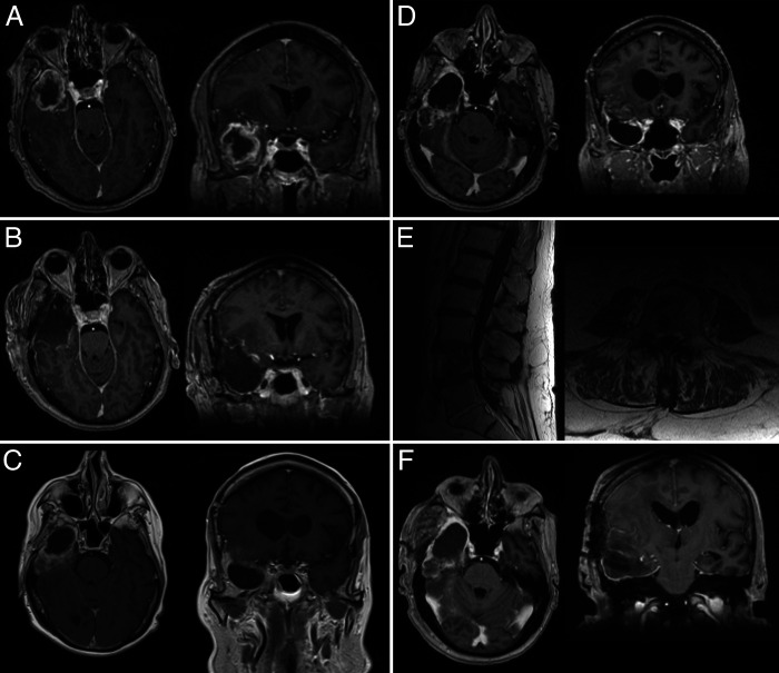
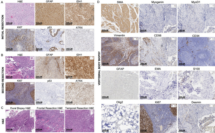
Observations: The authors present a series of 7 patients with previously diagnosed glioblastoma presenting with recurrent tumor and associated metastases with repeat tissue diagnosis consistent with gliosarcoma. The authors describe the clinical, imaging, and pathological characteristics in addition to carrying out a systematic review on metastases in secondary gliosarcoma.

Lessons: The present institutional series and the systematic review of the literature show that metastatic secondary gliosarcoma is a highly aggressive disease with a poor prognosis.

Keywords: cancer; malignant brain tumor; metastasis; secondary gliosarcoma.


Conflict of interest statement

Disclosures Dr. Liau reported being a member of the Board of Directors for ClearPoint Neuro, Inc. outside the submitted work.

Figures

FIG. 1.
FIG. 1.
A: Brain MRI shows a right temporal lesion with necrosis and edema. B: Brain MRI obtained postoperatively shows gross-total resection of the lesion. C: Brain MRI shows recurrent lesion toward the posterior aspect of the resection cavity. D: Brain MRI obtained 2 weeks following admission shows extensive lesions. E: Lumbar spine MRI shows lesions near the conus and cauda equina and in the vertebral bodies. F: Brain MRI shows total resection of the recurrent temporal lesion.
FIG. 2.
FIG. 2.
A: Pathologic stains from initial resection of the glioblastoma. B: Pathologic stains from resection of the recurrent lesion determined to be gliosarcoma. C: Hematoxylin and eosin (H&E) stains from various aspects of the recurrent lesion, including the frontal dural lesions, frontal part of the recurrent tumor, and temporal aspect of recurrent tumor. D: Various IHC stains performed on the vertebral biopsy sample obtained.
FIG. 3.
FIG. 3.
PRISMA flow diagram. The PubMed database was searched for studies of gliosarcoma and metastases. Seventy-five studies met initial criteria and were reviewed for inclusion/exclusion criteria. This review identified 8 cases for inclusion in the analysis.

References

    1. Lutterbach J, Guttenberger R, Pagenstecher A. Gliosarcoma: a clinical study. Radiother Oncol. 2001;61(1):57–64. - PubMed
    1. Cachia D, Kamiya-Matsuoka C, Mandel JJ, et al. Primary and secondary gliosarcomas: clinical, molecular and survival characteristics. J Neurooncol. 2015;125(2):401–410. - PMC - PubMed
    1. Zaki MM, Mashouf LA, Woodward E, et al. Genomic landscape of gliosarcoma: distinguishing features and targetable alterations. Sci Rep. 2021;11(1):18009. - PMC - PubMed
    1. Han SJ, Yang I, Ahn BJ, et al. Clinical characteristics and outcomes for a modern series of primary gliosarcoma patients. Cancer. 2010;116(5):1358–1366. - PubMed
    1. Han SJ, Yang I, Tihan T, Chang SM, Parsa AT. Secondary gliosarcoma: a review of clinical features and pathological diagnosis. J Neurosurg. 2010;112(1):26–32. - PubMed