Meta-Analysis
. 2023 Jun;307(5):e222639.
doi: 10.1148/radiol.222639. Epub 2023 May 23.

Standalone AI for Breast Cancer Detection at Screening Digital Mammography and Digital Breast Tomosynthesis: A Systematic Review and Meta-Analysis

Affiliations
Meta-Analysis

Standalone AI for Breast Cancer Detection at Screening Digital Mammography and Digital Breast Tomosynthesis: A Systematic Review and Meta-Analysis

Jung Hyun Yoon et al. Radiology. 2023 Jun.

Abstract

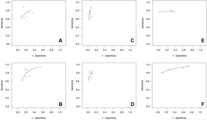
Background There is considerable interest in the potential use of artificial intelligence (AI) systems in mammographic screening. However, it is essential to critically evaluate the performance of AI before it can become a modality used for independent mammographic interpretation. Purpose To evaluate the reported standalone performances of AI for interpretation of digital mammography and digital breast tomosynthesis (DBT). Materials and Methods A systematic search was conducted in PubMed, Google Scholar, Embase (Ovid), and Web of Science databases for studies published from January 2017 to June 2022. Sensitivity, specificity, and area under the receiver operating characteristic curve (AUC) values were reviewed. Study quality was assessed using the Quality Assessment of Diagnostic Accuracy Studies 2 and Comparative (QUADAS-2 and QUADAS-C, respectively). A random effects meta-analysis and meta-regression analysis were performed for overall studies and for different study types (reader studies vs historic cohort studies) and imaging techniques (digital mammography vs DBT). Results In total, 16 studies that include 1 108 328 examinations in 497 091 women were analyzed (six reader studies, seven historic cohort studies on digital mammography, and four studies on DBT). Pooled AUCs were significantly higher for standalone AI than radiologists in the six reader studies on digital mammography (0.87 vs 0.81, P = .002), but not for historic cohort studies (0.89 vs 0.96, P = .152). Four studies on DBT showed significantly higher AUCs in AI compared with radiologists (0.90 vs 0.79, P < .001). Higher sensitivity and lower specificity were seen for standalone AI compared with radiologists. Conclusion Standalone AI for screening digital mammography performed as well as or better than radiologists. Compared with digital mammography, there is an insufficient number of studies to assess the performance of AI systems in the interpretation of DBT screening examinations. © RSNA, 2023 Supplemental material is available for this article. See also the editorial by Scaranelo in this issue.


Conflict of interest statement

Disclosures of conflicts of interest: J.H.Y. No relevant relationships. F.S. Grants or contracts from Region Stockholm and Swedish Breast Cancer Association; speaker fees from Lunit. P.A.T.B. Secretary general and executive board member of European Society of Breast Imaging (EUSOBI). E.F.C. Grants or contracts from iCAD, OM1, National Institutes of Health (NIH), and National Cancer Institute; consulting fees from iCAD; lecture payments from Hologic, MedScape, and Aunt Minnie; meeting and/or travel support from the RSNA; University of Pennsylvania patents planned, issued, or pending; leadership role at Society of Breast Imaging. F.J.G. Grants or contracts from Lunit, Bayer, Hologic, and GE Healthcare; consulting fees from Alphabet and Kheiron; lecture payment and receipt of equipment from GE Healthcare; president, EUSOBI. C.D.L. Grants or contracts from Breast Cancer Research Foundation, NIH, GE Healthcare, and Hologic; consulting fees from Clairity; meeting travel support from GE Healthcare, Hologic, and Clairity; co-founder of Clairity. E.A.M. Grants or contracts from Komen; lecture payments from Bayer and Guerbet; advisory board, Bracco and Kheiron; stockholder, Kheiron and Reveal Pharma. L.A.M. Research funding from IBM and Cepheid; consulting fees and/or honoraria from Hologic; chair of Diversity and Inclusion Committee, Maryland Radiological Society. R.M.N. Institutional grants or contracts from GE Healthcare, Koios Medical, Hologic, and iCAD; royalties from Hologic; consulting fees from maiData; advisory board, iCAD. N.S. Lecture payments from BARD and Hologic. I.V. No relevant relationships. L.M. Editor of Radiology; grants or contracts from Siemens Foundation, Gordon and Betty Moore Foundation, Mary Kay Foundation, and Google; consulting fees from Guerbet and iCAD; meeting and/or travel support from British Society of Breast Radiology and European Society of Breast Imaging; board member of Society of Breast Imaging and International Society for Magnetic Resonance in Medicine; stockholder, Lunit. R.M.M. Associate editor of breast imaging for Radiology; grants or contracts from Dutch Research Council, Dutch Cancer Society, European Union, Siemens Healthineers, Bayer Healthcare, Beckton-Dickinson, Medtronic, Screenpoint Medical, Lunit, Koning, and PA Imaging; royalties from Elsevier; consulting fees from Bayer, Guerbet, Siemens, Screenpoint Medical, and Beckton-Dickinson; lecture payments from Siemens, Bayer, and Beckton-Dickinson; data safety monitoring board for SMALL trial; executive board member, EUSOBI; research committee member, ESR Advisory; editorial board member for European Radiology, advisory board member, Dutch Cancer Society; clinical advisory board member, Oncode Institute.

Figures

None
Graphical abstract
Preferred Reporting Items for Systematic Reviews and Meta-Analyses
(PRISMA) flow diagram shows study inclusion and exclusion. One of the 16
included studies had data for both interpretation of digital mammography in
a historic cohort and interpretation of digital breast tomosynthesis (DBT).
AI = artificial intelligence.
Figure 1:
Preferred Reporting Items for Systematic Reviews and Meta-Analyses (PRISMA) flow diagram shows study inclusion and exclusion. One of the 16 included studies had data for both interpretation of digital mammography in a historic cohort and interpretation of digital breast tomosynthesis (DBT). AI = artificial intelligence.
Horizontal bar graphs show the assessment results for (A, B) risk of
bias and (C, D) concerns for application in (E) Quality Assessment of
Diagnostic Accuracy Studies 2 (QUADAS-2) and QUADAS Comparative (QUADAS-C)
according to the percentages of studies (n = 16) with low, unclear, and high
levels of risk or applicability concerns.
Figure 2:
Horizontal bar graphs show the assessment results for (A, B) risk of bias and (C, D) concerns for application in (E) Quality Assessment of Diagnostic Accuracy Studies 2 (QUADAS-2) and QUADAS Comparative (QUADAS-C) according to the percentages of studies (n = 16) with low, unclear, and high levels of risk or applicability concerns.
Forest plots show pooled estimates for the (A) six reader studies, (B)
seven studies using historic reads, and (C) four digital breast
tomosynthesis (DBT) studies. Reader studies are defined as studies in which
radiologists interpreted mammograms for the study, and historic reads are
defined as studies using retrospective interpretation in clinical practice.
AI = artificial intelligence, FN = false negative, FP = false positive, TN =
true negative, TP = true positive.
Figure 3:
Forest plots show pooled estimates for the (A) six reader studies, (B) seven studies using historic reads, and (C) four digital breast tomosynthesis (DBT) studies. Reader studies are defined as studies in which radiologists interpreted mammograms for the study, and historic reads are defined as studies using retrospective interpretation in clinical practice. AI = artificial intelligence, FN = false negative, FP = false positive, TN = true negative, TP = true positive.
Summary receiver operating characteristic curves show performances of
radiologists (top) and artificial intelligence (bottom) according to (A, B)
reader studies, (C, D) historic reads, and (E, F) digital breast
tomosynthesis (DBT) studies. Lines represent summary receiver operating
characteristic curves and circles represent individual study results. Reader
studies are defined as studies in which radiologists interpreted mammograms
for the study, and historic reads are defined as studies using retrospective
interpretation in clinical practice.
Figure 4:
Summary receiver operating characteristic curves show performances of radiologists (top) and artificial intelligence (bottom) according to (A, B) reader studies, (C, D) historic reads, and (E, F) digital breast tomosynthesis (DBT) studies. Lines represent summary receiver operating characteristic curves and circles represent individual study results. Reader studies are defined as studies in which radiologists interpreted mammograms for the study, and historic reads are defined as studies using retrospective interpretation in clinical practice.

Comment in

References

    1. Independent UK Panel on Breast Cancer Screening . The benefits and harms of breast cancer screening: an independent review . Lancet 2012. ; 380 ( 9855 ): 1778 – 1786 . - PubMed
    1. Gøtzsche PC , Jørgensen KJ . Screening for breast cancer with mammography . Cochrane Database Syst Rev 2013. ; 2013 ( 6 ): CD001877 . - PMC - PubMed
    1. Tabár L , Yen AM , Wu WY , et al. . Insights from the breast cancer screening trials: how screening affects the natural history of breast cancer and implications for evaluating service screening programs . Breast J 2015. ; 21 ( 1 ): 13 – 20 . - PubMed
    1. International Agency for Research on Cancer . Breast cancer screening . In: IARC handbooks of cancer prevention , Vol 15 . IARC Press; , 2015. .
    1. Hoff SR , Abrahamsen AL , Samset JH , Vigeland E , Klepp O , Hofvind S . Breast cancer: missed interval and screening-detected cancer at full-field digital mammography and screen-film mammography-- results from a retrospective review . Radiology 2012. ; 264 ( 2 ): 378 – 386 . - PubMed