. 2023 Dec;18(12):2307-2318.
doi: 10.1007/s11548-023-02929-8. Epub 2023 May 23.

Impact of bone and cartilage segmentation from CT and MRI on both bone forearm osteotomy planning

Affiliations

Impact of bone and cartilage segmentation from CT and MRI on both bone forearm osteotomy planning

Ruurd J A Kuiper et al. Int J Comput Assist Radiol Surg. 2023 Dec.

Abstract

Introduction: The use of MRI scans for pre-operative surgical planning of forearm osteotomies provides additional information of joint cartilage and soft tissue structures and reduces radiation exposure in comparison with the use of CT scans. In this study, we investigated whether using 3D information obtained from MRI with and without cartilage information leads to a different outcome of pre-operative planning.

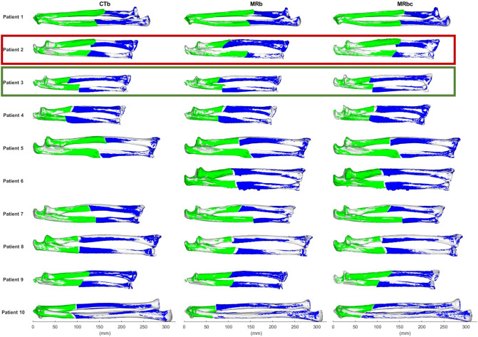
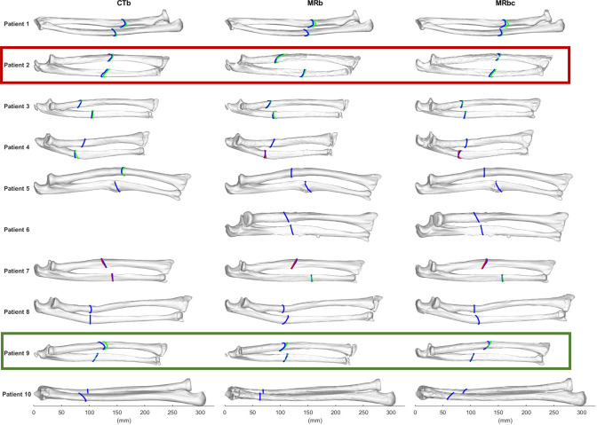
Methods: Bilateral CT and MRI scans of the forearms of 10 adolescent and young adult patients with a unilateral bone deformation were acquired in a prospective study. The bones were segmented from CT and MRI, and cartilage only from MRI. The deformed bones were virtually reconstructed, by registering the joint ends to the healthy contralateral side. An optimal osteotomy plane was determined that minimized the distance between the resulting fragments. This process was performed in threefold: using the CT and MRI bone segmentations, and the MRI cartilage segmentations.

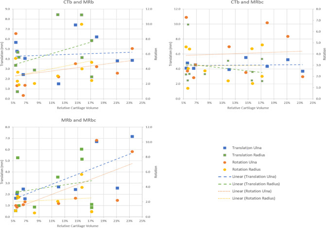
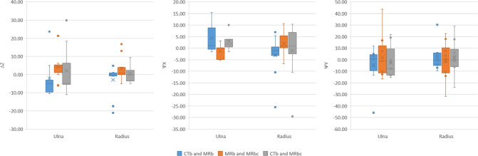
Results: Comparison of bone segmentation from MRI and CT scan resulted in a 0.95 ± 0.02 Dice Similarity Coefficient and 0.42 ± 0.07 mm Mean Absolute Surface Distance. All realignment parameters showed excellent reliability across the different segmentations. However, the mean differences in translational realignment between CT and MRI bone segmentations (4.5 ± 2.1 mm) and between MRI bone and MRI bone and cartilage segmentations (2.8 ± 2.1 mm) were shown to be clinically and statistically significant. A significant positive correlation was found between the translational realignment and the relative amount of cartilage.

Conclusion: This study indicates that although bone realignment remained largely similar when using MRI with and without cartilage information compared to using CT, the small differences in segmentation could induce statistically and clinically significant differences in the osteotomy planning. We also showed that endochondral cartilage might be a non-negligible factor when planning osteotomies for young patients.

Keywords: Bone; CT; MRI; Orthopaedic planning; Osteotomy.


Conflict of interest statement

This work was partially funded by the Netherlands Organization for Scientific Research (NWO) and is part of the Applied and Engineering Sciences research program, Project Number 15479. Peter Seevinck is minority shareholder and CSO at MRIguidance B.V. Harrie Weinans is minority shareholder at UPlanner B.V. Ralph Sakkers is minority shareholder at UPlanner B.V. and independent reviewer at MRIGuidance B.V. Max Viergever is Board Member, MICCAI Society. The other authors have no relevant interests to disclose.

Figures

Fig. 1
Fig. 1
Top row: example of the MRI (left) and CT (right) of lower right arm. Middle row: MRI and CT overlaid with radius (brown) and ulna (yellow) bone segmentation. Bottom row: MRI overlaid with bone and cartilage (blue) segmentation
Fig. 2
Fig. 2
Step-by-step overview of the automatic osteotomy planning workflow. The deformed radius and ulna (white) were aligned to the mirrored healthy contralateral radius and ulna (green) by registering the joints and subsequently optimizing the osteotomy plane to minimize the distance between the proximal and distal bone fragments. This example used a bone segmentation derived from MRI
Fig. 3
Fig. 3
Relative cartilage volume against the age of each patient at the time of the MRI scan. Both cartilage and bone segmentation were acquired from the MRI scan. An exponential regression line is fitted to show the decrease in relative cartilage volume by age
Fig. 4
Fig. 4
Translational and rotational realignment differences when planning the osteotomy using CTb, MRb, and MRbc, plotted against the relative cartilage volumes of the patients. Linear trendlines have been fitted
Fig. 5
Fig. 5
The rotational (top) and translational (bottom) differences between the CTb and MRb (left) and MRb and MRbc (right) in relative bone realignment after osteotomy are shown. The mean and standard deviation of the differences is compared to residual error after osteotomy surgery as reported by Vlachopoulos et al. [24].
Fig. 6
Fig. 6
Illustration of the difference in simulated bone reconstruction in the radius and ulna between CTb, MRb, and MRbc segmentations for all patients. The proximal (blue) and distal (green) fragment of the deformed bone after the planned reconstruction are overlayed on the mirrored healthy contralateral side (white). The CT scan of Patient 6 was incomplete and thus not included. Red rectangle: patient with largest relative rotational difference in ulna between MRb and MRbc. Green rectangle: patient with largest relative translational difference in ulna between MRb and MRbc
Fig. 7
Fig. 7
Illustration of the difference in optimal computed osteotomy location and orientation in the radius and ulna between CTb, MRb, and MRbc segmentations for all patients. The blue line indicates the primary osteotomy cut. Where necessary, the secondary osteotomy cut is visible. The secondary cut is shown in green if it does not intersect the primary cut, or in red if it does overlap the primary cut. The CT scan of Patient 6 was incomplete and thus not included. Red box: patient with largest difference in (radius) osteotomy location between MRb and Mbc. Green box: patient with highest RCV
Fig. 8
Fig. 8
Differences in location (ΔZ) and orientation (ψX and ψZ) of the osteotomy plane between the different methods of planning; on CTb, MRb, and MRbc

Similar articles

Cited by

References

    1. Richard MJ, Ruch DS, Aldridge JM. Malunions and nonunions of the forearm. Hand Clin. 2007;23:235–243. doi: 10.1016/j.hcl.2007.02.005. - DOI - PubMed
    1. Mulders MAM, D’Ailly PN, Cleffken BI, Schep NWL. Corrective osteotomy is an effective method of treating distal radius malunions with good long-term functional results. Injury. 2017;48:731–737. doi: 10.1016/j.injury.2017.01.045. - DOI - PubMed
    1. Saravi B, Lang G, Steger R, Vollmer A, Zwingmann J. Corrective osteotomy of upper extremity malunions using three-dimensional planning and patient-specific surgical guides: Recent advances and perspectives. Front Surg. 2021;8:1–7. doi: 10.3389/fsurg.2021.615026. - DOI - PMC - PubMed
    1. Bauer DE, Zimmermann S, Aichmair A, Hingsammer A, Schweizer A, Nagy L, Fürnstahl P. Conventional versus computer-assisted corrective osteotomy of the forearm: a retrospective analysis of 56 consecutive cases. J Hand Surg Am. 2017;42:447–455. doi: 10.1016/j.jhsa.2017.03.024. - DOI - PubMed
    1. de Muinck Keizer RJO, Lechner KM, Mulders MAM, Schep NWL, Eygendaal D, Goslings JC. Three-dimensional virtual planning of corrective osteotomies of distal radius malunions: a systematic review and meta-analysis. Strateg Trauma Limb Reconstr. 2017;12:77–89. doi: 10.1007/s11751-017-0284-8. - DOI - PMC - PubMed

LinkOut - more resources