. 2023 Jul 10;8(13):e134685.
doi: 10.1172/jci.insight.134685.

Specific regulation of mechanical nociception by Gβ5 involves GABA-B receptors

Affiliations

Specific regulation of mechanical nociception by Gβ5 involves GABA-B receptors

Mritunjay Pandey et al. JCI Insight. .

Abstract

Mechanical, thermal, and chemical pain sensation is conveyed by primary nociceptors, a subset of sensory afferent neurons. The intracellular regulation of the primary nociceptive signal is an area of active study. We report here the discovery of a Gβ5-dependent regulatory pathway within mechanical nociceptors that restrains antinociceptive input from metabotropic GABA-B receptors. In mice with conditional knockout (cKO) of the gene that encodes Gβ5 (Gnb5) targeted to peripheral sensory neurons, we demonstrate the impairment of mechanical, thermal, and chemical nociception. We further report the specific loss of mechanical nociception in Rgs7-Cre+/- Gnb5fl/fl mice but not in Rgs9-Cre+/- Gnb5fl/fl mice, suggesting that Gβ5 might specifically regulate mechanical pain in regulator of G protein signaling 7-positive (Rgs7+) cells. Additionally, Gβ5-dependent and Rgs7-associated mechanical nociception is dependent upon GABA-B receptor signaling since both were abolished by treatment with a GABA-B receptor antagonist and since cKO of Gβ5 from sensory cells or from Rgs7+ cells potentiated the analgesic effects of GABA-B agonists. Following activation by the G protein-coupled receptor Mrgprd agonist β-alanine, enhanced sensitivity to inhibition by baclofen was observed in primary cultures of Rgs7+ sensory neurons harvested from Rgs7-Cre+/- Gnb5fl/fl mice. Taken together, these results suggest that the targeted inhibition of Gβ5 function in Rgs7+ sensory neurons might provide specific relief for mechanical allodynia, including that contributing to chronic neuropathic pain, without reliance on exogenous opioids.

Keywords: G protein–coupled receptors; G proteins; Neuroscience; Pain.


Figures

Figure 1
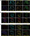
Figure 1. Knockout of Gnb5 from sensory neurons destabilizes its R7-RGS protein partners and impairs multiple modalities of nociception but not pruriception.
(AC) Sections through lumbar dorsal root ganglia (DRG) from mice harboring a conditional Gnb5-knockout allele (Gnb5fl/fl) alone (upper panels) or Advillin (Adv)-Cre+/– Gnb5fl/fl mice (lower panels). (A) DRG analyzed by dual immunohistochemistry (IHC) with antibodies against NF200 (Nefh) (green) or Gβ5 (red) as indicated. (B and C) DRG analyzed by combined DAPI staining (blue) and IHC with antibodies against Rgs7 (B) or Rgs9 (C) (green). The percentage of residual antibody signal following Adv-Cre–mediated knockout of Gnb5 is indicated below each image (mean ± SEM). (D and E) Cumulative scratching behavior in mice of the indicated genotypes induced by the intradermal administration of (D) compound 48/80 or (E) chloroquine (64, 65). (F and G) Cumulative scratching behavior in mice of the indicated genotypes induced by the intrathecal injection of gastrin-releasing peptide (GRP) (F) or Nppb (G). (HK) Behavioral testing of nociception in littermates with either the Gnb5fl/fl or Adv-Cre+/– Gnb5fl/fl genotypes, as indicated. (H) Hot plate testing (53°C). (I) Eye wipe testing using capsaicin. (J) Eye wipe testing of chemical nociception using mustard oil. (K) Von Frey filament behavioral testing of mechanical nociception. N values provided in Methods. For DK, the genotype of the mouse being tested was masked to the evaluator. For D, E, and K, the Mann-Whitney test was employed, and for FJ, the 2-tailed unpaired Student’s t test was employed, with bars indicating mean ± SEM. P values: D, P = 0.38; E, P = 0.52; F, P = 0.92; G, P = 0.67; H, **P = 0.0095; I, **P = 0.0098; J, *P = 0.012; K, *P = 0.02. For AC, DRG derived from 5 mice of each genotype were examined, with representative sections shown.
Figure 2
Figure 2. Expression of Gnb5 and nociceptive markers in sensory neurons.
Sections through lumbar dorsal root ganglia (DRG) harvested from wild-type mice and analyzed by immunohistochemistry with antibodies against Trpv1 (A), Trpa1 (B), or Mrgprd (C) (red) and following counterstaining with DAPI (purple) or costaining with additional antibodies against G5 (Gnb5) (blue), NeuN (gold), or NF200 (Nefh) (green) as indicated and as described in Methods. DRG from 4 to 5 mice of each genotype were examined, with representative sections shown.
Figure 3
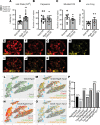
Figure 3. Knockout of Gnb5 from Rgs9+ neurons preserves chemical and mechanical nociception, as well as expression of Rgs9, Gnb5, and the nociceptors Trpv1 and Trpa1 in sensory neurons.
(AD) Behavioral testing of nociception in littermates with either the Gnb5fl/fl or else Rgs9-Cre+/– Gnb5fl/fl genotypes, as indicated. (A) Hot plate testing (53°C). (B) Eye wipe testing with capsaicin. (C) Eye wipe testing of chemical nociception with mustard oil. (D) Von Frey filament behavioral testing of mechanical nociception. For AD, the genotype of the mouse being tested was masked. (EK) Representative images from RNAscope analysis of transcripts for genes shown (EH): Gnb5, Trpa1, and Trpv1 are in red; Rgs9 is in yellow. The colocalization of Rgs9 transcript with that of Gnb5, Trpa1, and Trpv1 is shown (IK). (LO) Clustering analysis and UMAP visualization of 5,200 DRG expressing 1 or more of 4 gene transcripts above obtained from RNAscope experiments. The text colors in the lower right part of each figure correspond to individual cell coloring, indicating expression of that transcript, or combination of transcripts, in that cell. (P) Histogram illustrating the percentages of colocalized genes as shown. In AD, the 2-tailed unpaired Student’s t test of statistical significance was employed, with bars indicating mean ± SEM. For panels AD, number of mice from each genotype tested (for A, Gnb5fl/fl n = 11, Rgs9-Cre+/–; Gnb5fl/fl n = 13; for B, Gnb5fl/fl n = 11, Rgs9-Cre+/–; Gnb5fl/fl n = 13; for C, Gnb5fl/fl n = 7, Rgs9-Cre+/–; Gnb5fl/fl n = 7; for D, Gnb5fl/fl n = 6, Rgs9-Cre+/–; Gnb5fl/fl n = 5). P values: A, **P = 0.006; B, P = 0.88; C, P = 0.38; D, P = 0.20.
Figure 4
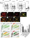
Figure 4. Knockout of Gnb5 from Rgs7+ neurons specifically impairs mechanical nociception and expression of Rgs7, Gnb5, Mrgprd, and Th in sensory neurons.
(AD) Behavioral testing of nociception in littermates with either the Gnb5fl/fl or Rgs7-Cre+/– Gnb5fl/fl genotypes, as indicated. (A) Hot plate testing (53°C). (B) Eye wipe testing with the Trpv1 agonist capsaicin. (C) Eye wipe testing of chemical nociception with mustard oil. (D) Von Frey filament behavioral testing of mechanical nociception. (EK) Representative images of RNAscope analysis of transcripts for genes shown (EH): Gnb5, Th, and Mrgprd are in red; Rgs7 is in yellow. The colocalization of Rgs7 mRNA with that of Gnb5, Th, and Mrgprd is shown (IK). (LO) Clustering analysis and UMAP visualization of 5,200 DRG expressing 1 or more of the 4 genes above obtained from RNAscope experiments. The text colors in the lower right part of each figure correspond to individual cell coloring, indicating expression of that transcript, or combination of transcripts, in that cell. (P) Histogram illustrating the percentages of colocalized genes as shown. For AD, the genotype of the mouse being tested was masked. For AC, the 2-tailed unpaired Student’s t test of statistical significance was employed, and for D the Mann-Whitney test was employed, with bars indicating mean ± SEM. For AD, n = number of mice from each genotype tested (for A, Gnb5fl/fl n = 8, Rgs7-Cre+/–; Gnb5fl/fl n = 6; for B, Gnb5fl/fl n = 8, Rgs7-Cre+/–; Gnb5fl/fl n = 8; for C, Gnb5fl/fl n = 8, Rgs7-Cre+/–; Gnb5fl/fl n = 8; for D, Gnb5fl/fl n = 8, Rgs7-Cre+/–; Gnb5fl/fl n = 8). P values: A, P = 0.69; B, P = 0.21; C, P = 0.59; D, ***P = 0.0008.
Figure 5
Figure 5. Gnb5-dependent and Rgs7-related regulation of mechanical nociception in sensory neurons is abolished by GABA-B receptor antagonism.
(AC) Behavioral testing of nociception in littermates with Gnb5fl/fl or Advillin-Cre+/– Gnb5fl/fl genotypes, as indicated, without and with the systemic administration of the GABA-B receptor antagonist 2-hydroxysaclofen (2HS). (A) Hot plate testing (53°C). (B) Eye wipe testing with capsaicin. (C) Von Frey filament testing of mechanical nociception. (D) Von Frey filament testing of mechanical nociception in Gnb5fl/fl or Rgs7-Cre+/– Gnb5fl/fl littermate mice, as indicated, without and with the systemic administration of 2HS. N values provided in Methods. For AD, the evaluator was masked to the genotype of the mouse being tested and to drug treatment. For A, C, and D, the Kruskal-Wallis (KW) test with Dunn’s multiple comparisons (Dunn’s) testing was employed, and for B, 1-way ANOVA with Tukey’s multiple comparisons testing was employed, with bars indicating mean ± SEM. P values: A, KW test **P = 0.004; A, Advillin-Cre+/– Gnb5fl/fl without versus with 2HS, Dunn’s test NS P > 0.99; B, 1-way ANOVA **P = 0.002; B, Advillin-Cre+/– Gnb5fl/fl without versus with 2HS, Tukey’s test NS P = 0.957; C, KW test **P = 0.0076; C control Dunn’s test *P = 0.02, C 2HS Dunn’s test NS P > 0.99, C Advillin-Cre+/– Gnb5fl/fl without versus with 2HS, Dunn’s test *P = 0.021; D, KW test ***P = 0.0004; D control Dunn’s test **P = 0.002, D 2HS Dunn’s test NS P > 0.99; D, Rgs7-Cre+/– Gnb5fl/fl without versus with 2HS, Dunn’s test **P = 0.007.
Figure 6
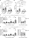
Figure 6. Analgesic effects of GABA-B agonist potentiated by loss of Gnb5 from sensory cells and from Rgs7+ cells in a model of subacute inflammatory pain.
(A and B) Mechanical nociception before and 24 hours following (24hf) the intraplantar injection of complete Freund’s adjuvant (CFA). (A) Control and Advillin (Adv)-Cre+/– Gnb5fl/fl littermates tested before and after CFA. (B) Control and Rgs7-Cre+/– Gnb5fl/fl littermates tested before and after CFA. (C) (Left) Control and Adv-Cre+/– Gnb5fl/fl littermates tested 24hf CFA and 30 minutes following (30mf) the systemic administration of baclofen. (Right) Comparison of control Gnb5fl/fl mice only. (D) (Left) Control and Rgs7-Cre+/– Gnb5fl/fl littermates tested 24hf CFA and 30mf baclofen. (Right) Comparison of control Gnb5fl/fl mice only. N values provided in Methods. For AD, evaluator was masked to both genotype and drug treatment. For A and B, Kruskal-Wallis (KW) testing with Dunn’s multiple comparisons testing (Dunn’s) employed, with bars indicating mean ± SEM. For C and D (left), Mann-Whitney test was employed. For C and D (right), KW testing was employed. P values: A, KW ****P < 0.0001, A Gnb5fl/fl mice pre- vs. post-CFA, Dunn’s **P = 0.0045, A Adv-Cre+/– Gnb5fl/fl mice pre- vs. post-CFA, Dunn’s **P = 0.0014, A Gnb5fl/fl vs. Adv-Cre+/– Gnb5fl/fl mice post-CFA Dunn’s NS; B, KW ****P < 0.0001, B Gnb5fl/fl mice pre- vs. post-CFA, Dunn’s NS, B Rgs7-Cre+/– Gnb5fl/fl mice pre- vs. post-CFA, Dunn’s ***P = 0.0002, B Gnb5fl/fl vs. Rgs7-Cre+/– Gnb5fl/fl post-CFA Dunn’s NS; C left, 0.5 mg/kg dose **P = 0.009; C left, 2 mg/kg dose ***P = 0.0003; C left, 5 mg/kg dose **P = 0.001; C right, *P = 0.01; D left, 0.5 mg/kg dose *P = 0.01; D left, 1 mg/kg dose **P = 0.003; D left, 2.5 mg/kg dose ****P < 0.0001; D right, NS.
Figure 7
Figure 7. Functional response of primary sensory neurons to activation by the Mrgprd agonist β-alanine before and after treatment with the GABA-B agonist baclofen.
Primary cultures of sensory neurons derived from DRG harvested from Rgs7-Cre+/– and Rgs7-Cre+/– Gnb5fl/fl mice and transduced in vitro with recombinant adeno-associated virus expressing tdTomato in a Cre-dependent fashion were loaded with the green-fluorescence calcium indicator Fluo-4. Rgs7+ cells were identified and studied for responsiveness to treatment with 10 mM of the Mrgprd receptor agonist β-alanine, before and after the addition of 1 μM of the GABA-B receptor agonist baclofen. Experiments were performed in pairs, using primary neurons representing each genotype. (A) Tracings of calcium-activated fluorescence from a representative pair of single neurons (1 each from Rgs7-Cre+/– and Rgs7-Cre+/– Gnb5fl/fl mice) with the time course and sequence of drug treatment shown. At 240 seconds, 50 mM KCl was added to all cells to induce maximum depolarization. Note that baclofen remained in the bath during the second β-alanine and KCl applications. (B) The average peak calcium-activated fluorescence response to the sequential treatments with 10 mM β-alanine, before and after the addition of 1 μM baclofen, derived from 4 experiments are shown. In B, the total number of neurons analyzed from Rgs7-Cre+/– mice was 10 (from n = 4 mice), and from Rgs7-Cre+/–; Gnb5fl/fl mice the total number of neurons analyzed was 18 (from n = 4 mice). In B, the Kruskal-Wallis (KW) test with Dunn’s multiple comparisons testing was employed for all comparisons, with bars indicating mean ± SEM of n = 10 neurons (Rgs7-Cre+/– mice) or n = 18 neurons (Rgs7-Cre+/– Gnb5fl/fl mice). P values: KW **P = 0.0014; without baclofen, first β-alanine treatment, Rgs7- Cre+/– control vs. Rgs7-Cre+/– Gnb5fl/fl, Dunn’s test NS P = 0.581; with baclofen, second β-alanine treatment, Rgs7- Cre+/– control vs. Rgs7-Cre+/– Gnb5fl/fl, Dunn’s test **P = 0.0097.

Similar articles

Cited by

References

    1. Davies M, et al. The prevalence, severity, and impact of painful diabetic peripheral neuropathy in type 2 diabetes. Diabetes Care. 2006;29(7):1518–1522. doi: 10.2337/dc05-2228. - DOI - PubMed
    1. Abbott CA, et al. Prevalence and characteristics of painful diabetic neuropathy in a large community-based diabetic population in the U.K. Diabetes Care. 2011;34(10):2220–2224. doi: 10.2337/dc11-1108. - DOI - PMC - PubMed
    1. Gerber KJ, et al. Roles for regulator of G protein signaling proteins in synaptic signaling and plasticity. Mol Pharmacol. 2016;89(2):273–286. doi: 10.1124/mol.115.102210. - DOI - PMC - PubMed
    1. Anderson GR, et al. The R7 RGS protein family: multi-subunit regulators of neuronal G protein signaling. Cell Biochem Biophys. 2009;54(1–3):33–46. doi: 10.1007/s12013-009-9052-9. - DOI - PMC - PubMed
    1. Slepak VZ. Structure, function, and localization of Gβ5-RGS complexes. Prog Mol Biol Transl Sci. 2009;86:157–203. doi: 10.1016/S1877-1173(09)86006-7. - DOI - PMC - PubMed

Publication types