Multiplexed Nanopore-Based Nucleic Acid Sensing and Bacterial Identification Using DNA Dumbbell Nanoswitches
- PMID: 37220424
- PMCID: PMC10251517
- DOI: 10.1021/jacs.3c01649
Multiplexed Nanopore-Based Nucleic Acid Sensing and Bacterial Identification Using DNA Dumbbell Nanoswitches
Abstract
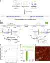
Multiplexed nucleic acid sensing methods with high specificity are vital for clinical diagnostics and infectious disease control, especially in the postpandemic era. Nanopore sensing techniques have developed in the past two decades, offering versatile tools for biosensing while enabling highly sensitive analyte measurements at the single-molecule level. Here, we establish a nanopore sensor based on DNA dumbbell nanoswitches for multiplexed nucleic acid detection and bacterial identification. The DNA nanotechnology-based sensor switches from an "open" into a "closed" state when a target strand hybridizes to two sequence-specific sensing overhangs. The loop in the DNA pulls two groups of dumbbells together. The change in topology results in an easily recognized peak in the current trace. Simultaneous detection of four different sequences was achieved by assembling four DNA dumbbell nanoswitches on one carrier. The high specificity of the dumbbell nanoswitch was verified by distinguishing single base variants in DNA and RNA targets using four barcoded carriers in multiplexed measurements. By combining multiple dumbbell nanoswitches with barcoded DNA carriers, we identified different bacterial species even with high sequence similarity by detecting strain specific 16S ribosomal RNA (rRNA) fragments.
Conflict of interest statement
The authors declare no competing financial interest.
Figures


Similar articles
-
Programmable DNA Nanoswitch Sensing with Solid-State Nanopores.ACS Sens. 2019 Sep 27;4(9):2458-2464. doi: 10.1021/acssensors.9b01053. Epub 2019 Aug 26. ACS Sens. 2019. PMID: 31449750
-
Split G-Quadruplexes Enhance Nanopore Signals for Simultaneous Identification of Multiple Nucleic Acids.Nano Lett. 2022 Jun 22;22(12):4993-4998. doi: 10.1021/acs.nanolett.2c01764. Epub 2022 Jun 7. Nano Lett. 2022. PMID: 35730196 Free PMC article.
-
Nanopore extended field-effect transistor for selective single-molecule biosensing.Nat Commun. 2017 Sep 19;8(1):586. doi: 10.1038/s41467-017-00549-w. Nat Commun. 2017. PMID: 28928405 Free PMC article.
-
DNA nanotechnology assisted nanopore-based analysis.Nucleic Acids Res. 2020 Apr 6;48(6):2791-2806. doi: 10.1093/nar/gkaa095. Nucleic Acids Res. 2020. PMID: 32083656 Free PMC article. Review.
-
The Utility of Nanopore Technology for Protein and Peptide Sensing.Proteomics. 2018 Sep;18(18):e1800026. doi: 10.1002/pmic.201800026. Epub 2018 Aug 5. Proteomics. 2018. PMID: 29952121 Free PMC article. Review.
Cited by
-
Brownian motion data augmentation: a method to push neural network performance on nanopore sensors.Bioinformatics. 2025 Jun 2;41(6):btaf323. doi: 10.1093/bioinformatics/btaf323. Bioinformatics. 2025. PMID: 40439147 Free PMC article.
-
DNA Carrier-Assisted Molecular Ping-Pong in an Asymmetric Nanopore.Nano Lett. 2023 Dec 13;23(23):11145-11151. doi: 10.1021/acs.nanolett.3c03605. Epub 2023 Nov 30. Nano Lett. 2023. PMID: 38033205 Free PMC article.
-
Numerical Modeling of Anisotropic Particle Diffusion through a Cylindrical Channel.Molecules. 2024 Aug 10;29(16):3795. doi: 10.3390/molecules29163795. Molecules. 2024. PMID: 39202873 Free PMC article.
-
Nanopore detection of single-nucleotide RNA mutations and modifications with programmable nanolatches.Nat Nanotechnol. 2025 Jun 27. doi: 10.1038/s41565-025-01965-6. Online ahead of print. Nat Nanotechnol. 2025. PMID: 40579472
References
Publication types
MeSH terms
Substances
Grants and funding
LinkOut - more resources
Full Text Sources
Other Literature Sources
Miscellaneous

