. 2022 Dec;34(12):1913-1927.
doi: 10.1002/elan.202100580. Epub 2021 Dec 31.

Separation and Detection of Tyrosine and Phenylalanine-derived Oxidative Stress Biomarkers Using Microchip Electrophoresis with Electrochemical Detection

Affiliations

Separation and Detection of Tyrosine and Phenylalanine-derived Oxidative Stress Biomarkers Using Microchip Electrophoresis with Electrochemical Detection

Dhanushka B Weerasekara et al. Electroanalysis. 2022 Dec.

Abstract

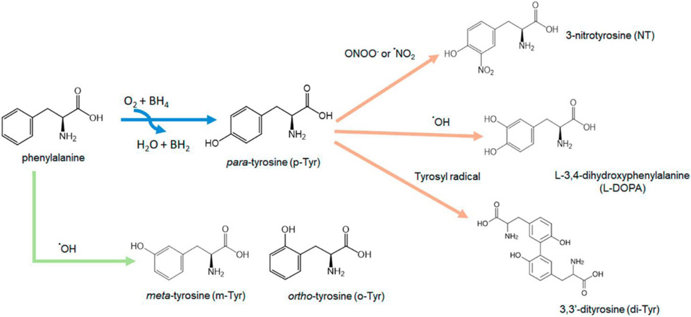
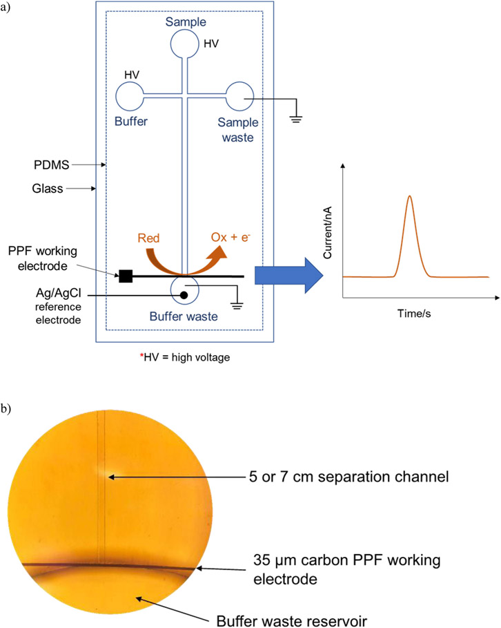
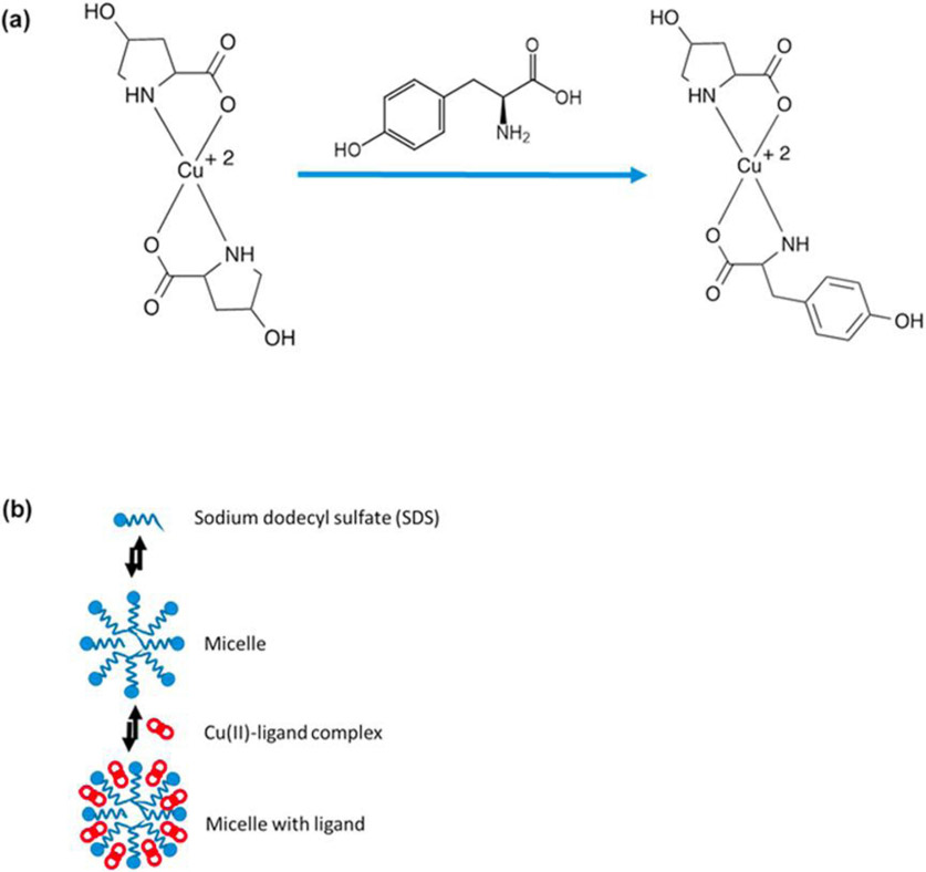
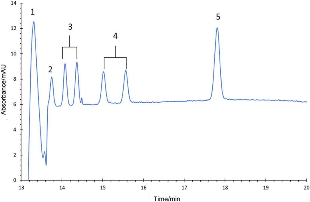
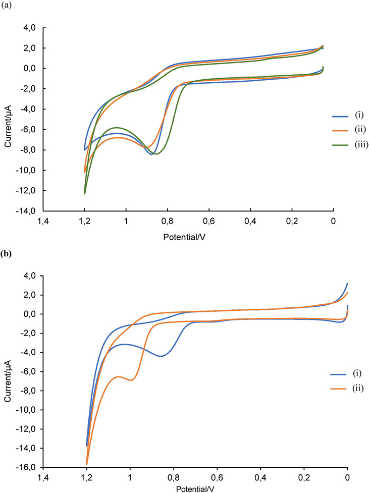
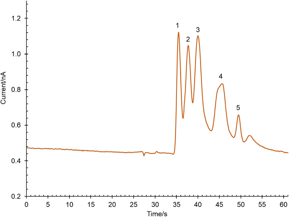
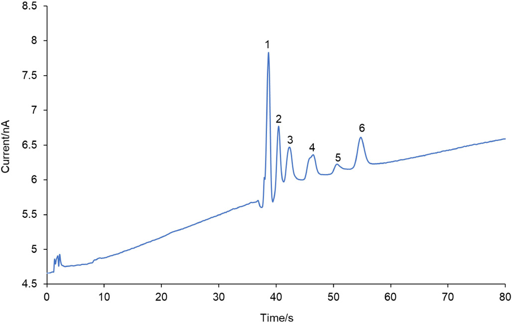
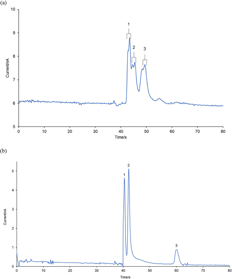
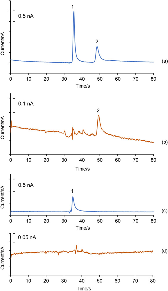
A method for the determination of selected aromatic amino acid biomarkers of oxidative stress using microchip electrophoresis with electrochemical detection is described. The separation of the major reaction products of phenylalanine and tyrosine with reactive nitrogen and oxygen species was accomplished using ligand exchange micellar electrokinetic chromatography with a PDMS/glass hybrid chip. Electrochemical detection was achieved using a pyrolyzed photoresist film working electrode. The system was evaluated for the analysis of the products of the Fenton reaction with tyrosine and phenylalanine, and the reaction of peroxynitrite with tyrosine.

Keywords: hydroxyl radical; microchip electrophoresis with electrochemical detection; peroxynitrite; reactive nitrogen and oxygen species; tyrosine.


Figures

Fig. 1.
Fig. 1.
Production of modified tyrosine and phenylalanine products due to the reactions of reactive nitrogen and oxygen species with phenylalanine and tyrosine.
Fig. 2.
Fig. 2.
a) Diagram of the ME-EC detection system consisting of a PDMS/glass hybrid chip with pyrolyzed photoresist film (PPF) working electrode. b) An image under an inverted microscope (10x magnification) of the 35 μM carbon PPF electrode aligned with the separation channel of the PDMS chip in a pseudo-end channel configuration.
Fig. 3.
Fig. 3.
Separation mechanism for ligand exchange micellar electrokinetic chromatography (LE-MEKC) (a) 4-hydroxyproline complexes with copper. Tyrosine and its derivatives replace the 4-hydroxyproline leading to a new complex and a change in mobility. (b) Illustration of the partitioning of Cu(II)-ligand complex with micelles, adapted from Chen et al. [39].
Fig. 4.
Fig. 4.
Electropherogram of CE-UV detection of L–DOPA, tyrosine isomers and 3-nitrotyrosine standards. The concentrations of each standard were 100 μM. The BGE consisted of 50 mM 4-hydroxyproline and 25 mM Cu(II) at pH 4.5 with 10 mM SDS. A capillary with an effective length of 47.5 cm and a separation voltage of 14 kV were used. UV detection was performed at 208 nm. Peak identities: (1) L–DOPA, (2) p-Tyr (3) DL-m-Tyr, (4) DL-o-Tyr and (5) NT.
Fig. 5.
Fig. 5.
a) Cyclic voltammograms of 200 μM p-Tyr dissolved in (i) SE1 = 0.1 M acetate buffer at pH 4.0 (ii) SE2 = 25 mM 4-hydroxyproline and 12.5 mM CuSO4 at pH 4.0 (iii) SE3 = 25 mM 4-hydroxyproline and 12.5 mM CuSO4 at pH 4.0 with 8 mM SDS. A glassy carbon working electrode, Pt auxiliary electrode, and Ag/AgCl reference electrode were used at a scan rate of 100 mV/s. b) Cyclic voltammograms of (i) 200 μM p-Tyr and (ii) 200 μM NT in 25 mM 4-hydroxyproline, 12.5 mM CuSO4 and 15 mm acetate buffer at pH 4.0 with 16.5 mM SDS. A glassy carbon working electrode, Pt auxiliary electrode, and Ag/AgCl reference electrode were used at a scan rate of 100 mV/s.
Fig. 6.
Fig. 6.
ME-EC electropherogram of a mixture of (1) L–DOPA, (2) DL-p-Tyr, (3) DL-m-Tyr, (4) DL-o-Tyr and (5) NT (final concentration = 25 μM for each standard). The BGE consisted of 25 mM 4-hydroxyproline and 12.5 mM Cu(II) at pH 4.5 with 10 mM SDS. A PDMS/glass chip with a 5 cm separation channel and a PPF working electrode were used. Separation voltages: + 1900 V at buffer reservoir and +1600 V at sample reservoir. Applied working electrode (WE) potential was +1.1 V vs. Ag/AgCl.
Fig. 7.
Fig. 7.
Electropherogram for the separation of a mixture containing of all the selected RNOS biomarkers (final concentration = 25 μM for each standard) by ME-EC. The separation buffer consisted of 25 mM 4-hydroxyproline, 12.5 mM Cu(II) and 15 mM ammonium acetate at pH 4.0 with 16.5 mM SDS. A PDMS/glass chip with a 7 cm separation channel was used. Separation voltages: + 2700 V at buffer reservoir & + 2400 V at sample reservoir. Applied WE potential was +1.1 V vs. Ag/AgCl. Peak identities: (1) L–DOPA, (2) L-p-Tyr, (3) DL-m-Tyr, (4) DL-o-Tyr, (5) NT and (6) di-Tyr.
Fig. 8.
Fig. 8.
Electropherogram for (a) the reaction mixture of the Fenton reaction with 5 mM DL-Phe (Peak identities: (1) DL-p-Tyr (2) DL-m-Tyr and (3) DL-o-Tyr) and (b) the reaction mixture of the Fenton reaction with 1 mM L-p-tyrosine (Peak identities: (1) L–DOPA (2) L-p-Tyr and (3) unknown). Both reaction mixtures were diluted 2-fold with 10 mM acetate buffer at pH 4.0 and injected into microchip with 7 cm separation channel containing 25 mM 4-hydroxyproline, 12.5 mM Cu(II) and 15 mM acetate buffer at pH 4.0 with 16.5 mM SDS. Separation voltages and applied WE potentials were similar to those used in Figure 7.
Fig. 9.
Fig. 9.
Electropherograms of (a) a standard mixture of 50 μM L-p-Tyr and 100 μM NT in BGE (25 mM 4-hydroxyproline, 12.5 mM Cu(II) and 15 mM acetate buffer at pH 4.0 with 16.5 mM SDS) at an applied working electrode potential of +1.1 V vs. Ag/AgCl (b) Sample from the reaction of peroxynitrite with L-p-Tyr (diluted 2-fold with BGE) at an applied working electrode potential of +1.1 V vs. Ag/AgCl, and same standard (c) and reaction mixture (d) at an applied working electrode potential of + 0.9 V vs. Ag/AgCl. Peak identities: (1) L-p-Tyr and (2) NT. Separation voltages were similar to those used in Figure 7.

Similar articles

References

    1. Ozcan A, Ogun M in Basic principles and clinical significance of oxidative stress,1st ed., Vol.3 (Ed.:Gowder SJT), IntechOPen, Croatia, 2015, 37–58.
    1. Ye Z-W, Zhang J, Townsend DM, Tew KD, Biochim. Biophys. Acta Gen. Subj 2015, 1850, 1607–1621. - PMC - PubMed
    1. Lichtenberg D, Pinchuk I, Biochem. Biophys. Res. Commun 2015, 461, 441–444. - PubMed
    1. Pisoschi AM, Pop A, Iordache F, Stanca L, Predoi G, Serban AI, Eur. J. Med. Chem 2020, 112891. - PubMed
    1. Senoner T, Dichtl W, Nutrients 2019, 11, 2090. - PMC - PubMed

LinkOut - more resources