

Sarcoidosis of the lacrimal gland: the prominence of the differential diagnosis
- PMID: 37221006
- PMCID: PMC10230888
- DOI: 10.1136/bcr-2022-253880
Sarcoidosis of the lacrimal gland: the prominence of the differential diagnosis
Abstract
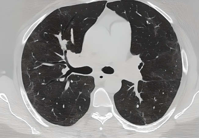
Sarcoidosis is a multisystem disease that can involve any organ; lungs, lymph nodes and skin are the most involved. Compatible clinical and imaging features, the identification of non-caseous granulomas on biopsy and the exclusion of other causes of granulomatous disorders help formulate the diagnosis of sarcoidosis. A bilateral symmetrical hilar lymphadenopathy together with the typical perilymphatic distribution of nodules is typically visible on high-resolution CT.The average age is 48 years. Ocular sarcoidosis is not rare, it is reported in 25% of cases. Half of the sarcoidosis patients resolve spontaneously; treatment is only indicated in cases with severe symptoms or signs of organ damage. Classical treatments are based on the use of corticosteroids and immunosuppressive therapies, sometimes combined.
Keywords: COVID-19; Interstitial lung disease; Oral and maxillofacial surgery.
© BMJ Publishing Group Limited 2023. No commercial re-use. See rights and permissions. Published by BMJ.
Conflict of interest statement
Competing interests: None declared.
Figures


Similar articles
-
Less Common Respiratory Conditions: Sarcoidosis.FP Essent. 2021 Mar;502:18-22. FP Essent. 2021. PMID: 33683850
-
Clinical presentation of sarcoidosis and diagnostic work-up.Semin Respir Crit Care Med. 2014 Jun;35(3):336-51. doi: 10.1055/s-0034-1381229. Epub 2014 Jul 9. Semin Respir Crit Care Med. 2014. PMID: 25007086 Review.
-
Recognition of distinctive patterns of gallium-67 distribution in sarcoidosis.J Nucl Med. 1990 Dec;31(12):1909-14. J Nucl Med. 1990. PMID: 2266386
-
Pulmonary Sarcoidosis: Diagnosis and Differential Diagnosis.Diagnostics (Basel). 2021 Aug 28;11(9):1558. doi: 10.3390/diagnostics11091558. Diagnostics (Basel). 2021. PMID: 34573900 Free PMC article. Review.
-
A case of lacrimal sarcoidosis following interstitial pneumonia: imaging and management.Nihon Rinsho Meneki Gakkai Kaishi. 2015;38(3):164-8. doi: 10.2177/jsci.38.164. Nihon Rinsho Meneki Gakkai Kaishi. 2015. PMID: 26213195
References
Publication types
MeSH terms
LinkOut - more resources
Full Text Sources
Medical